https://doi.org/10.14734/PN.2020.31.3.151 pISSN 2508-4887•eISSN 2508-4895
Sun-Young Lee, MD1, Chae-Ku Jo, MD1, Seo-Hee Rha, MD2, Myo-Jing Kim, MD1
Departments of 1Pediatrics and
2Pathology, Dong-A University College of Medicine, Busan, Korea
Food protein-induced enterocolitis (FPIES) is not well documented in newborns. Although FPIES is rare in premature infants, it may occasionally be confused with necrotizing enterocolitis (NEC). This is particularly true for low birth weight infants admitted to the neonatal intensive care unit. We report a case of cow’s milk protein-induced enterocolitis mimicking recurrent NEC in a premature infant. The patient presented bloody stools, abdominal distension, pneumatosis intestinalis, peripheral eosino- philia, and successful resolution of the symptoms upon modification of the diet to an amino acid- based formula. We aim to highlight that although the prevalence of FPIES is relatively rare in premature infants, clinicians should lead to consideration of FPIES in NEC-like illness.
Key Words: Food allergy, Milk proteins, Necrotizing enterocolitis, Premature infant
Introduction
Food protein-induced enterocolitis (FPIES), a non-immunoglobulin E (IgE)-mediated synd rome resulting in hypersensitivity to food antigens, is an inflammatory gastrointestinal process that affect 3% to 7% of healthy infants during the first year of life.1,2 The most common food triggers are cow’s milk and soy proteins.3 Although FPIES is rare in newborns, more than 20 cases who were diagnosed to FPIES during neonatal period have been reported.4 The incidence and clinical manifestations of FPIES in premature infants with comorbidities remains poorly identified.1 Reports in premature infants described cases of hematochezia and enterocolitis while receiving intact milk protein that resolved with its elimination and recurred with its reintroduction.5-9 Necrotizing enterocolitis (NEC) is the most common life- threatening medical and surgical emergency in premature infants.10,11 Therefore, NEC is the first concern when premature infants present with hematochezia or feeding intolerance. In this article we report a case of FPIES mimicking NEC in a 27-week-old premature infant whose symptoms resolved by the elimination of milk protein in the diet.
Case
A male preterm infant was born at 27 weeks and one day of gestation with a birth weight of 1,100 g. The medical issues during hospitalization included a respiratory distress syndrome, apnea of prematurity, hyperbilirubinemia, bronchopulmonary dysplasia, feeding intolerance, and total parenteral nutrition associated cholestasis. There was no intraventricular hemorrh- age, and patent ductus arteriosus was closed after medical treatment at 15 days of birth. Me- conium passage occurred within 24 hours of birth. Trophic feeding was started on the 1st day Received: 27 April 2020
Revised: 12 June 2020 Accepted: 15 June 2020 Correspondence to Myo-Jing Kim, MD
Department of Pediatrics, Dong-A University College of Medicine, 32 Daesingongwon-ro, Seo-gu, Busan 49201, Korea
Tel: +82-51-240-2589 Fax: +82-51-242-2765 E-mail: [email protected]
Copyright© 2020 by The Korean Society of Perinatology
This is an Open Access article distributed under the terms of the Creative Com- mons Attribution Non-Commercial License (http://creativecommons.org/
license/by-nc/4.0/), which permits unrestricted non-commercial use, distribution, and reproduction in any
Food Protein-induced Enterocolitis Mimic-
king Necrotizing Enterocolitis in a Premature
Infant: Case Report
age, PMA), DOL 24 (30 weeks and 4 days PMA), and DOL 31 (31 weeks and 4 days PMA), the patient developed large gastric residuals and had episodes of abdominal distension after feeding with the preterm formula and expressed breast milk (EBM). At each episode, the first clinical impression was NEC, therefore enteral feeding was stopped and broad spectrum antibiotics were treated repeatedly after multiple sepsis workups were conducted.
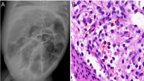
On DOL 45 (33 weeks and 4 days PMA), the patient was re- ferred to the neonatal intensive care unit (NICU) of our institu- tion for an emergent surgical operation on the marked abdominal distension (Fig. 1A). Intraoperatively, a 10 cm ileo-ileal intussu- sception was discovered. It required a small bowel resection and anastomosis, approximately 30 cm proximal to the ileocecal valve. Other parts of the intestine were grossly non-necrotic and viable. The biopsy report showed a transmural hemorrhagic infarction and increased eosinophilic infiltration at the mucosa (Fig. 1B).
On DOL 52 (34 weeks and 4 days PMA), enteral feeding was restarted with either a preterm formula or EBM at 30 mL/kg/
day. Unfortunately, abdominal distension and emesis developed.
After two days of bowel rest, continuous infusion of a preterm formula was restarted. However, the patient, once again, de- veloped emesis.
Transpyloric tube was inserted in NICU by neonatologist and
X-ray. Transpyloric feeding was initiated on DOL 60 (35 weeks and 5 days PMA) with an extensively hydrolyzed milk formula (HA®; Maeil Dairies Co. Ltd., Seoul, Korea) for feeding intolerance. Full enteral feeding was performed on DOL 70.
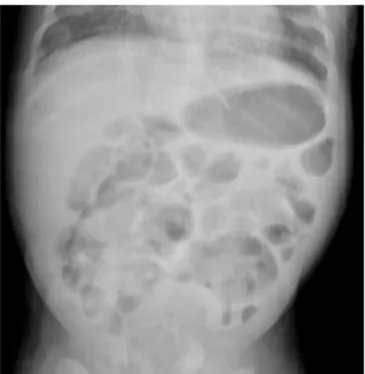
On DOL 75 (37 weeks and 6 days PMA), the patient developed abdominal distension again. This was now associated with large, grossly bloody stools. An abdominal X-ray and an abdominal ultrasonography were performed. These revealed pneumatosis intestinalis, distended bowel loops, wall thickening, and portal venous gas formation (Fig. 2). The laboratory findings included a white blood cell count of 4,520/mm3, hemoglobin of 8.5 g/dL, a platelet count of 298,000/mm3, albumin of 2.4 g/dL, C-reactive protein of 0.78 mg/dL (0.5 mg/dL, cut-off value), and a fecal cal- protectin of 846.7 mg/kg (50 mg/kg, cut-off value). The initial clinical impression was NEC. However, the C-reactive protein was consistently negative at all short-term evaluations. No leu- kopenia, thrombocytopenia, and metabolic acidosis developed during the follow-up evaluations. The baseline peripheral eosi- nophil count was 4%, and then showed a gradual increase with time to 11.3% at 3 days of illness. Clinically, the patient appeared well and had stable vital signs. Evaluations for bacterial and viral sepsis were performed repeatedly. These all came out as nega- tive. We noted that the patient’s clinical condition was not com- patible with NEC. The case described had clinical findings which were more consistent with the diagnosis of FPIES. The serum
Fig. 1. Abdominal radiography showing marked bowel dilatation on the 45th day of life (33 weeks and 4 days of postmenstrual age) (A). High power view of the small intestine shows increased eosinophilic infiltration (12 eosinophils/HPF, arrows) at the mucosa. Hematoxylin and eosin stain [H&E, x400] (B).
progressed despite feeding with an extensively hydrolyzed for- mula.
FPIES is triggered by a non-IgE-mediated food hypersensi- tivity.12 The exact underlying mechanisms of FPIES, however, total IgE <2.00 kU/L and the specific IgE tests (milk, α-lactal-
bumin, β-lactoglobulin, casein, soy bean) were all negative.
After three days of bowel rest, a change in the diet to an amino acid-based formula (Neocate®; Nutricia Co. Ltd, Auckland, New Zealand) caused the prompt resolution of symptoms and impro- vement of radiologic findings within 72-96 hours (Fig. 3). After seven days of feeding with the amino acid-based formula, the patient eventually reached full enteral feeding. The peripheral eosinophil count was decreased to 2% after amino acid-based formula feeding. Thereafter, the patient had no further episodes of hematochezia nor feeding intolerance and was subsequently discharged. At the 18th months visit, the growth and development of the patient were within normal limits. He no longer had any signs of food intolerance.
Discussion
This report presents a case of cow’s milk protein-induced enterocolitis in a preterm infant with laboratory and radiologic findings mimicking recurrent NEC-like illness. Clinical diagnosis of FPIES was made with a successful resolution of symptoms after the modification of the diet with an amino acid-based for- mula. This case was a severe form of FPIES because the disease
Fig. 2. Diffuse bowel dilatation with pneumatosis (black arrows) on the 75th day of life (37 weeks and 6 days of postmenstrual age) at abdominal radiography (A) and abdominal ultraso no gra phy (B). The portal vein gas (white arrows) is seen through the abdominal ultrasound (C).
Fig. 3. Improvements of the diffuse bowel dilatation and pneumatosis are observed after modification of the diet to an amino acid-based formula.
by intussusception in the literature.17,18 However, FPIES pre- senting as intussusception has not been reported in neonates so far. Intussusception in premature infants has been reported primarily in relation to NEC.19 In this case, the recurrent clinical symptoms, acute onset of intussusception, and increased eosi- nophilic infiltration at the mucosa initially caused the misdiag- nosis of NEC. However, the diagnosis of FPIES was eventually reached after further evaluation. These findings led us to hypo- thesize that the mucosal edema caused by the allergic inflam- matory reaction could have resulted to the formation of an intus - susception. Based on this premise, further researches about atypical cases of intussusception in preterm infants need to be conducted.
In conclusion, although the prevalence of FPIES is relatively rare in premature infants, clinicians should lead to consideration of FPIES in NEC-like illness with atypical clinical course, a late- onset, episodic recurrences, the presence of peripheral eosi- nophilia, and resolution with dietary modifications.
Conflict of Interests
No potential conflict of interest relevant to this article was reported.
References
1) Lenfestey MW, de la Cruz D, Neu J. Food protein-induced enterocolitis instead of necrotizing enterocolitis? A neonatal intensive care unit case series. J Pediatr 2018;200:270-3.
2) Yang M, Geng L, Xu Z, Chen P, Friesen CA, Gong S, et al. Severe food pro- tein-induced enterocolitis syndrome to cow's milk in infants. Nutrients 2015;8:1.
3) Nowak-Węgrzyn A, Katz Y, Mehr SS, Koletzko S. Non-IgE-mediated gas- trointestinal food allergy. J Allergy Clin Immunol 2015;135:1114-24.
4) Aktaş S, Ergenekon E, Ünal S, Türkyılmaz C, Hirfanoğlu IM, Atalay Y.
Different presentations of cow’s milk protein allergy during neonatal period. Turk J Pediatr 2017;59:322-8.
5) Cordova J, Sriram S, Patton T, Jericho H, Gokhale R, Weinstein D, et al.
Manifestations of cow's-milk protein intolerance in preterm infants. J Pediatr Gastroenterol Nutr 2016;62:140-4.
6) Yee WH, Soraisham AS, Shah VS, Aziz K, Yoon W, Lee SK, et al. Incidence and timing of presentation of necrotizing enterocolitis in preterm mune system and T cells become activated once exposed to
food antigens.12 Increased levels of tumor necrosis factor-α and decreased expression of transforming growth factor-β receptors have also been noted in the intestinal mucosa of patients with FPIES.13-15 The most common food triggers in FPIES are cow’s milk and soy proteins.3 Thus, reactions may occur with infant formulas. FPIES typically presents in first months of life. A patient may have vomiting, diarrhea, abdominal distension, bloody stools, or lethargy within 1-4 weeks after initial exposure to a trig gering antigen.1,16 The incidence of FPIES in neonates-es- pecially in preterm infants- is not well documented.5-9 Similar symptoms are sometimes associated with the initial presenta- tions of NEC in preterm infants. NEC may occur at any time;
however, it is mostly commonly observed near 32 weeks of gestation. On the other hand, symptoms associated with FPIES rarely develop before 6 weeks of life.10,11 Although an oral food challenge is the most conclusive diagnostic method, it has been associated with risk of systemic reactions.1,2,16 Thus, the diagnosis is suspected based on the clinical presentations as vomiting, diarrhea, bloody stool, abdominal distension, pneu- matosis intestinalis, and clinical resolution of symptoms to elimi- nation of dietary protein.3-5 These clinical suspicions match the findings of this case. Although laboratory findings alone cannot confirm the diagnosis of FPIES, peripheral eosinophilia is a com mon hematologic abnormality observed.2 A recent study reported that cases initially diagnosed as NEC may be more consis tent with FPIES if these cases have less leukopenia, more eosinophilia, less thrombocytopenia, and less CRP elevation.1,4,7 Gradual increase of peripheral eosinophilia (11.3%) was the only abnormal laboratory finding in this case. It developed together with the clinical symptoms and correspondingly decreased (2%) after modification of the diet to an amino acid-based formula.
The clinical course of FPIES is relatively benign. On the other hand, NEC has a poor prognosis. It is therefore important to discern FPIES from NEC because the treatment strategies of these two diseases are very different. With NEC, treatment mo- dalities have focused mostly on stopping enteral feeding, broad spectrum antibiotics.10,11 In contrast, the management of FPIES is dietary modification.16
With regards to the occurrence of intussusception before the patient was diagnosed with FPIES, there have been a few docu-
infants. Pediatrics 2012;129:e298-304.
7) Srinivasan P, Brandler M, D'Souza A, Millman P, Moreau H. Allergic ente- rocolitis presenting as recurrent necrotizing enterocolitis in preterm neonates. J Perinatol 2010;30:431-3.
8) Bosa L, Martelossi S, Tardini G, Midrio P, Lago P. Early onset food protein induced enterocolitis syndrome in two breastfed newborns masque- rading as surgical diseases: case reports and literature review. J Matern Fetal Neonatal Med 2019 Apr [cited 2020 Mar 31];1-5.
9) Daza W, Dadan S, Uribe MC. Two case reports of food protein induced enterocolitis. Rev Colomb Gastroenterol 2013;28:240-5.
10) Lin PW, Nasr TR, Stoll BJ. Necrotizing enterocolitis: recent scientific ad- vances in pathophysiology and prevention. Semin Perinatol 2008;32:70- 82.
11) Fell JM. Neonatal inflammatory intestinal diseases: necrotising entero- colitis and allergic colitis. Early Hum Dev 2005;81:117-22.
12) Leonard SA, Pecora V, Fiocchi AG, Nowak-Wegrzyn A. Food protein- induced enterocolitis syndrome: a review of the new guidelines. World Allergy Organ J 2018;11:4.
13) Merras-Salmio L, Kolho KL, Pelkonen AS, Kuitunen M, Mäkelä MJ, Savilahti E. Markers of gut mucosal inflammation and cow’s milk specific immu- noglobulins in non-IgE cow’s milk allergy. Clin Transl Allergy 2014;4:8.
14) Caubet JC, Nowak-Węgrzyn A. Current understanding of the immune mechanisms of food protein-induced enterocolitis syndrome. Expert Rev Clin Immunol 2011;7:317-27.
15) Goswami R, Blazquez AB, Kosoy R, Rahman A, Nowak-Węgrzyn A, Berin MC. Systemic innate immune activation in food protein–induced en- terocolitis syndrome. J Allergy Clin Immunol 2017;139:1885-96.
16) Leonard SA, Nowak-Węgrzyn A. Food protein–induced enterocolitis syndrome: an update on natural history and review of management.
Ann Allergy Asthma Immunol 2011;107:95-162.
17) Aydin E, Beşer OF, Ozek E, Sazak S, Duras E. Is there a causal relationship between intussusception and food allergy? Children (Basel) 2017;4:89.
18) Aydın E, Beşer ÖF. Food allergy: a rare cause of recurrent intussusception.
APSP J Case Rep 2017;8:2.
19) Avansino JR, Bjerke S, Hendrickson M, Stelzner M, Sawin R. Clinical fea- tures and treatment outcome of intussusception in premature neonates.
J Pediatr Surg 2003;38:1818-21.