A stereotactic core needle biopsy using a prone type device is a widely used technique for the diagnosis of nonpalpable breast lesions discovered by mammogra- phy (1-3). An add-on device attached to a regular mam- mography unit can be used for a stereotactic breast biop-
sy. Its advantages are that it is less expensive than a prone table and extra space for the equipment is unnec- essary (3).
There is a paucity of reports on the effectiveness of the use of stereotactic biopsies in Korea (4, 5). One report dealt with a 14G core needle biopsy of non-mass calci- fications using a prone-type dedicated stereotactic mam- mography unit (4), and another study examined a core needle biopsy of a mass or calcifications using an add-on type stereotactic device (5). Neither report dealt with a vacuum-assisted breast biopsy.
Stereotactic Vacuum-Assisted Biopsy of
Microcalcifications Using an Upright Add-On Type Stereotactic Mammography Unit
1Sung Hun Kim, M.D., Jae Hee Lee, M.D., Byoung Joo Song, M.D.2, Sang Seol Jung, M.D.2
1Departments of 1Radiology and 2General Surgery, Kangnam St. Mary’s Hospital, College of Medicine, The Catholic University of Korea Received May 30, 2007 ; Accepted July 19, 2007
Address reprint requests to : Jae Hee Lee, M.D., Department of Radiology, Kangnam St. Mary’s Hospital, The Catholic University of Korea, 505 Banpo-dong, Seocho-gu, Seoul 137-040, Korea
Tel. 82-2-590-2944 Fax. 82-2-599-6771 E-mail: [email protected]
Purpose: This study examined the yield of mammographically detected calcifications following a stereotactic vacuum-assisted biopsy (SVAB) using an upright add-on type stereotactic device.
Materials and Methods: Ninety-two women underwent a SVAB between April 2002 and December 2005, and 90 calcifications obtained by the SVAB were evaluated retro- spectively. The calcification retrieval rate was examined. The false negative and under- estimation rates were determined by comparing the biopsy results with the results from surgery and follow-up mammography.
Results: The calcification retrieval rate was 97.8% (90/92). The histopathology of the 90 lesions was benign for 66 (73.3%), borderline for 3 (3.3%) and malignant for 21 (23.3%). A total of 22 malignancies were confirmed surgically. SVAB had a false nega- tive rate of 4.5 % (1/22). The underestimation rate of a surgically excised atypical duc- tal hyperplasia (ADH) and of ductal carcinoma in situ (DCIS) was 50% (1/2) and 10%
(2/20), respectively.
Conclusion: The use of SVAB with an upright add-on type stereotactic device is an effi- cient biopsy method for mammographically detected microcalcifications, with low false negative and high calcification retrieval rates.
Index words :Biopsy, needle Breast neoplasms Stereotaxic techniques Calcification, physiological
We report our 3-year experience of performing biop- sies using a combination of a vacuum-assisted device (VAD) and an upright add-on type stereotactic mammo- graphic unit for mammographically detected microcalci- fications.
Materials and Methods
Between April 2002 and December 2005, 92 women (age 27-79 years, mean 50 years) underwent a stereo- tactic vacuum-assisted biopsy (SVAB) for microcalcifica- tions. None of the lesions was either palpable or associ- ated with masses on mammography. The categories ac- cording to the American College of Radiology Breast Imaging Report and Data System (ACR BI-RADS) on the pre-biopsy mammographic reports were Category 3 for 4 cases, Category 4 for 84 cases, and Category 5 for 4 cases.
One radiologist (J.H.L) examined 60 cases (65%), and two radiologists, who had expertise in breast imaging and intervention, examined the remaining cases.
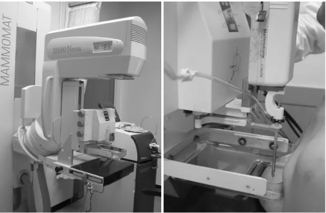
Stereotactic guidance was obtained using an Opdima Digital Stereotactic add-on attached to an upright mam- mography unit (Mammomat 3000, Siemens, Solna, Sweden) (Fig. 1A). An 11-gauge VAD Mammotome probe (Ethiocon Endo-Surgery, Cincinnati, OH U.S.A.) was used as the biopsy device. Before scheduling an SVAB, the mammographic images were reviewed in or- der to determine the feasibility of a SVAB with special consideration given to the breast size as well as to the lo- cation and conspicuity of the calcifications. Written in- formed consent was obtained from all patients. The pa- tients were advised of the indications, contraindications, benefits, and risks such as a hematoma and vasovagal reactions, as well as the possibility of insufficient sam- pling requiring re-biopsy or surgery. The patients were also recommended not to take aspirin or any other anti- coagulant drugs for three days before the procedure date. We did not routinely examine the blood profile for coagulation function.
Before the procedure, both the stereotactic mammo- graphic unit and the VAD were checked carefully.
Before compressing the breast for the biopsy, the patient was advised to keep calm and not move after targeting the calcifications. The patient sat in a special chair, which could be adjusted forward and backward and re- cline like a bed. The radiologist and radiographer that were present during the procedure attempted to make the patient feel as comfortable as possible. In order to
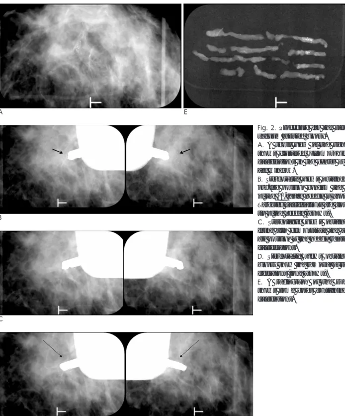
prevent the patient from being startled or frightened by the sound of the VAD during the procedure, the radiog- rapher allowed the patient to hear the VAD in its active state before the procedure. The craniocaudal (CC) posi- tion was preferred if the calcifications were located in the upper portion of the breast as that procedure was easier for the radiologist to perform, and there was less chance of the downward spilling of blood. If the calci- fications were located at a level that was lower than the nipple, either the lateromedial (LM) or the mediolateral (ML) position was chosen according to the location of the calcifications. A single spot mammogram was taken and the position was adjusted accordingly to move the calcifications to the center of the field (Fig. 2A). If the calcifications were in a favorable position, stereotactic double mammograms of the lesion were taken at ±10°
from the zero position. Calcifications were targeted on the stereotactic images on a digital monitor, and numeri- cal coordinates were calculated to localize the lesion in three dimensions. The skin in the window of the com- pressed paddle was disinfected with povidone and alco- hol. An 11G VAD needle was attached to the add-on type stereotactic device and the location of the needle was adjusted according to the numerical coordinates on the digital screen (Fig. 1B). Local anesthesia was provid- ed without administering any medicine for the sedative effects. For skin anesthesia, 2% lidocaine was used. For the deeper portions, a 5-10 cc mixture of 1% lidocaine was infused with 0.1 cc of a 1:100,000 epinephrine solu- tion. Before the skin incision, the stereotactic views were obtained in order to verify the location of the nee- dle. A 3-4 mm skin incision was then made with a scalpel. An 11G needle was inserted to the level of z=0.
Stereotactic views were again obtained (Fig. 2B) and the needle was fired if the location was favorable (Fig. 2C).
Ten to 12 samples were obtained. The direction of the needle insertion was controlled with special considera- tion being given to the needle and the calcifications.
After sampling, stereotactic views were obtained in or- der to ensure that the targeted calcifications had been re- moved (Fig. 2D). If the biopsy of the targeted calcifica- tions were unsatisfactory, more cores were further ob- tained in consideration of the locations of the needle and the calcifications. If the calcifications were removed completely, a microclip (MicroMark; Ethicon Endo- Surgery) was placed at 5 mm above the biopsy site. If the calcifications remained, we did not place a micro- clip. Subsequently, the compression paddle was re- leased and the biopsied breast was compressed for ap-
proximately 10-20 minutes for hemostasis. Specimen radiography was also performed (Fig. 2E) and speci- mens containing calcifications were placed in a separate container. In case the calcifications were not shown on specimen mammography, we did not retry performing the SVAB and recommended a surgical excisional biop- sy.
A surgical excision was performed if the biopsy result revealed a malignancy or a borderline lesion. Atypical ductal hyperplasia (ADH) and an intraductal papilloma with atypism were included in the borderline category.
Patients with benign pathology were advised to undergo a close mammographic follow-up.
The retrieval rate for a calcification observed at speci- men mammography was examined. The false negative and underestimation rates for the SVAB were also eval- uated. A benign or borderline pathology on a SVAB was regarded as a “false-negative” if it was found to be a ma- lignant lesion after surgery. An SVAB diagnosis of a bor- derline lesion with a surgical diagnosis of an in situ or invasive carcinoma, or an SVAB diagnosis of an in situ carcinoma with a surgical diagnosis of invasive carcino- ma was regarded as a “histological underestimation”.
We also evaluated the procedure-related complications
such as bleeding and vasovagal reactions.
Results
Calcifications were observed in 90 of the 92 cases (97.8%) by specimen mammography. The calcifications could not be retrieved in two cases as one calcification was too close to the chest wall (n = 1) and the other le- sion was too superficial for SVAB (n = 1). The latter le- sion was followed-up for 1 year at 6-month-intervals and did not show any interval changes. The former lesion underwent a surgical excision after needle localization due to an interval increase in the number of calcifica- tions on the follow-up mammograms that was found to be due to fibrocystic disease.
Of the 90 cases where retrieval of the calcifications was successful, the pathological diagnoses were malig- nant in 21 cases (23.3%), borderline in 3 cases (3.3%), and benign in 66 cases (73.3%). All pathological results were concordant to the imaging findings. Surgery was performed in 24 cases. Table 1 gives a comparison of the pathological diagnoses by SVAB and surgery.
Of the 21 cases of malignancy diagnosed by SVAB, 20 cases (95%) were due to a ductal carcinoma in situ
A B
Fig. 1. The instruments used for the stereotactic vacuum assisted biopsy
A. The Opdima Digital Stereotactic Add-on unit is attached to the film-screening mammography.
B. A vacuum assisted device with an 11-gauge needle is set on the Mammomat 3000, and the numeric coordinates are displayed.
(DCIS) and only 1 case (5%) was an invasive ductal car- cinoma. Of the 21 cases, 17 patients underwent subse- quent definitive surgical treatment at our institution.
Four patients underwent surgery in another hospital, and final pathologies were obtained by contacting the patients as well as the radiologists of the other hospitals.
Of the 20 DCIS cases, two (10%) were upgraded to an
invasive ductal carcinoma after surgery (Table 1): one le- sion was a 0.3 cm-sized invasive component with a 3.4 cm-sized intraductal component and the other lesion re- vealed a 0.3 cm-sized invasive component with a 1 cm diameter intraductal component. Both lesions contained mainly intraductal components. One invasive ductal carcinoma on biopsy showed the same result on a surgi-
A E
B
C
D
Fig. 2. Procedure for the stereotactic yacuum assisted biopsy.
A. A scout view of the right breast shows clustered pleomorphic micro- calcifications in the center of the im- age window.
B. Stereotactic views obtained in the pre-fire position confirm the location of the 11-gauge needle is appropriate.
Targeted calcifications are close to the tip of the needle (arrows).
C. Stereotactic views obtained after firing also demonstrate the appropri- ate position of the needle related to the calcifications.
D. Stereotactic views obtained after biopsy show the removal of some cal- cifications (long arrows).
E. A radiograph of the specimens shows some cores containing micro- calcifiations.
cal pathology (Table 1). The three borderline cases in- cluded two instances of an atypical ductal hyperplasia (ADH) and one instance of an intraductal papilloma with atypism. Of the three cases diagnosed as borderline by SVAB, one patient having an intraductal papilloma with atypism had a mammographic follow-up and showed no change over a 24-month period. Excisional biopsies were performed on two patients with an atypi- cal ductal hyperplasia that revealed fibrocystic changes with ductal hyperplasia in one patient and a ductal car- cinoma in situ in the other patient (Table 1).
A total of 22 malignancies were confirmed by surgery.
The false negative rate of SVAB for diagnosing a malig- nancy was 4.5 % (1/22). The underestimation rate of sur- gically excised borderline lesions and for a ductal carci- noma in situ was 50% (1/2) and 10% (2/20), respectively.
Of the 66 benign cases, 62 cases, 2 cases, 1 case and 1 case showed fibrocystic changes, ductal hyperplasia, stromal fibrosis, and an intraductal papilloma, respec- tively. Of the benign cases, an excisional biopsy was performed on one patient because the patient wanted to remove the calcifications. The diagnosis from that biop- sy was fibrocystic disease. The mammographic follow- up in 34 of the 66 benign patients (52.3%) for a period of 6-48 months (mean of 9.5 months) revealed no interval increase in the number or size of calcifications or the de- velopment of new areas of calcifications.
The procedure-related complications included bleed- ing and vasovagal reactions. Profuse bleeding during the procedure required compression for more than 30 min- utes in 8 cases (8.9%) with hemostasis being successful- ly achieved in all cases using manual compression.
Hematomas observed on the post biopsy mammogra- phy were detected in only three cases and none re- quired surgical intervention. Vasovagal reactions devel- oped in 6 cases (6.7%). The symptoms of a vasovagal re-
action included nausea, shortness of breath, numbness of the extremities, and fainting. These symptoms gener- ally occurred at the end of the procedure and we ended the procedure. Vasovagal reactions were relieved com- pletely after rest in the supine position while engaging in deep respiration.
Discussion
Calcifications have been shown to be a component in up to 50% of malignant lesions and in 84% of ductal car- cinomas in situ (DCIS) (6). Calcifications detected by mammography remain a diagnostic problem, as precise differentiation between benign and malignant lesions is difficult with clusters of microcalcifications (7).
Therefore, image-guided biopsy is essential for mammo- graphically detected suspicious microcalcifications. A stereotactic guided core needle biopsy is used preferen- tially in the United States (3). However, in Korea, dedi- cated prone-type stereotactic equipment is not as popu- lar due to its high cost. An stereotactic breast biopsy can be performed with a dedicated prone table with its main advantage being that it can hold the breast rigidly and the patient can be biopsied in the prone position, which is more comfortable than the sitting position (3).
However, its drawbacks include a very high cost and in- compatibility with routine mammography. An add-on device attached to a regular mammography unit can be used for a stereotactic breast biopsy. Its advantages are that it is less expensive than a prone table and extra space for the equipment is unnecessary. However, pa- tient movement and anxiety can be a problem.
Nevertheless, by using VAD, it is now possible to obtain a large volume of tissue with only a single needle inser- tion, which makes the pathological diagnosis easier and faster than the use of an automated gun biopsy. In addi- Table 1. Comparison of the Diagnoses by SVAB and Surgery
SVAB diagnosis Number Surgical Diagnosis
Benign ADH DCIS IDC
Benign 66 1
ADH 02 1 01
Papilloma with atypism 01
DCIS 20 18 2
IDC 01 1
Note - Surgery was not performed in one case of papilloma with atypism.
SVAB = Stereotactic vacuum assisted biopsy ADH = Atypical ductal hyperplasia DCIS = Ductal carcinoma in situ IDC = Invasive ductal carcinoma
tion, VAD requires less precision to localize the needle (8, 9), and has excellent sensitivity and specificity with a very low rate of false negative results (10). For correctly diagnosing the nature of microcalcifications, the sensi- tivity of SVAB was found to be better than that of a core needle biopsy (11).
These results were compared with other results of VAB for microcalcifications using upright add-on type and prone type stereotactic equipment. The combina- tion biopsy method (upright add-on type SVAB) for mi- crocalcifications was both simple to perform and demonstrated a very high success rate.
The calcification retrieval rate in this study was 97.8
%, which is higher than reported in other studies using the upright add-on type (96.3 and 86%) SVAB (12, 13).
However, the reported calcification retrieval rate of the prone type SVAB was approximately 99%, which is slightly higher than that reported here (14, 15).
The false negative rate of this study was 4.5 %, which is similar to 6.4% reported using an upright add-on type SVAB (13) and 2.8 % using prone SVAB (14).
In this study, the underestimation rates of surgically excised DCIS and borderline pathology were 10% and 50%, which are also similar to 20.8 % and 66.6 % report- ed in another study using upright add-on type SVAB (13). The reported underestimation rates of DCIS and ADH using the prone type SVAB vary considerably:
15.2 % and 8.3 % for DCIS, 52.1 % and 22.2 % for ADH (14, 15).
Compared with the prone type SVAB, the upright add- on type SVAB appears to have a slightly lower rate of calcification retrieval but similar false negative and un- derestimation rates.
In the patients with microcalcifications, the vast ma- jority (86.3%) of malignant lesions were DCIS. This find- ing indicates that the detection and biopsy of microcalci- fications are important for diagnosing breast malignan- cies in their early stage.
The complications related to the prone-type procedure are bleeding, an infection, clip migration and an abnor- mal mammographic density along the track (16). The complications related to the upright-type procedure are similar to those of the prone-type procedure. However, the vasovagal reaction is thought to be more frequent and important complication of the upright-type proce- dure (5,12,13). The vasovagal reaction is mainly due to the psychological stress experienced by patients that ei- ther were in pain or could see the wound and blood (12).
Vasovagal symptoms in other studies of upright-type
procedure were reported from 3.7% (5) to 15% of cases (12). In this study, six patients suffered from vasovagal reactions (6.5%).
There are some limitations in this study. We did not obtain specimen mammography before release of the compression paddle because the other mammography unit was located on another hospital floor. However, we could determine the retrieval of calcifications by obtain- ing post biopsy stereotactic views and confirm the result by obtaining specimen mammography after the proce- dure. The follow-up mammograms were available in only 52% of cases with a benign pathology and the fol- low-up duration was not long enough. Some studies have reported that additional cancers were discovered during the follow-up period in patients whose stereotac- tic biopsy results were benign (4, 14). In order to avoid this problem in the future, it will be necessary to inform the clinicians and patients of the importance of follow- up examinations after a biopsy, even if the biopsy proves to be benign.
In conclusion, SVAB with an upright add-on type stereotactic device is an efficient biopsy method for mammographically detected microcalcifications with low false negative and high calcification retrieval rates.
References
1. Caines JS, McPhee MD, Konok GP, Wright BA. Stereotaxic needle core biopsy of breast lesions using a regular mammographic table with an adaptable stereotaxic device. AJR Am J Roentgenol 1994;163:317-321
2. Elvecrog EL, Lechner MC, Nelson MT. Nonpalpable breast le- sions: correlation of stereotaxic large-core needle biopsy and surgi- cal biopsy results. Radiology 1993;188:453-455
3. Parker SH, Lovin JD, Jobe WE, Burke BJ, Hopper KD, Yakes WF.
Nonpalpable breast lesions: stereotactic automated large-core biopsies. Radiology 1991;180:403-407
4. Han BK, Choe YH, Ko YH, Nam SJ, Kim JH, Yang JH. Stereotactic core-needle biopsy of non-mass calcifications: outcome and accu- racy at long-term follow-up. Korean J Radiol 2003;4:217-223 5. Seo MR, Park JM, Gong GY, Ahn SH. Results with Add-on
Stereotactic Core Biopsy (ASCB) of the Breast Lesions. J Korean Radiol Soc 2000;43:245-250
6. Stomper PC, Connolly JL, Meyer JE, Harris JR. Clinically occult ductal carcinoma in situ detected with mammography: analysis of 100 cases with radiologic-pathologic correlation. Radiology 1989;172:235-241
7. Spencer NJ, Evans AJ, Galea M, Sibbering DM, Yeoman LJ, Pinder SE, et al. Pathological-radiological correlations in benign lesions ex- cised during a breast screening programme. Clin Radiol 1994;49:853-856
8. Parker SH, Klaus AJ. Performing a breast biopsy with a direction- al, vacuum-assisted biopsy instrument. Radiographics 1997;17:
1233-1252
9. Reynolds HE, Poon CM, Goulet RJ, Lazaridis CL. Biopsy of breast
microcalcifications using an 11-gauge directional vacuum-assisted device. AJR Am J Roentgenol 1998;171:611-613
10. Kettritz U, Rotter K, Schreer I, Murauer M, Schulz-Wendtland R, Peter D, et al. Stereotactic vacuum-assisted breast biopsy in 2874 patients: a multicenter study. Cancer 2004;100:245-251
11. Liberman L, Gougoutas CA, Zakowski MF, LaTrenta LR, Abramson AE, Morris EA, et al. Calcifications highly suggestive of malignancy: comparison of breast biopsy methods. AJR Am J Roentgenol 2001;177:165-172
12. Nisbet AP, Borthwick-Clarke A, Scott N. 11-gauge vacuum assist- ed directional biopsy of breast calcifications, using upright stereo- tactic guidance. Eur J Radiol 2000;36:144-146
13. Ohsumi S, Takashima S, Aogi K, Ishizaki M, Mandai K. Breast
biopsy for mammographically detected non-palpable lesions using a vacuum-assisted biopsy device (mammotome) and an upright- type stereotactic mammography unit. Jpn J Clin Oncol 2001;31:
527-531
14. Kettritz U, Morack G, Decker T. Stereotactic vacuum-assisted breast biopsies in 500 women with microcalcifications: radiologi- cal and pathological correlations. Eur J Radiol 2005;55:270-276 15. Cangiarella J, Waisman J, Symmans WF, Gross J, Cohen JM, Wu
H, et al. Mammotome core biopsy for mammary microcalcifica- tion: analysis of 160 biopsies from 142 women with surgical and radiologic followup. Cancer 2001;91:173-177
16. Parkinson BT. Stereotactic biopsy, Prone. In Berg WA, Birdwell RL.
Diagnostic Imaging: Breast Salt Lake City: Amirsys, 2006:28-31
대한영상의학회지 2007;57:291-297
석회화 유방 병소에 대한 부착식 입체정위기계를 이용한 흡입 보조 조직 생검
11가톨릭대학교 강남성모병원 영상의학과
2가톨릭대학교 강남성모병원 일반외과
김성헌・이재희・송병주2・정상설2
목적: 저자들은 유방촬영술에서 나타난 석회 병소에 대한 부착식 입체정위기계를 이용한 흡입 보조 조직 생검 (Stereotactic Vacuum-assisted biopsy, SVAB)의 유용성을 알아보고자 하였다.
대상과 방법: 2002년 4월부터 2005년 12월까지 입체정위기계를 이용해 석회화 병소 대한 SVAB를 받은 92명의 환자에서 석회화 검출률, SVAB 조직검사 결과와 조직검사 후 시행된 수술결과나 추적 유방촬영술을 검토하여 SVAB 의 유방암 진단에 대한 위음성률과 조직학적 저평가율을 후향적으로 분석하였다.
결과: 석회의 획득은 90명에서 성공하여 석화화 검출률은 97.8% 이었다. 석회화 획득에 성공한 90예의 조직검사 는 66예(73.3%)에서 양성, 3예(3.3%)에서 고위험병변, 21예(23.3%)에서 악성을 보였다. 수술로 확진된 악성 병 소 22예 중 21예가 SVAB로 진단되어 유방암진단의 위음성률은 4.5%이었다. 비정형상피증식에 대한 저평가율은 50%(1/2), 상피내암의 저평가율은 10%(2/20)이었다.
결론: 부착식 입체정위기계를 이용한 SVAB는 높은 석회화 검출율과 유방암 진단에 대한 낮은 위음성률을 보여 유 방촬영술에서 나타난 석회화 병소에 대해 유용한 조직 검사 방법으로 생각된다.