253
254
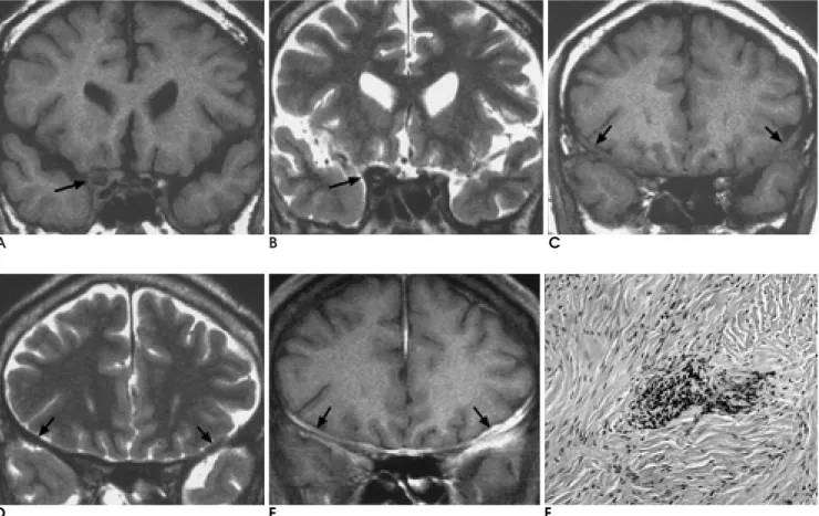
A B C
D E F
Fig. 1. A, B. Coronal T1-weighted image (A) shows iso-signal intensity mass (arrow) in right cavernous sinus, and the mass is hy- pointense (arrow) on coronal T2-weighted image (B).
C-E. The thickened dura matter in both frontal lobes (arrows) is isointense on coronal T1-weighted image (C), and hypointense (ar- rows) on coronal T2-weighted image (D). Coronal postcontrast T1-weighted image (E) shows diffuse well enhancement (arrows).
F. The meningeal lesion represents dense proliferation of thick collagen and patchy infiltration of lymphoplasma cells (H & E, 100).
1. Deprez M, Born J, Hauwaert C, Otto B, Reznik M. Idiopathic hy- pertrophic crainal pachymeningitis mimicking multiple meni- gioma: case report and review of the literature. Acta Neuropathol 1997;94:385-389
2. Goyal M, Malik A, Mishra NK, Gaikwad SB. Idiopathic hyper- trophic pachymeningitis: spectrum of the disease. Neuroradiology 1997;39:619-623
3. Kioumehr F, Rooholamini SA, Yaghmani I, Verma R. Idiopathic hypertrophic cranial pachymeningitis: a case report. Neuroradiol- ogy 1994;36:292-294
4. Uemura K, Matsumura A, Kobayashi E. Idiopathic chronic hyper- trophic craniocervical pachymeningitis: case report. Neurosurgery 1995;37:358
5. Martin N, Masson C, Henin D, Momptint D, Marsult C, Nahum H. Hypertrophic cranial pachymenigitis: assessment with CT and MR imaging. AJNR Am J Neuroradiol 1989;10:477-484
6. Wild T, Strotzer M, Volk M, Feuerbach S. Idiopathic hypertrophic cranial pachymenigitis associated with an orbital pseudotumor.
Eur Radiol 1999;9:1401-1403
7. Goyal M, Sharma A, Mishra NK, Gaikwad SB, Sharma MC.
Imaging appearance of pachymenigeal tuberculosis. AJR Am J Roentgenol 1997;169:1421-1424
255
Address reprint requests to : Won Chul Hong, M.D., Department of Diagnostic Radiology, Yeungnam University College of Medicine, 317-1, Daemyungdong, Namgu, Daegu 705-717, Korea.
Tel. 82-53-620-3030 Fax. 82-53-653-5484 E-mail: [email protected]
Idiopathic Hypertrophic Cranial Pachymeningitis: Case Report1
Won Chul Hong, M.D., Weon Kyu Park, M.D., Woo Mok Byun, M.D., Dong Sug Kim, M.D.
21Department of Diagnostic Radiology, Yeungnam University College of Medicine
2Department of Pathology, Yeungnam University College of Medicine
Idiopathic hypertrohpic cranial pachymeningitis is rare, and is essentially a diagnosis of exclusion. A 53- year-old man presented with headache and visual loss in the right eye, first experienced a month earlier. MR images depicted a mass in the right cavernous sinus. At T1-weighted imaging, both the mass and the thickened dura mater present in both fromted lobes were isointense, while at while T2-weighted imaging, the signal in- tensity of both the mass and the dura mater was low. After the injection of contrast medium, pachymeningeal enhancement was observed. We report the radiologic findings in a case of idiopathic hypertrophic cranial pachymeningitis, confirmed surgically and pathologically.
Index words :