A wide variety of pulmonary disorders may occur in hemodialysis patients, with variable radiologic manifes- tations, though the most common abnormality demon- strated at chest radiograph is pulmonary edema due to volume overload. In patients with chronic renal failure, many factors such as uremic toxin, parathyroid hor- mone, abnormal hemostasis and immunologic dysfunc- tion, may cause various disorders involving the lung, pleura, and pericardium, and hemodialysis also cause
thoracic complications, mainly related to vascular ac- cess. In patients with pulmonary-renal syndrome due to systemic vasculitis, pulmonary lesions give rise to chest radiographic abnormalities. The radiologic findings of pulmonary disorders in hemodialysis patients are some- times complex, and multiple concomitant such disor- ders are frequently disclosed, making differential diag- nosis difficult. An awareness of the various pulmonary disorders which can arise in hemodialysis patients may thus be helpful both for differential diagnosis and timely patient management.
Pulmonary abnormality associated with Chronic Renal Failure
Pulmonary Edema
In uremic patients undergoing dialysis, pulmonary edema is the most frequent complication (Fig. 1). Its common causes are volume-overload and left ventricu- lar failure secondary to systemic hypertension, uremic
Variable Pulmonary Manifestations in Hemodialysis Patients
1Yookyung Kim, M.D., Sung Shine Shim, M.D., Jung Hee Shin, M.D., Gyu Bock Choi, M.D.2, Kyung Soo Lee, M.D.3, Chin-A Yi, M.D.3, Yu-Whan Oh, M.D.4
A wide variety of pulmonary disorders related to hemodialysis or pre-existing renal disease occurs in hemodialysis patients. The disorders may be classified as 1) pul- monary abnormalities associated with chronic renal failures; 2) pulmonary complica- tions arising during hemodialysis; 3) pulmonary infection; or 4) pulmonary-renal syn- drome. An awareness of the various possible pulmonary disorders arising in hemodial- ysis patients may be helpful for the proper and timely management of such patients.
We describe and illustrate various radiographic and CT findings of variable pulmonary disorders in hemodialysis patients.
Index words : Hemodialysis Lung, edema Lung, infection Vasculitis
1Department of Radiology, College of Medicine, Ewha Womans University
2Department of Internal Medicine, College of Medicine, Ewha Womans University
3Department of Radiology, Samsung Medical Center, Sungkyunkwan University School of Medicine
4Department of Diagnostic Radiology, College of Medicine, Korea University
Received April 14, 2003 ; Accepted June 11, 2003
Address reprint requests to : Yookyung Kim, M.D., Department of Radiology, Ewha Womans University Mokdong Hospital
911-1 Mokdong YangCheon-gu, Seoul 158-710, South Korea.
Tel. 82-2-2650-5174 Fax. 82-2-2650-5071 E-mail: [email protected]
cardiomyopathy, and coronary disease. In most cases differentiation between renal and cardiogenic pul- monary edema at chest radiography is difficult. The typ- ical pattern of renal edema includes batwing central dis- tribution (1) (Fig. 2), but not all cases demonstrate this, and cardiogenic edema may also present with this pic- ture.
Cardiovascular disease is the most frequent cause of death in patients with end-stage renal disease.
Atherosclerosis of the coronary artery is accelerated by hypertension and lipid abnormalities and metastatic vascular calcification also promote coronary artery dis- ease. Acute exacerbation of pulmonary edema in pa- tients who have been undergoing hemodialysis may lead to the development of ischemic heart disease (Fig.
3).
Uremic Pericarditis
If present, uremic pericarditis usually appears late in the course of renal failure, though with the advent of dialysis therapy its occurrence has declined dramatical- ly, and it is now most common in patients who have un- dergone inadequate hemodialysis. It is usually dry, fibri- nous, and is associated with fever and chest pain, and in a minority of cases, varying amounts of exudative or he- morrhagic effusion are present. In the majority of cases, uremic pericarditis is cured by dialysis, but at times it evolves into a chronic constrictive form. A triangular shaped “water bottle”heart may be present at chest ra- diography, and the CT findings include pericardial effu- sion (Fig. 4) and, in chronic cases pericardial thickening, fibrous adhesions, or pericardial calcification (2).
Uremic Pleuritis
Pleural effusion is a common problem in patients with uremia; its causes include volume overload and cardiac failure, uremic pleuritis, severe hypoproteinemia in nephrotic syndrome, and pulmonary infection. The di- agnosis of uremic pleuritis is one of exclusion; clinical signs and symptoms of dry and fibrinous pleuritis, with- out appreciable effusion, are more common. If pleural effusion is present, it takes the form of an exudates which is at times hemorrhagic, often unilateral, and which disappears with dialysis (Fig. 5) or evolves into a
A B
Fig. 1. A 76-year-old woman with end stage renal disease and pulmonary interstitial edema.
A. Chest radiograph shows cardiomegaly, peribronchovascular cuffing, and Kerley’s B lines.
B. High-resolution CT scan coronal reconstruction image shows peribronchial interstitial and interlobular septal thickenings and patch areas of ground-glass attenuation.
Fig. 2. A 44-year-old man with end stage renal disease and vol- ume-overload pulmonary edema (renal edema). Chest radi- ograph shows pulmonary consolidation with “batwing” central distribution.
A B
Fig. 3. A 70-year-old woman with end stage renal disease and cardiogenic pulmonary edema induced by acute myocardial infarction.
A. Chest radiograph obtained 1 month ago shows cardiomegaly.
B. Chest radiograph obtained at emergency room shows peribronchovascular cuffings and Kerley’s B lines in both lungs with slight interval aggravation of cardiomegaly. He was diagnosed as having acute myocardial infarction.
A
C
B
Fig. 4. A 62-year-old man with uremic serositis (uremic peri- carditis and peritonitis) induced by inadequate hemodialysis.
A. Chest radiograph shows enlarged heart with “leather bottle appearance”.
B, C. CT scan at level of inferior pulmonary vein (B) and at level of renal vein (C) show pericardial effusion and ascites. Also not- ed are shrinked both kidneys.
fibrothorax. An ultrasonogram may depict septa, fibri- nous bands, and debris (2).
Pulmonary Hemorrhage
Abnormal hemostasis is common in chronic renal dis- ease and its most common hemorrhagic manifestations are prolonged bleeding from puncture sites, epistaxis,
A B
Fig. 5. A 75-year-old man with uremic pleuritis induced by inadequate hemodialysis.
A. Initial chest radiograph shows bilateral pleural effusions and mild cardiomegaly.
B. Follow-up chest radiograph 1 week after adequate hemodialysis shows improvement of pleural effusion.
A
C B
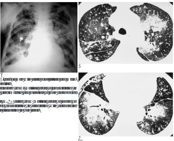
Fig. 6. A 41-year-old man with end stage renal disease and pul- monary hemorrhage.
A. Chest radiograph shows cardiomegaly and diffuse bilateral pulmonary consolidation.
B, C. High-resolution CT scans show mixed areas of consolida- tion and ground-glass attenuation in both lungs. Interlobular septal thickenings, fissural thickenings, and bilateral pleural ef- fusions are also noted, which are consistent with pulmonary in- terstitial edema.
and gastrointestinal, retroperitoneal, and intracranial bleeding. Pulmonary hemorrhage can also occur. A chest radiograph demonstrates air space consolidation, which may be widespread or show a perihilar or mid- dle- to- lower zone predominance, and tends to be more pronounced centrally. Costophrenic angles and apices are usually spared. The CT findings consist of bilateral areas of ground-glass attenuation or consolidation (3) (Fig. 6).
Metastatic Pulmonary Calcification
Pulmonary calcification associated with chronic renal failure is rare, and regarded as a sign of secondary hy-
perparathyroidism. In most cases, calcification is asymptomatic, but in some patients there may be dysp- nea and even severe respiratory distress. Bone scintigra- phy is the most sensitive study, and in the correct clini- cal setting, pulmonary uptake of the radionuclide, even in patients whose chest radiographic findings are nega- tive is sufficient for diagnosis. Chest radiography may reveal diffuse alveolar opacity, reticulation, or calcified nodules (4), while the HRCT findings include ground- glass attenuation nodules in centrilobular regions, with or without macroscopic calcification, a reflection of metastatic calcification in the alveolar septa and walls of the bronchioles and arterioles (5) (Fig. 7).
A B C
Fig. 7. A 45-year-old man with end stage renal disease and metastatic pulmonary calcification.
A. Chest radiograph shows ill-defined haziness and small nodules in both lungs.
B. High-resolution CT scan of lung window setting shows multiple small nodules and areas of ground-glass attenuation.
C. High-resolution CT scan of mediastinal window setting shows calcification in small nodules.
A B
Fig. 8. A 60-year-old woman with high-output cardiac failure induced by arterivenous fistula.
A, B. Angiograph obtained with contrast injection into the left axillary artery shows arteriovenous fistula between radial artery (open arrow) and vein. Markedly dilated draining vein from fistula is noted (arrow).
Pulmonary Complications accruing during Hemodialysis
Thoracic complications in patients undergoing he- modialysis are commonly related to the malpositioning or malfunctioning of the vascular catheters used, or arise iatrogenically for other reasons. Other complica- tions related to these maneuvers are common to all cen- tral venous catheterizations, and include pneumothorax,
hemothorax, mediastinal hemorrhage, sepsis, and steno- sis of the subclavian vein. Other rare complications in- clude pulmonary, air, and septic embolism, high-output cardiac failure due to the arteriovenous fistula (Fig. 8), and pulmonary hemorrhage or hemothorax due to sys- temic heparinization and platelet dysfunction (6).
Pulmonary Infection
Infection is the second-leading cause of death among
A B
Fig. 9. A 31-year-old woman undergoing hemodialysis with candidiasis.
A, B. High-resolution CT scans show multiple small nodules with surrounding ground-glass attenuation halo scattered through both lungs, predominantly in peripheral lung.
A B
Fig. 10. A 59-year-old man with end stage renal disease and Legionella pneumonia.
A. Chest radiograph shows round areas of consolidation in right upper lobe.
B. Chest CT scan shows ill-defined round consolidation in right upper lobe.
patients with chronic renal failure, following cardiovas- cular disease, and sepsis (Fig. 9) and pulmonary infec- tions account for 75% and 20% of such deaths, respec- tively. Exposure to infectious agents can occur in a he- modialytic setting, butin a uremic patient, fundamental abnormalities of the phagocytic system, including defec- tive cellular immunity, neutrophil function, and com- plement activation, also exist. Infections are usually due to common bacterial pathogens, and the radiologic man- ifestations are indistinguishable from the usual patterns of pulmonary infection (7) (Fig. 10).
Pulmonary-renal Syndrome
The term ‘pulmonary-renal syndrome’is used to de- scribe disorders associated with pulmonary hemorrhage and glomerulonephritis, and includes Wegener’s granu-
lomatosis (or related systemic vasculitides such as mi- croscopic polyangiitis and Churg-Strauss syndrome), Goodpasture disease, Henoch-Schonlein purpura, and systemic lupus erythematosus (8). Diagnostic markers can be used and include 1) anti-glomerular basement membrane antibodies, which are essentially diagnostic of Goodpasture disease; 2) anti-neutrophil cytoplasmic antibodies (ANCA), which are suggestive of Wegener’s granulomatosis, Churg-Strauss syndrome (C-ANCA) or microscopic polyangiitis (P-ANCA); 3) anti-nuclear anti- bodies in patients in whom lupus is suspected.
The radiologic manifestations of pulmonary disease in systemic vasculitis are varied. In the angiitis-granulo- matosis group, including Wegener’s granulomatosis and Churg-Strauss syndrome, multiple nodules or masses that may demonstrate cavitation are usually seen, while diffuse multifocal air space opacities with or without
A B
C D
Fig. 11. A 68-year-old woman with progressive renal failure and Wegener’s granulomatosis.
A. Chest radiograph shows ill-defined opacity and poorly-defined nodules in both lungs.
B. Water’s view shows diffuse haziness in both maxillary sinuses.
C, D. High-resolution CT scans show multiple nodules and large areas of ground-glass attenuation in both lungs.
cavitation are less frequent (9) (Fig. 11). The diffuse pul- monary hemorrhage with capillaritis group, including microscopic polyangiitis and Goodpasture disease, shows focal or diffuse air-space consolidation (10) (Fig.
12).
References
1. Milne E, Pistolesi M, Miniati M, Giuntini C. The radiologic distinc- tion of cardiogenic and noncardiogenic edema. AJR Am J Roentgenol 1985;144:879-894
2. Schwartz E. Thoracic manifestations of chronic renal disease.
Contemporary Diagn Radiol 1989;12:1-6
3. Cheah FK, Sheppard MN, Hansell DM. Computed tomography of diffuse pulmonary haemorrhage, with pathological correlation.
Clin Radiol 1993;48:89-93
4. Firooznia H, Pudlowski R, Golinbu C, Rafii M, McCauley D.
Diffuse interstitial calcification of the lungs in chronic renal fail- ure, mimicking pulmonary edema. AJR Am J Roentgenol 1997;129:
1103-1105
5. Hartman T, Muller N, Primack SL, et al. Metastatic pulmonary calcification in patients with hypercalcemia: findings on chest radi- ographs and CT scans. AJR Am J Roentgenol 1994;162:799-802 6. Gavelli G, Zompatori M. Thoracic complications in uremic pa-
tients and in patients undergoing dialytic treatment: state of the art. Eur J Radiol 1997;7:708-717
7. Armstrong P, Dee P. Infections of the lung and pleura. In: Armstrong P, Wilson AG, Dee P, Hansell DM, ed. Imaging of diseases of the chest, 2nd ed. St. Louis: Mosby, 1995:159-160
8. Von Vigier RO, Trummler SA, Laux-End R, Sauvain MJ, Truttmann AC, Bianchetti MG. Pulmonary renal syndrome in childhood: a report of twenty-one cases and a review of the litera- ture. Pediatr Pulmonol 2000;29:382-388
9. Seo JB, Im JG, Chung JW. Pulmonary vasculitis: imaging features.
J Korean Radiol Soc 1999; 40:263-273
10. Hansell DM. Small-vessel diseases of the lung: CT-pathologic cor- relates. Radiology 2002;225:639-653
A
C B
Fig. 12. A 63-year-old man with acute renal failure and micro- scopic polyangiitis.
A. Chest radiograph shows multiple areas of consolidation in both lungs, cardiomegaly, and bluntings of both costophrenic angles.
B, C. Chest HRCT scans show multiple areas of round or wedge-shaped consolidation in association with surrounding ground-glass attenuation in both lungs.
대한방사선의학회지 2003;49:89-97
혈액투석환자에서 관찰되는 다양한 폐병변의 영상소견1
1이화여자대학교 의과대학 방사선과학교실
2이화여자대학교 의과대학 내과학교실
3성균관대학교 의과대학 서울삼성병원 방사선과학교실
4고려대학교 의과대학 진단방사선과학교실
김유경・심성신・신정희・최규복2・이경수3・이진아3・오유환4
혈액투석 환자에서 혈액투석이나 기존의 신장질환과 관계된 다양한 폐병변이 나타날 수 있다. 이러한 폐병변은 크게 1) 만성신부전과 관련된 병변; 2) 혈액투석의 합병증; 3) 폐감염; 4) 폐-신장 증후군 등으로 나누어 볼 수 있다. 저자 들은 혈액투석 환자에서 발생할 수 있는 다양한 폐병변에 대한 인식은 빠른 진단과 치료에 도움이 되리라 생각하고, 이 러한 다양한 폐병변의 방사선학적 소견을 보여주고자 한다.