ISSN 2234-3806 • eISSN 2234-3814
379
http://dx.doi.org/10.3343/alm.2013.33.5.379 www.annlabmed.org
Ann Lab Med 2013;33:379-382
http://dx.doi.org/10.3343/alm.2013.33.5.379
Letter to the Editor
Clinical Microbiology
16S Ribosomal RNA Identification of Prevotella nigrescens from a Case of Cellulitis
John Jeongseok Yang, M.D.1,2, Tae Yoon Kwon, M.D.3, Mi Jeong Seo, M.T.2, You Sun Nam, M.S.4, Chung Soo Han, M.D.3, and Hee-Joo Lee, M.D.2
Department of Medicine1, Graduate School, Kyung Hee University; Departments of Laboratory Medicine2 and Orthopedic Surgery3, School of Medicine, Kyung Hee University, Kyung Hee University Hospital, Seoul; Department of Biomedical Science4, Graduate School, Kyung Hee University, Seoul, Korea
Gram-negative anaerobes of the Prevotella intermedia/nigrescens group are usually isolated from polymicrobial infections of peri- odontal origin, although recovery from healthy individuals is also possible [1, 2]. Only a limited number of isolates have been identified from extraoral infections, which causes difficulty in the assessment of the frequency of extraoral infections caused by P.
intermedia and P. nigrescens. A previous study of 154 isolates of P. intermedia/P. nigrescens described a low frequency of extra- oral infections recovered from sites of foot ulcer, blood stream infection, and lung aspirates [3]. Here we report a case of extra- oral infection where P. nigrescens was identified at the strain level by 16S ribosomal RNA (rRNA) sequencing.
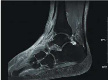
A 52-yr-old man with a history of diabetes and hypertension was referred to the department of orthopedics on December 18 complaining of cellulitis of the left ankle. The patient initially re- ceived acupuncture for his sprained ankle during a hiking trip in November 2012. As the symptoms did not improve, he visited a secondary hospital where he was diagnosed with cellulitis. Mag- netic resonance imaging (MRI) taken on December 1 showed cellulitis with talus osteochondral lesions and a low probability of abscess or osteomyelitis (Fig. 1). In spite of a week of antibiotic treatment with cefazedone followed by another week with cef- tazidime, the patient was referred to our hospital with symptoms of swelling, redness, and aggravated pain. Ultrasound sonogra-
phy (US) performed on December 18 revealed an abscess on the left ankle, as suggested by fluid accumulation and diffuse inflammation of soft tissue. Complete blood counts were as fol- lows: hemoglobin, 12.7 g/dL; white blood cells, 12.48×10⁹/L;
platelets, 514×109/L; and erythrocyte sedimentation rate (ESR), 75 mm/hr. The C-reactive protein (CRP) level was 4.68 mg/dL and the procalcitonin level was 0.068 µg/L (reference range:
~0.046 µg/L).
Initial treatment with aminoglycoside was ineffective; the symptoms were aggravated and the CRP level rose to 7.46 mg/
dL. US-guided aspiration and drainage were performed and the antibiotic regimen was changed to ampicillin/sulbactam, as the first set of culture results recovered very low levels of Strepto- coccus viridians. The symptoms of pain and swelling of the left ankle were persistent. MRI on January 8 revealed diffuse soft tissue edema, in addition to tenosynovitis with fluid collection and debris, which were signs of progressive infection. As the re- sponse to treatment was poor, surgical management was con- sidered. On Jan 14, incision and drainage (I&D) with debride- ment of severely infected tissue was required, and a closed pus specimen was obtained during the procedure for follow-up cul- ture.
Black pigment-producing colonies with gram-negative rod- shaped anaerobes were recovered (Fig. 2), in which positive
Received: April 12, 2013 Revision received: May 20, 2013 Accepted: July 3, 2013
Corresponding author: Hee-Joo Lee
Department of Laboratory Medicine, School of Medicine, Kyung Hee University, Kyung Hee University Hospital, 23 Kyunghee-daero, Dongdaemun-gu, Seoul 130-872, Korea
Tel: +82-2-958-8674, Fax: +82-2-958-8609, E-mail: [email protected]
© The Korean Society for Laboratory Medicine.
This is an Open Access article distributed under the terms of the Creative Commons Attribution Non-Commercial License (http://creativecommons.org/licenses/by-nc/3.0) which permits unrestricted non-commercial use, distribution, and reproduction in any medium, provided the original work is properly cited.
Yang JJ, et al.
Prevotella nigrescens from a case of cellulitis
380 www.annlabmed.org http://dx.doi.org/10.3343/alm.2013.33.5.379 biochemical reactions were seen in α-galactosidase (αGAL),
α-glucosidase (αGLU), mannose fermentation (MNF), and raffi- nose fermentation (RAF). The colonies were initially identified as Prevotella spp. using API rapid ID32A (API-bioMérieux UK Ltd., Basingstoke, UK). The reference biochemical profile used for comparison with the API rapid ID32A result is described in Ta- ble 1. The results of antimicrobial susceptibility testing are de- scribed in Table 2.
The sequence obtained from the cultured isolates was a match to the P. nigrescens gene for 16S rRNA (Strain: JCM 6322, GenBank: AB547697.1). Capillary sequencing was performed by Macrogen Inc. (Seoul, Korea) using two primers (518F-forward:
5’-CCAGCAGCCGCGGTAATACG-3’ and 800R-reverse 5’-TAC-
CAGGGTATCTAATCC-3’). The sequencing service included qual- ity control of template DNA, library preparation and filtering of low quality reads post-sequencing.
It was important to note the polymicrobial tendency of Pre- votella infection, which in this case was present with S. viridans.
Table 1. Biochemical identification of Prevotella intermedia/ni- grescens*
P. intermedia P. nigrescens Present case Production of
Pigment + + +
Indole + + +
Lipase + + N/A
α-Fucosidase + + +
N-Acetyl-
β-glucosaminidase - - N/A
Hydrolysis of
Gelatin + + N/A
Esculin - - -
Fermentation of
Arabinose - - N/A
Cellobiose - - N/A
Lactose - - -
Salicin - - -
Sucrose + + +
Mannose v + +
Raffinose v + +
*Reference [12].
Abbreviations: P. intermedia, Prevotella intermedia; P. nigrescens, Prevotella nigrescens; v, variable; N/A, not available.
Fig. 1. Magnetic Resonance Imaging: T1-weighted image showing cellulitis associated with talus osteochondral lesions.
Fig. 2. Recovery of black-pigmented anaerobe under anaerobic condition from blood agar plate. Black-pigmented Bacteroides in- cluding Prevotella nigrescens can be suspected.
Table 2. Antimicrobial susceptibility testing result
Antibiotics MIC (μg/mL) Interpretation*
Piperacillin 8 S
Piperacillin-tazobactam ≤0.06 S
Cefoxitin 2 S
Cefotetan 4 S
Imipenem ≤0.03 S
Meropenem ≤0.03 S
Clindamycin ≤0.06 S
Moxifloxacin 0.5 S
Chloramphenicol 1 S
Metronidazole 0.5 S
Tigecycline 0.25
*Interpretations using breakpoints suggested by CLSI guidelines: antimicro- bial susceptibility testing standards M02 and M07, 2012.
Abbreviations; MIC, minimal inhibitory concentration; S, susceptible.
Yang JJ, et al.
Prevotella nigrescens from a case of cellulitis
381
http://dx.doi.org/10.3343/alm.2013.33.5.379 www.annlabmed.org
Specifically, in all four results in which Prevotella was recovered, there was co-infection with S. viridians.
The antibiotic regimen was changed to tazobactam and piper- acillin, as soft tissue inflammation was persistently observed on radiological evaluation with computed tomography (CT) and X- ray imaging. On February 6, a second I&D with curettage of the previously infected site was performed. The antibiotic regimen was reverted to ampicillin and sulbactam. Discharge gradually decreased and the patient was discharged on February 15.
Prevotella spp. is a clinically important pathogen, although it is rarely recovered from clinical specimens other than perioral locations [1, 2]. Although most previously reported extraoral in- fections involving the P. intermedia/nigrescens group are based on biochemical identification and morphological features of characteristic black pigment-producing gram-negative anaer- obes, differentiation of P. intermedia and P. nigrescens should be based on molecular methods such as 16S rRNA sequencing [4, 5]. Importantly, the biochemical profiles of P. intermedia and P. nigrescens are almost identical, which complicates the differ- entiation between the two organisms (Table 1). Therefore, the API rapid ID32A system is unable to properly identify P. ni- grescens, and indications of P. intermedia presence using this system should be further verified with additional tests [4, 6, 7].
As infections with Prevotella spp. are polymicrobial in the major- ity of cases (including the present case), careful identification of the causative organism(s) will be helpful in the treatment of in- fections [3].
In the present case, P. nigrescens was identified at the spe- cies level by 16S rRNA sequencing of an aspirated specimen and pus obtained by US-guided aspiration and two procedures of I&D, respectively. The clinical course of infection was inces- sant, which may be attributed to several factors. The first is the rather distal and deep location of infection site, which likely ren- dered conventional therapeutic doses of antibiotics ineffective.
Secondly, the underlying hypertension and diabetes of this pa- tient were adverse factors for treatment. Furthermore, patient compliance was also unfavorable, as he persistently ambulated and kept smoking throughout the admission period. Finally, polymicrobial infection, with a synergistic effect of P. nigrescens and S. viridans, may have been present. Prevotella spp. infec- tions are often polymicrobial, which is frequently more patho- genic than infections involving a single organism [3, 8]. The pre- cise cause of infection is difficult to identify, although both a his- tory of acupuncture and aspiration are possible causes. Indeed, there is one case report of Prevotella spp. infection in which a history of acupuncture was also present, and early detection
and aggressive drainage were important for treatment in that case [9].
Despite retrospective antimicrobial susceptibility testing, anti- microbial treatment using piperacillin and tazobactam seemed sufficient, as both agents were found to be effective, and had activity in this case with a low minimal inhibitory concentration.
A high percentage of Prevotella spp. are known to produce β- lactamase [10], and therefore, antimicrobial susceptibility test- ing may be helpful in such cases, although it is not currently recommended as a routine procedure.
Although P. nigrescens is a phenotypically similar pathogen to P. intermedia with an indistinguishable biochemical profile, a greater likelihood of suppurative infection has been suggested [11]. Distinction by PCR-based methods such as 16S rRNA gene sequencing for detecting genotypic differences should be helpful in the management of such anaerobic infections.
Authors’ Disclosures of Potential Conflicts of Interest
No potential conflicts of interest relevant to this article were re- ported.
Acknowledgements
We express our sincere gratitude to professor Kyungwon Lee, M.D., Ph.D. of Yonsei University for arrangements and conduct- ing antimicrobial susceptibility testing.
REFERENCES
1. Finegold SM, Strong CA, McTeague M, Marina M. The importance of black-pigmented gram-negative anaerobes in human infections. FEMS Immunol Med Microbiol 1993;6:77-82.
2. Jousimies-Somer H, Savolainen S, Mäkitie A, Ylikoski J. Bacteriologic findings in peritonsillar abscesses in young adults. Clin Infect Dis 1993; 16(S4):S292-8.
3. Mättö J, Asikainen S, Väisänen ML, Rautio M, Saarela M, Summanen P, et al. Role of Porphyromonas gingivalis, Prevotella intermedia, and Pre- votella nigrescens in extraoral and some odontogenic infections. Clin In- fect Dis 1997;25(S2):S194-8.
4. Frandsen EV, Poulsen K, Kilian M. Confirmation of the species Prevotella intermedia and Prevotella nigrescens. Int J Syst Bacteriol 1995;45:429- 35.
5. Gharbia SE, Haapasalo M, Shah HN, Kotiranta A, Lounatmaa K, Pearce MA, et al. Characterization of Prevotella intermedia and Prevotella ni- grescens isolates from periodontic and endodontic infections. J Peri- odontol 1994;65:56-61.
6. Mättö J, Saarela M, von Troil-Lindén B, Alaluusua S, Jousimies-Somer H, Asikainen S. Similarity of salivary and subgingival Prevotella intermedia and Prevotella nigrescens isolates by arbitrarily primed polymerase
Yang JJ, et al.
Prevotella nigrescens from a case of cellulitis
382 www.annlabmed.org http://dx.doi.org/10.3343/alm.2013.33.5.379 chain reaction. Oral Microbiol Immunol 1996;11:395-401.
7. Shah HN and Gharbia SE. Biochemical and chemical studies on strains designated Prevotella intermedia and proposal of a new pigmented spe- cies, Prevotella nigrescens sp. nov. Int J Syst Bacteriol 1992;42:542-6. 8. Brook I. Microbiology of polymicrobial abscesses and implications for
therapy. J Antimicrob Chemother 2002;50:805-10.
9. Kim SS and Ha GI. Mediastinitis caused by Prevotella intermedia/ni- grescens occurred after acupuncture: a case report. Korean J Thorac Cardiovasc Surg 2000;33:440-4.
10. Lee Y, Park Y, Kim MS, Yong D, Jeong SH, Lee K, et al. Antimicrobial
susceptibility patterns for recent clinical isolates of anaerobic bacteria in South Korea. Antimicrob Agents Chemother 2010;54:3993-7.
11. Stubbs S, Park SF, Bishop PA, Lewis MA. Direct detection of Prevotella intermedia and P. nigrescens in suppurative oral infection by amplifica- tion of 16S rRNA gene. J Med Microbiol 1999;48:1017-22.
12. Könönen E, Wade WG, and Citron DM. Bacteroides, Porphyromonas, Prevotella, Fusobacterium, and other anaerobic gram-negative rods. In:
Versalovic J, ed. Manual of clinical microbiology. 10th ed. Washington:
ASM Press, 2011:858-80.