Various shapes of congenital abnormalities of the menisci have been reported. Among them, the double-layered meniscus is extremely uncommon. Anomalies of the menisci tend to affect the lateral meniscus more commonly and are reportedly more prevalent in Asians than Americans or European populations. There have been a few reports of double-layered lateral meniscus. We report a case of double-layered medial meniscus which have never been reported yet.
CASE REPORT
A 67-year-old female with no history of trauma complained of left knee pain. She had felt pain and discomfort in the left knee for 5 years. On physical examination, she had posteromedial side joint line tenderness, a positive McMurray test on the medial side and normal gait. There were no signs of locking, clicking, ligamentous laxities. She had normal gait, full range of motion, and no giving way sensation.
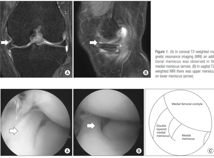
Neurovascular examination was normal. There was no bony anomaly. On coronal, saggital magnetic resonance images posterior portion of radial and horizontal tear of medial meniscus was ob- served. Double layered medial meniscus was observed anterior to the middle portion of medial meniscus (Fig. 1).
On arthroscopy, complex tear of posterior portion of medial me- niscus was observed. The medial meniscus had another meniscus overlying a normal one. The upper meniscus extended from anterior portion to mid portion. Its anterior edge was attached to the lower meniscus. Its periphery was connected to the joint capsule. The pos- terior portion of upper accessory meniscus was connected to joint capsule (Fig. 2). The accessory meniscus was not mobile on probing.
So we think that it is not an origin of symptom, impingement of meniscus.
Only the partial meniscectomy was done on complex tear of pos- terior portion of medial meniscus. Double-layered medial meniscus was not resected. Postoperatively, ambulation with partial weight bearing was permitted, and the preoperatively pain was alleviated.
Three months after surgery, the patient could walk freely and re- turned to normal life without any discomfort.
DISCUSSION
Various anomalies of meniscus have been reported. Among those discoid meniscus was the most common anatomic variant of the meniscus.1) Other less common anomalies include incomplete dis- coid meniscus, Wrisberg meniscus, ring-shaped meniscus, accessory meniscus, congenital separation meniscus, congenital partial defi- ciency and absence of the menisci.2)
Double-layered meniscus have been reported with a prevalence of 0.06% to 0.09%.3) Suzuki et al.1) reported two cases of double- layered lateral meniscus with one meniscus overlying the other.
pISSN : 1226-2102, eISSN : 2005-8918
96
Copyright © 2016 by The Korean Orthopaedic Association
“This is an Open Access article distributed under the terms of the Creative Commons Attribution Non-Commercial License (http://creativecommons.org/licenses/by-nc/4.0/) which permits unrestricted non-commercial use, distribution, and reproduction in any medium, provided the original work is properly cited.”
The Journal of the Korean Orthopaedic Association Volume 51 Number 1 2016 Received May 29, 2015 Revised September 25, 2015 Accepted September 30, 2015 Correspondence to: Sang-Yup Lee, M.D.
Department of Orthopedic Surgery and Traumatology, Cheju Halla General Hospital, 65 Doryeong-ro, Jeju 63127, Korea
TEL: +82-64-740-5555 FAX: +82-64-740-5412 E-mail: [email protected]
Case Report
J Korean Orthop Assoc 2016; 51: 96-99 • http://dx.doi.org/10.4055/jkoa.2016.51.1.96 www.jkoa.orgDouble-Layered Medial Meniscus
Seong-Tae Kim, M.D., Ph.D., Sang-Yup Lee, M.D. , and Min-Suk Park, M.D.
Department of Orthopedic Surgery and Traumatology, Cheju Halla General Hospital, Jeju, Korea
We report on the case of double-layered medial meniscus, which was overlying anterior to mid portion of the medial meniscus. The upper accessory meniscus was connected to the anterior portion of the normal medial meniscus. And its periphery was connected to the joint capsule. The posterior portion of the upper accessory meniscus was connected to the joint capsule. This case demonstrates an interesting and rare anatomical abnormality of the medial meniscus. We report on the case with a review of the literature.
Key words: meniscus abnormality, medial meniscus, double-layered meniscus
97
Double-Layered Medial Meniscus
Double-layered medial meniscus was an extremely rare anatomical abnormality more than lateral meniscus.
Double-layered lateral meniscus was thin, mobile in the previ- ous case report.1,4) But in this case, double-layered medial meniscus was cord like round shape. And it was not mobile. We think that it was not an origin of pain. These findings were different from the double-layered lateral meniscus which was reported previously.1-3,5) Komatsu et al.6) reported double-layered medial meniscus. The anterior portion of the medial meniscus had two layers, attached to the anterior surface of the tibia. In this case the anterior portion of the medial mesniscus had two layers but it was attached to lower normal medial meniscus.
Lee and Min7) reported abnormal band of the lateral meniscus. It was characteristic that upper abnormal band was loose and serpen- tine. In this case upper accessory meniscus was not loose, nor mobile.
Regarding the differentiation of double layered meniscus from
a horizontal tear, the cleavage of a horizontal tear is sharp margin, irregular in shape.3) In this case, the margin was smooth, regular in both upper and lower menisci. In addition, there were no problems with respect to the volume of the residual meniscus suggesting that the anomaly was not due to a horizontal tear (Fig. 2). Based on these findings, we concluded that this anomaly was different from acquired changes induced by degeneration or trauma.
Menisci differentiate directly from blastemal cells connected to the capsule.4,8) The causes of such variations are multifactorial, including congenital and developmental influences, but clinical presentations, pathology, and epidemiology of variations, altered biomechanics of medial compartment are still unclear.2)
After surgery, the patient had no discomfort or pain. We think that double-layered medial meniscus was not the cause of pain. It is unclear how double-layered medial meniscus influences knee joint biomechanics and also double-layered medial meniscus can be a
C
Medial femoral condyle
Medial meniscus Double
layered medial meniscus
A B
Figure 2. Arthroscopic findings. (A) An additional semicircular meniscus was observed over the normal medial meniscus (arrow). (B) The upper meniscus was cord like round shape (arrow). (C) The upper meniscus extended from anterior portion to mid portion. Its anterior edge was attached to the lower meniscus. Its periphery was connected to the joint capsule and the lower meniscus. Its posterior edge was attached to joint capsule.
A B
Figure 1. (A) In coronal T2-weighted m a- gnetic resonance imaging (MRI) an addi- tional meniscus was observed in the me dial meniscus (arrow). (B) In sagital T2- wei ghted MRI there was upper meniscus on lower meniscus (arrow).
98
Seong-Tae Kim, et al.
cause of meniscus tear.
Double-layered medial meniscus was an extremely rare ana- tomical abnormality. We thought that abnormal band of meniscus,7) double-layered meniscus,1-3) separated meniscus6) were similar me- niscus abnormalites. But they were a little different. It was unclear that morphological classification, its characteristics. It is expected to clarify its characteristics, altered biomechanics, influence to original meniscus and causes of double-layer medial meniscus.
CONFLICTS OF INTEREST
The authors have nothing to disclose.
REFERENCES
1. Suzuki S, Mita F, Ogishima H. Double-layered lateral menis- cus: a newly found anomaly. Arthroscopy. 1991;7:267-71.
2. Takayama K, Kuroda R, Matsumoto T, et al. Bilateral double-
layered lateral meniscus: a report of two cases. Knee Surg Sports Traumatol Arthrosc. 2009;17:1336-9.
3. Okahashi K, Sugimoto K, Iwai M, Oshima M, Fujisawa Y, Takakura Y. Double-layered lateral meniscus. J Orthop Sci.
2005;10:661-4.
4. Karataglis D, Dramis A, Learmonth DJ. Double-layered lat- eral meniscus. A rare anatomical aberration. Knee. 2006;13:
415-6.
5. Wang Q, Liu XM, Liu SB, Bai Y. Double-layered lateral menis- cus. Knee Surg Sports Traumatol Arthrosc. 2011;19:2050-1.
6. Komatsu N, Yamamoto K, Chosa E. Bilateral congenital separation of the lateral meniscus. A case report. Knee.
2008;15:330-2.
7. Lee BI, Min KD. Abnormal band of the lateral meniscus of the knee. Arthroscopy. 2000;16:11.
8. Hosea TM, Tria AJ Jr, Bechler JR. Evolution and embryology of the knee. In: Insall JN, Scott WN, ed. Surgery of the knee.
3rd ed. New York: Churchill-Livingstone; 2001. 3-12.
내측 이중 반월상 연골
김성태 • 이상엽 • 박민석
제주한라병원 정형외과
본 저자들은 내측 이중 반월상 연골에 대하여 보고한다. 내측 이중 반월상 연골은 내측 반월상 연골의 전방부와 중간부위에 위치하고 있었다. 위층 반월상 연골은 전방부에서 정상 내측 반월상 연골의 앞부분과 연결되어 있었고 측면부와 후방부에서는 관절낭에 연결 되어 있었다. 내측 이중 반월상 연골은 매우 드문 해부학적 변이로 문헌 고찰과 함께 보고하는 바이다.
색인단어: 반월상 연골 기형, 내측 반월상 연골, 내측 이중 반월상 연골
접수일 2015년 5월 29일 수정일 2015년 9월 25일 게재확정일 2015년 9월 30일 책임저자 이상엽
63127, 제주시 도령로 65, 제주한라병원 정형외과
TEL 064-740-5555, FAX 064-740-5412, E-mail [email protected]
pISSN : 1226-2102, eISSN : 2005-8918
99
Copyright © 2016 by The Korean Orthopaedic Association
“This is an Open Access article distributed under the terms of the Creative Commons Attribution Non-Commercial License (http://creativecommons.org/licenses/by-nc/4.0/) which permits unrestricted non-commercial use, distribution, and reproduction in any medium, provided the original work is properly cited.”
대한정형외과학회지:제 51권 제 1호 2016