• 검색 결과가 없습니다.

견관절의 정상 초음파 소견

N/A
N/A
Protected

Academic year: 2021

Share "견관절의 정상 초음파 소견"

Copied!
7
0
0

로드 중.... (전체 텍스트 보기)

전체 글

(1)

접수일 : 2008611, 게재승인일 : 2008717

책임저자 : 김준성 , 경기도 수원시 팔달구 지동 93

󰂕 442-729, 가톨릭대학교 성빈센트병원 재활의학과

Tel: 031-249-7650, Fax: 031-251-4481 E-mail: svpmr@chol.com

Table 1. Pathological Conditions Detectable by Ultrasound Joints

Synovitis Bone erosion Osteophytes Subluxation Peri-articular soft tissue

Tenosynovitis

LHB tendon subluxation/dislocation Tendon tears

Tendinitis Tendinosis Calcification Bursitis
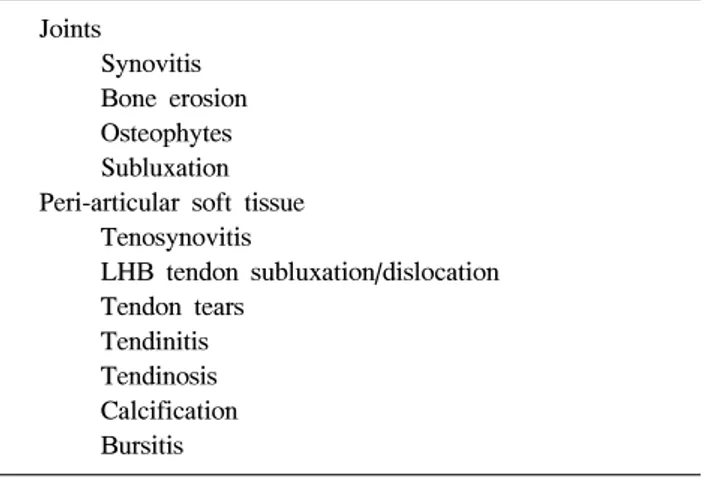
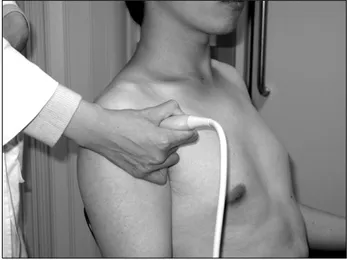
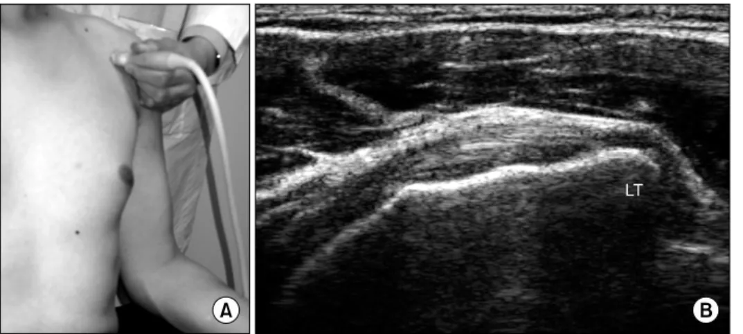
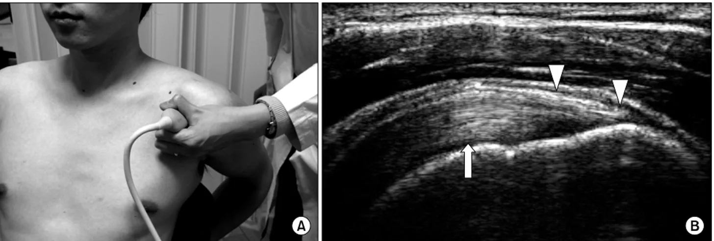
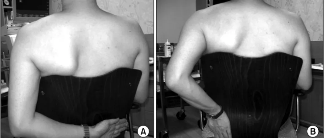
견관절의 정상 초음파 소견

가톨릭대학교 의과대학 성빈센트병원 재활의학교실

김 준 성

Ultrasonography of the Shoulder: Normal Findings

Joon-Sung Kim, M.D.

Department of Rehabilitation Medicine, St. Vincent Hospital, The Catholic University of Korea College of Medicine, Suwon, Korea

The shoulder is one of the most common applications of musculoskeletal ultrasonography due to the high incidence of rotator cuff disorders related to aging and sports activities. Ultrasonography has proved to be a useful diag- nostic tool with shoulder pain. It allows careful assessment of a wide range of changes involving many different ana- tomic structures of the shoulder girdle, such as the rotator cuff tendons, the long head of the biceps tendon, the bur- sae, and the soft tissues of the gleno-humeral and acromio- clavicular joints. (J Korean Assoc Pain Med 2008;7:

55-61)

Key Words: Ultrasonography, Shoulder, Position

서 론

견관절의 통증은 요통과 목의 통증에 이어 세 번째로 흔 한 근골격계 통증이다.

1)

견관절 통증의 원인으로는 관절내 (intra-articular) 병변과 관절주위(peri-articular) 병변으로 나눌 수 있다. 관절내 병변의 질환으로는 관절염과 동결견 (유착성관절낭염)이 있으며 관절주위 병변의 질환으로는 회전근개 건염, 충돌증후근, 회전근개 파열, 점액낭염, 이두 박근 건염, 석회성 건염 등이 있다.

2)

이러한 질환을 진단할 때 이학적 검사만으로는 정확한 평가가 어렵다.

3)

정확한 진 단 및 치료를 위해 견관절 질환에서 초음파 이용은 반드시 필요한 수단으로 인식되고 있다. 최근에는 발단된 초음파 장비 및 기술로 인하여 보다 편리하게 초음파를 이용한 검

사를 할 수 있다. 그러나 견관절 초음파 검사는 충분한 지식 과 경험이 없는 경우 잘못된 진단을 하기 쉽다.

4)

따라서 해 부학 및 초음파 소견에 대한 정확한 지식 및 경험이 매우 중요하다.

적응증

관절내 병변 및 관절주위 병변의 진단에 사용한다(Table 1).

1. 장비

정확한 진단을 위해서는 고성능의 장비가 필수적이다.

선형 탐색자를 사용하며 탐색자(prove)의 주파수(frequen- cy)는 7.5 Hz 이상이 요구된다. 대게 10 MHz 이상의 탐색 자가 사용된다. 컬라도플러와 파워도플러가 가능한 초음파 기종이 유용하다.

2. 검사 자세

견관절 초음파 검사할 때 환자는 눕거나 앉은 상태에서

실시한다. 환자가 앉는 상태로 검사할 때 검사자는 환자의

앞 또는 뒤에서 실시한다. 외래에서 진료와 연결해서 검사

하는 경우 시간적 편의성 및 환자의 이해를 돕기 위해서,

필자의 경우 환자가 의자에 앉은 상태에서 환자의 뒤 쪽에

위치해서 시행한다(Fig. 1). 이 경우 환자의 오른쪽 어깨를

(2)

Fig. 2. Transverse scan of bicipital groove. (A) Transverse imaging over the bicipital groove. (B) The hyperechoic biceps brachii long head tendon (arrow) within the bicipital groove and subscapularis tendon (sb), transverse humeral ligament (arrow head) and deltoid (d).

Table 2. Position of Examination Operator position

In front of or behind the patient.

A comfortable position is recommended to obtain stability.

Patient position Sitting position

Upper limb in neutral position with 90° flexed elbow Hand in supination and on the thigh

Other position

Shoulder in hyperextension, internal rotation and adduction with the arm placed behind the back (for the suprasupinatus) Shoulder in external rotation (for the subscapularis tendon) Raised arm or shoulder in 90

o

abduction (axillary scan for

the gleno-humeral joint)

Shoulder movements for dynamic study

External and internal rotation of the humerus over the full range of motion with adducted arm and 90

o

flexed elbow (for LHB subluxation)

Abduction and adduction of the humerus for assessing the supraspinatus tendon

Fig. 1. Position of examination.

검사할 때에는 검사자가 우측 손을 이용하고 환자의 왼쪽 어깨를 검사할 때에는 검사자가 좌측 손을 사용해야 한다.

좌측 어깨를 검사할 경우 환자의 앞쪽에서 우측 손을 이용 하여 검사 할 수도 있다. 검사 하는 동안 자세 유지가 어려 운 환자를 위해서는 등받이가 있는 의자를 사용하는 것이 편리하다. 검사시 환자의 팔의 위치는 보고자 하는 구조물 에 따라 다르게 위치시킨다(Table 2).

3. 탐색자의 방향에 따른 명칭

탐색자를 물체에 수평으로 위치시키는 것을 가로스캔 (transverse view)라 하며 세로로 위치시키는 것을 세로스캔 (longitudinal view; sagittal view)라 한다. 탐색자의 위치와

관계없이 근육의 주행방향에 대하여 수평으로 탐색자를 위 치시키는 경우 long axis view라 하며 수직으로 위치시키는 경우 short axis view라 한다.

4. 이두박근의 가로스캔 (Transverse bicipital groove view; short-axis view)

환자는 앉은 채로 이두박근이 앞쪽으로 회전하게 하기

위해 검사 받을 어깨 쪽의 손을 손바닥이 위로 향하게 하여

무릎 위에 올려놓는다. 탐색자를 가로로 앞쪽에 위치하면

(3)

Fig. 4. Transverse subscapularis view. (A) Transverse imaging of subscapularis. (B) Normal hyper- echoic subscapularis tendon inserts at the lesser tuberosity (LT).

Fig. 3. Longitudinal biceps view. (A) Longitudinal imaging of the biceps tendon. (B) The biceps tendon (arrow head) demonstrates a well-defined fibrillar echotextrure made up of multiple parallel linear echoes.

이두박근 고랑(groove)에서 이두박근의 건(tendon)이 잘 보 인다. 건의 모양은 약간 타원형이다. 초음파에서 정상적인 건은 고에코의 결체조직에 둘러 쌓인 저음영의 건섬유 (tendon fibril)의 특징적인 모양이 나타난다. 초음파 빔의 방향이 비스듬하게 되면 비등방성(anisotropy)에 의해 음영 이 감소하여 이상이 있는 것처럼 보인다. 이두박근 건막 안 에는 흔히 소량의 액체(2 mm 미만)가 보이며 하부에서 더 잘 관찰된다. 그러므로 반대측과 비교하는 것이 중요하다.

팔을 내회전, 외회전 시키면서 이두박근 건의 아탈구나 탈 구를 관찰한다(Fig. 2).

5. 이두박근의 세로스캔 (Longitudinal biceps view;

long-axis view)

가로 스캔 후 탐색자를 90

o

회전하여 이두박근 건을 검사 한다. 세로 스캔이 비후(thickening)와 같은 형태학적 변화 를 관찰하는데 유리하다. 스캔시 아래 쪽을 살짝 누르면서

검사하면 건의 주행을 잘 볼 수 있다(Fig. 3).

6. 견갑하근의 가로스캔 (Transverse subscapularis view; Long-axis view)

탐색자를 가로로 하여 이두박근고랑의 내측에 위치시키 고 팔을 외회전하여 관찰한다. 팔을 외회전 시킨 상태로 검 사하지 않으면 비등방성에 의해 저에코의 음영으로 나타난 다. 동적 검사로서 팔을 내회전, 외회전 시키면서 관찰한다 (Fig. 4).

7. 견갑하근의 세로스캔 (Longitudinal subscapularis view; short-axis view)

탐색자를 세로로 하여 이두박근고랑의 내측에 위치시키

고 팔을 외회전하여 관찰한다. 견갑하근이 multipanate 근

이어서 건과 근육이 섞인 영상이 나타나므로 3∼4개의 근

육 다발과 그사이에 저에코의 틈(cleft)이 보인다. 견갑하근

(4)

Fig. 6. Coronal transverse view of acromioclavicular joint. (A) Coronal imaging of acromiclavicular joint. (B) Hyperechoic distal end of the clavicle (C) and acromion (Acr) separated by a hypoechoic space reflecting the joint cavity.

Fig. 5. Longitudinal subscapularis view. (A) Longitudinal imaging of subscapularis. (B) Hyperechoic ten- don bundle and hypoechoic cleft (arrow head; muscle).

건의 폭이 넓으므로 탐색자를 위, 아래로 움직이며 관찰한 다. 상부에 병변이 있는 경우가 많다(Fig. 5).

8. 견봉 - 쇄골 관절 영상 (Acromio-clavicular joint view) 쇄골의 장축에 따라 탐색자를 위치시킨다. 쇄골측이 견 봉보다 위에 위치한다. 표층의 쇄골-견봉인대는 얇은 에코 띠로 보인다. 관절낭의 팽윤정도와 관절 간격을 측정하며 반드시 반대측과 비교하여 평가해야 한다. 초음파에서 측 정되는 뼈의 테두리에서 관절막까지의 평균거리는 21∼32 세가 2.2±0.5 mm이고 37∼81세가 2.9±0.7 mm이다. 보통 3 mm 이내이다. 정상 평균 관절간격은 각각 4.1±0.9 mm와 3.5±0.9 mm이다.

5)

동적 검사로서 팔을 외전 시키면서 관절 간격의 변화를 보거나 반대측 어깨에 손을 얹은 상태에서 관절간격의 변화를 측정하여 관절의 부딪힘(impingement) 등을 평가한다(Fig. 6).

9. 극상근의 세로스캔 (Longitudinal supraspinatus view; long-axis view)

중립위치에서 세로스캔을 하면 극상근이 가려져서 잘 보

이지 않으므로 팔을 뒤로해서 반대측 엉덩이에 위치시켜서

(Crass position) 검사한다. 이 자세는 팔을 내회전시켜 대결

절(greater tuberosity)이 쇄골 앞쪽에 위치한다. 극상근의

건은 견봉 하방부터 볼록한(convex) 모양으로 대결절에 붙

어 새 부리 모양(beak-shape)의 형태를 나타낸다. 이때 건의

주행방향에 따라 대결절 부위에서 비등방성에 의한 저에코

음영이 나타날 수 있으므로 탐색자의 위치를 변화시키면서

검사해야 한다. 극상근의 상방에는 삼각근이 위치하며 그

사이에 가는(thin) 저에코의 견봉하-삼각근하 점액낭(suba-

cromial subdeltoid bursa)이 있으며 고에코의 점액낭주위지

방(peribursal fat)에 의해 쌓여있다. 정상에서 약간의 액체

가 점액낭에 있을 수 있으며 이 경우 두께는 2 mm 미만이

(5)

Fig. 8. Transverse supraspinatus view (short-axis view). (A) Transverse imaging of supraspinatus. (B) The supraspinatus tendon lies between the humeral cartilage (arrow head) and the deltoid (D). (C) Transverse image of the intra-articular portion of the biceps brachii long head (arrow) shows the coracohumeral ligament and joint capsule (arrow head).

Fig. 7. Longitudinal (long-axis) supraspinatus view. (A) Longitudinal imaging of supraspinatus. The long axis of the tendon is oriented approximately 45° between the sagittal and coronal planes. (B) The thick echogenic supraspinatus tendon insert into the greater tuber- osity (GT). The subacromial, subdeltoid bursa (arrow head) overlies the tendon, covered by hyperechoic peribursal fat, and may extend laterally to the tuberosity. Hypoechoic hyaline cartilage (arrow) of the humerus is noted.

다(Fig. 7).

6)

10. 극상근의 가로스캔 (Transverse supraspinatus view;

short-axis view)

극상근을 세로스캔 한 후 탐색자를 90

o

회전시켜 검사한 다. 관절강 내 부위부터 스캔을 시작하여 대결절까지 시행 한다. 상완골 골두의 유리연골(hyaline cartilage)이 저에코 음영으로 잘 나타나며 그 위로 균일하게 중간 에코의 극상 근이 보인다. 극상근의 평균 두께는 6±1 mm

7)

이다. 극상근 과 극하근은 부착부위에서 근섬유의 일부가 서로 교차하므 로 탐색자의 위치에 따라 모양이 다르게 보일 수 있다(Fig.

8). 회전근개 간극(rotator cuff interval) 부위도 반드시 검사 를 해야 하며 이때 팔을 뒤로 해서 손을 검사하는 쪽의 뒤 주머니에 대는 자세(modified Crass position)가 유용하다

(Fig. 9).

11. 극하근의 가로스캔 (Transverse infraspinatus view;

long-axis view)

극하근을 스캔하기 위해서는 손을 다리에 올려 놓는 자

세에서 하거나 또는 손을 반대쪽 어깨에 놓는 자세에서 환

자의 후방에서 극상근과 나란히 탐색자를 위치시킨다. 첫

번째 방법을 이용하는 것이 극상근과 분리하여 관찰하기가

편리하다. 이때 후방 견관절(posterior glenohumeral joint)

및 소원근(teres minor)도 관찰한다. 극하근의 건이 새 부리

모양(beak-shaped)으로 상완골에 부착된다. 소원근은 극하

근에 비해 작으며 비스듬하게 위치하므로 탐색자의 방향을

조정해야 한다(Fig. 10).

(6)

Fig. 11. Longitudinal infraspinatus scan (short-axis view). (A) Longi- tudinal imaging of infraspinatus.

(B) Normal infraspinatus (InfraS) and teres minor (Tm) muscles. Hy- perechoic aponeuroses (arrow) are noted.

Fig. 9. Position for supraspinatus view. (A) Crass position. (B) Mo- dified Crass position.

Fig. 10. Transverse infraspinatus scan (long-axis view). (A) Transverse imaging of infraspinatus. (B) The infraspinatus tendon appears as a thick beak-shaped structure coursing deep to the deltoid muscle (D). (C) Posterior glenoid labrum (arrow) is noted between hume- rus (H) and bony glenoid (GL).

12. 극하근의 세로스캔 (Longitudinal infraspinatus view;

short-axis view)

견갑골 극돌기(scapula spine; 어깨뼈 가시)를 확인한 후 탐색자를 아래쪽으로 이동시켜 중간 단면(middle facet)에 위치하는 극하근과 하방 단면(inferior facet)에 위치하는 소

원근을 관찰한다. 극하근은 소원근에 비해 두 배 이상의 단 면적을 가지고 있다(Fig. 11).

13. 동적 견관절 검사 (Dynamic shoulder examination)

초음파 검사의 장점 중 하나가 동적 검사를 할 수 있는 것

이다. 다양한 각도의 회전 및 압력을 근육 및 점액낭에 가하

(7)

면서 검사를 할 수 있으며, 팔을 움직이면서 검사를 시행하면 서 병변을 찾을 수 있다. 이두박근 건의 탈구, 견봉하 관절에 서의 건의 부딪힘(impingement), 동결견(frozen shoulder)에 서 극상간 건의 움직임 제한 등을 확인할 수 있다.

결 론

견관절 초음파 검사는 견관절 질환의 진단을 위해 매우 유용한 검사로 근골격계 질환의 환자를 진찰할 때 이학적 검사와 동시에 실시함으로써 정확한 진단 및 치료 계획을 세우는데 없어서는 안 될 중요한 검사이다. 그러나 검사자 의 숙련도에 따라 진단의 정확도에 차이가 있으며, 이상소 견으로 오인할 수 있는 많은 허상(artifact)이 있으므로 이에 대한 철저한 준비가 필요하다.

참 고 문 헌

1. Bland TH, Merrit JA, Boushey DR. The painful shoulder.

Semin Arthritis Rheum 1977;7:1-47.

2. Andrew JR. Diagnosis and treatment of chronic painful shoulder: review of nonsurgical intervention. Arthroscopy 2005;21:333-47.

3. Dalton S. The shoulder. In: Klippel JH, Dieppe PA, editors.

(Des.): Rheumatology. London: Mosby; 1998. p.4.7.1-4.7.14.

4. Filippucci E, Unlu Z, Farina A, Grassi W. Sonographic training in rheumatology: a self teaching approach. Ann Rheum Dis 2003;62:565-7.

5. Alasaarela E, Tervonen O, Takalo R, Lahde S, Suramo I.

Ultrasound evaluation of the acromiclavicular joint. J Rheum 1997;24:1959-63.

6. Naredo E, Aguado P, Demiguel E. Painful shoulder: compar- ison of physical examination and ultrasonographic findings.

Ann Rheum Dis 2002;61:132-6.

7. Bretzke AA, Crass JR, Craig EV, Feinber SB. Ultrasonog- raphy of the rotator cuff: normal and pathologic anatomy.

Invest Radiol 1985;20:311-5.

수치

Table  1.  Pathological  Conditions  Detectable  by  Ultrasound Joints
Fig.  2.  Transverse  scan  of  bicipital  groove.  (A)  Transverse  imaging  over  the  bicipital  groove
Fig.  3.  Longitudinal  biceps  view.  (A)  Longitudinal  imaging  of  the  biceps  tendon
Fig.  6.  Coronal  transverse  view  of  acromioclavicular  joint.  (A)  Coronal  imaging  of  acromiclavicular  joint
+3

참조

관련 문서

 Stroke하기 전단계로 치은열구와 치아표면 사이에서 기구가 사용될 수 있도록 절단날을 위치시키는 것(작동부 날의 1/3(tip

Chest X-ray: Posterio-anterior (PA) View - Left Anterior Oblique

Animating View s (via their ViewModifier s which can implement the Animatable protocol) Transitions (animating the appearance/disappearance of View s by specifying

View The Invisible Know The

주택형별 2순위까지 전체 신청자수가 일반공급 세대수의 140%에 미달하는 경우 낙첨자 모두를 추첨의 방법으로 예비입주자로 선정하고 남는 경우 추첨의 방법으로

The definitive fixation using Steinmann pin & 4.0, 6.5 cannulated screws was performed(A-D), Postoperative X-ray (A : ankle lateral view, B : Calcaneal axial view, C

본 열화상 이미지를 통해 서 초음파 가진시 인공적인 결함이 있는 Spot2에서는 다른 정상정인 부위보다 온 도가 상승하는 Hot spot이 발생하는 것을 확인하였으며, 결함이

본 논문에서는 기존의 영상검색 기법들의 단점인 칼라의 수축,팽창등과 같은 크 기 변화에 관한 문제를 개선하기 위해 칼라의 색상과 명암 정보를 이용해서 정규