大韓放射線톨흩學슐誌 第25 卷 第3 號 pp. 476-481, 1989 Journal of Korean Radiological Society, 25131 476-481, 1989
악성변화 신경섬유종의
-3예 보고-
다발성
고려대학교 의과내학 방사선과학교실
이 종 문·강 은 영 ·이 남 준·정 규 병 ·서 원 혁 - Abstract-
Malignant Degeneration in Neurofibromatosis 3 Case Report
Jong Moon Lee
,
M.D.,
Eun Young Kang,
M.D.,
Nam Joon Lee,
M.D.,
Kyoo Byung Chung
,
M.D.,
Won Hyuck Suh,
M.D.Department of Radiolog, College of Medicine, Korea University
Von Recklinghausen’s nuerofibromatosis is a kind of neurocutaneous syndrome characterized by skin, nervous system, neuromusculoskeletal manifestations
Most central and peripheral nervous neoplastic lesions in neurofibromatosis are benign but have a significant potential for malignant transformation such as neurofibrosarcoma, neurogenic sarcoma, malignant schwannoma
And it is a relatively common disorder but malignant change with radiologic findings has been reported rarely in Korea‘
Recently we experienced three cases of malignant degeneration in neurofibromatosis and confirmed by pathology
So we report the cases with review including radiologic findings
다발성 신경 섬유종은 비교적 흔한 질환으로 많은 저 자들에 의하여 여러 뱅변이 보고 된 바 있다. 그러나 신경섬유종의 악성 변화에 대한 방사선파학적 소견을 보고한 예는 국내에 많지 않다. 이에 최근 고려대학교 부속영원에서 초음파 검사 및 전산화 단충 촬영을 시 행 하고 수술 또는 초음파 유도하에 조직 검 사를 하여 확진된 다발성 신경섬유종의 악성변화 3례를 경험하 였기에 문헌 고찰파 함께 보고하는 바이다.
려|
;ξ
n.
ε5론
다발성 신경섬유종은 일종의 신경피부증후군 (neu
rocutaneous syndrome) 으로써 피 부 색 소 침 착파 중추 및 말초신경제의 다발성 종양 그러고 혈판계 및 내부 장기의 명변을 특정으로 한다.
다발성 신경섬유종에서 영발된 중추 및 말초 신경종 양은 대개가 양성이지만 2_4 %-29 %에서 신경섬유 육종 (neurofi brosarcoma) 이 나 신경성섬유육종
(neurogenic sarcoma), 아니연 악성신경초종 (mal
ignant schwannoma) 둥으로 악성화 할 수 있다1) 1. 서
증례 1
오른쪽 가숭의 통증파 호흡곤란을 주소로 내 원한 29
…
”
이 논문은 1989년 2월 28일 접 수하여 1989년 3월 7 일에 채 택되었음
- 이종문 외 : 다발성 신경섬유종의 악성변화 -
세 남자 환자로써 이학적 검사상 전신에 다알성 피부 색 소의 침 착 (cafe-au-!ait spot) 과 피 하 결절 종양이 촉 지 되었마.
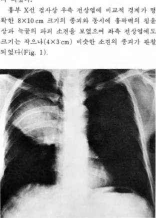
흉부 X선 검사상 우측 천상엽에 매교적 경계가 영 확한 8XIOcm 크기의 종괴와 동시에 흉팍벽의 칩윤 상과 늑골의 파괴 소견을 보였으며 좌측 천상엽에도 크기는 작A나(4X3 cm) 바슷한 소견의 종괴가 판창 되 었마 (Fig. 1).
Fig. 1. Chest PA: A relatively well defined homogen- eous soft tissue mass in RUL.A smaller mass in L UL
우측 전상엽의 종괴는 흉부 전산화 단층 촬영상 우 측 상페엽에 주변조직파 경계가 잘 되지 않은 7X8X9 cm 크기의 연부조직 종괴였으며, 우측 종격동 및 흉 팍벽에 고착 되었고 조영 증강후에 이 종괴의 내부에 는 불규칙한 조영증강이 판찰 되었다 (Fig. 2).
흉부 초음파 검사상 혼합펀 에코를 보였으여 이어 시행한 조직 생검상 신경섬유육종 (neurofi brosarcoma) 으호 확진 되었마 (Fig.3)
증례 2
좌측 옆구리의 통증파 종괴가 만져지는 것을 주소로 내원한 58세 여자 환자로써 이학적 검사상 북부에서 다발성의 펴부색소의 침착과 피하 결절 종양이 촉지 되였다.
소창바륭 조영상 종피에 의해 소장이 밀런 형태의 소견이 보였다 (Fig.4)
Fig. 2. Chest CT scan: A ill defined heterogeneous soft tissue mass with anterior chest wall invasion and rib destruction
Fig. 3. Chest US: Well defined bulky heterogeneous soft tissue mass
복부 초음파 검사상 7X8XIO cm 크기의 비교적 주 위와 경계가 분영하고 혼합된 에코를 보이는 종괴가 좌상복부에서 판찰되었다 (Fig. 5).
이 종괴는 복부 전산화 단층 촬영상 비교적 경계가 불분영한고형의 종피였고, 조형 증강후이 종괴는뚜 렷한 외벽의 조영 증강과 더불어 내부에 불규칙한 조 영 증강 형태로 판찰되었다 (Fig. 6).
종괴 제거술에 이은조직 영리학 검사상 악성신경초 종 (malignant schwannoma) 으로 확진되 었다.
증례 3
오른쪽 둔부에 크기 가 점차 증가하는 종괴를 주소로 내원한 30세 남자환자로써 이학적 검사상 역시 피부 색소의 침착 및 마발성의 결절 둥이 촉진되었다. 단순 골반 사진상 연부조직 종괴음영이 우측 영덩이에 보였
찌
- 大韓放射線뿔學會픔 ; 第 25 卷 第 3 號 1989 -
Fig. 4. Small bowel series: Displaced small bowel by mass
Fig. 5. Abdominal US: Well defined mixed echo pat- tern mass
고 (Fig. 7), 우측 둔부의 초음파 검사상 불엽성의
(10 bu lated) 혼합된 에코를 나타내는 종피가 판찰되었 다(Fig.8)
골안 전산화 단층 촬영상 6X 10 cm 크기의 불규칙 석으로 나타냐는 음영의 연부 조직 종괴가 우측 둔부 에 있었으며, 또한 좌측 골반강내에도 5X8cm정도의 연부 조직 종괴가 판찰되었다.
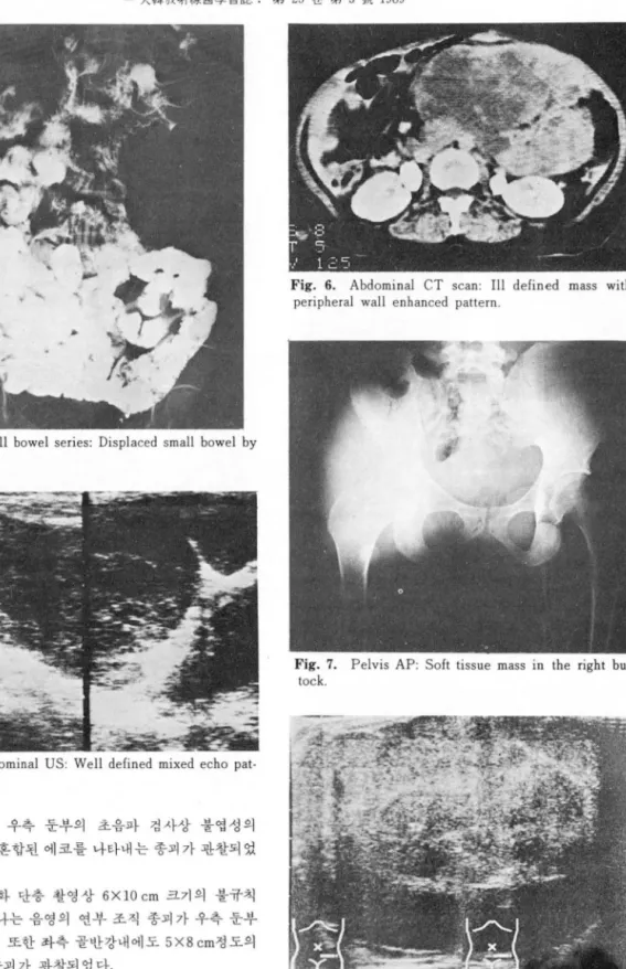
Fig. 6. Abdominal CT scan: III defined mass with peripheral wall enhanced pattern
Fig. 7. Pelvis AP: Soft tissue mass in the right but- tock
조영 증강후 외 벽 의 조영 증강과 함께 종괴 의 내 부 Fig. 8. Right gluteal US: Lobulated echogenic mass - 478 -
이종운 외 : 마발성 신경섬유종의 악성연화
에는 불규칙적인 조영증강 형태를 관창할 수 있었다 (Fig.9).
초음파 유도하에 시행된 우측 둔부 종괴의 조직 생 검 상 악성 신경 초종 (ma1ignant schwannoma) 으로 확 진되었마 (Fig. 10).
m.
고 찰다발성 신경 섬 유종은 18c초 처 음으로 이 질환에 대 한 기술이 있었 A여 1882년에 Von Reckling hausen disease 란 명 칭 이 붙게 되 었 다2)
다발성 신경 섬 유종의 반도는 1 : 2000-1 : 3000이 며
Fig. 9. Pelvic CT scan: A large irregular enhancing soft tissue mass in the left pelvic area, and in the ri- ght buttock.
Table 1. Areas and Types of Involvement in Neurofibromatosis
Skin Ophthalmic
Neurofibromas Pulsating exophthalmos
Cafe au lait spots Sinuous deformity of eyelid border
Axillary freckling Glaucoma
Skeleton and soft tissue Schwannoma and malignant melanoma of choroid
Kyphoscoliosis Optic nerve glioma
Posterior vertebral body scalloping Sectoral retinal pigmentation
Skull defects Intrathoracic
Bony erosion from tumor Pesterior mediastinal neurofibromas and mening- Ribbon deformity and notching of ribs oceles
Primary marginal defects of long bones Interstitial pulmonary fibrosis
Intraosseous cystic lesions Idiopathic hypertrophic subaortic stenosis Congenital bowing and pseudoarthrosis of tibia Gastrointestinal
Elephantiasis neuromatosa, verrucous or villous N eurofibromas
skin hypertrophy Carcinoid
N eurofibromas
J
uvenile polyposis coliSubperiosteal hemorrhage and cysts Urinary tract
Central nervons ststem Neurofibromas of bladder
Intracranial tumors Neurogenic bladder
Intraspinal tumors Retroperitoneal neurofibromas affecting ureters
Aqueductal stenosis and hydrocephalus Endocrine system
Syringomyelia Pheochromocytoma
Vascular ststem Sipple’s syndrome
Vessel-wall hyperplasia Reduced fertility
Microaneurysms Hyperparathyroidism
Renal artery stenosis and/or abdominal aortic Hypoglycemia from insulinoma coarctation with hypertension Myxedema
Retroperitoneal hemorrhage Obesity
Mesenteric arterial insufficiency Precocious or retarded sexual development Intracranial arterial occlusive disease Goiter
Cerebrovascular developmental anomalies Diabetes insipidus
- 479 -
- 大韓放射線쩔學會誌 : 第 25 卷 第 3 號 1989 -
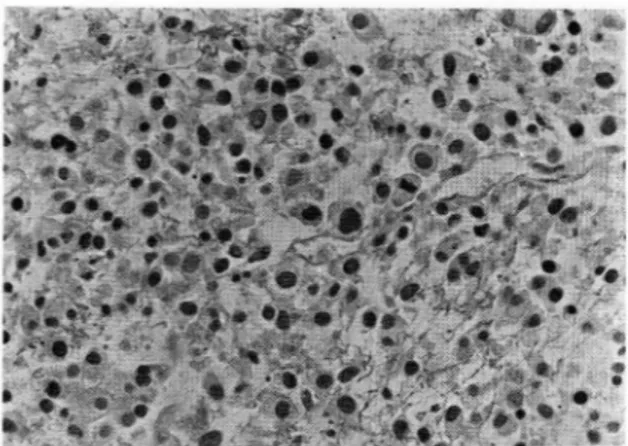
Fig. 10. Micro: Hapazardly arranged pleomorphic round to oval shaped tumor cell and moderate amount of acidophilic cytoplasm in the background of severe necrosls
주로상염색체 우성으로유전되나 일부는새로운돌연 변이 유전자에 의하여 발생되기도 한다3)
진단은 특정적인 피부 영뻔과 다발성 피하결절이 있 는경우에는어렵지 않으냐피부영변이 없는경우에는 피하 결철의 조직 검사를 함으로써 확진된다. 다발성 신경섬유종은임상양상이 다양하여 여러 장기를동시 에 청엄할 수 있고 여러가지 질환을 동안할 수 있다.
그 부위 및 유형은 마음과 같다(표 1)3,4,5,6),
다발성 신경성유종에서는 신경섬유종이 아닌 다른 종류의 뇌종양, 즉 다발성 수막종, 시신경 및 시신경 교차의 신경교종, 대뇌 및 소뇌반쿠 척추 신경 교종 둥의 발생 빈도가 정 상에 서 보다 높마7)
마말성 신경섬유종의 악성변화는 동통이 있거나 기 왕에 존재했던 종양이 급격히 성장한 경우, 또는 심부 에 존재한 경우에서 악성화를 의싱할 수 있 A며 악성 화 율이 2.4 %-29 %나 보고되고 았다7)
악성 종양은 신경파 판련없이 연부 조직에서도 발생 될 수도 있다9)
그렇지만 대부분의 경우는 증식중인 간엽조직 (proliferating mesenchymal tissue)파 덩 굴모양의 신 경섬유종 (plexiform neurofibroma) 의 종괴에 판련되 어 발생한다8)
영리학적으로 신경섬유육종 (neurofi brosarcoma), 섬유육종(fi brosarcoma), 방추체세포육종(spindle ce- 11 sarcoma), 황운근육종 (rhabdomyo sarcoma), 정액 육종 (myxosacoma) 또는 악성 신경 초종 (malignant schwannoma)둥이 보고 된 바 있 다7, 10)
다발성 신경섬유종의 대표적안 양성 종괴인 신경섬 유종 (neurofi broma) 전산화 단층 촬영 상 마양한 크기 의 경 계 가 분영 하고 둥근 모양의 30-40 Hu의 균일한 농도를 보이는 종괴로 조영 증강후에도 뚜렷한 조영 증강 소견이 보이지 않은 것으로 보고 되었마10, 11)
안연 악성 변화인 신경 섬유육종( neurofibrosar- coma) 에서는 종괴는 불규칙하여 경계를 냐타냈고 바 교적 농도가증가된 형태이었으여 종괴의 내부에는중 앙부나주변 부위에 둥글거나선모양의 불규칙한저농 도저농도부위가존재하였으며, 조영 증강후에 이 종 괴 내부의 농도 차이는 더 욱 증가한 것 A로 보고 되 었 다10) 이는 저자들의 세 증례에서 보이는 전산화 단층 촬영 소견파 유사한 형태로 판찰되었마.
N.
걸 론저자들은 최근 고려대학교 의파내학 부속병원에 내 원하여 천산화 단충 촬영을 시행하였고 영리 조직 검 사에서 확진펀 다발성 신경섬유종의 악성변화 3 례를 경험하였기에 문헌 고찰과 함께 보고하는 바이다.
REFERENCES
1. Taveras ]M, Ferrucci ]T: radiology Diabno- sis-Imaging-Intervention. Vol 5‘93‘1-10, 1987 2. Von Recklinghausen K: Uber die multiplen fibrome
der Haut und ihre Beziehung zu den multiplen Neuromen. Berlin. A. Hirschwald. 1882
3. Barone DA: Neurofibromatosis. A Clinical ovinical overview. Postgraduate Medicine. 66:73-82, 1979 4. Holt ]F: Neurofibromatosis in children. AJR
130:615-635, 1978
5. Holt ]F, Wright EM‘ Radiologic features of neuro- fibromatosis. Radiology 51:647-663, 1948
6 하 성규, 임 승걸, 김 성규 N eurofibromafosis 환자 에서 명딸된 상출성 흉악영 및 우측흉벽에 생긴 종 괴 . 결핵 및 호흉기 질환, Vol‘29. No 4. Dec. 1982 7. Knight WA III, Murphy UK, Gottlieb ]A: Neuro
fibromatosis associated with malignant neuro
“
br-oma. Arch, dermatol. 107:747-750, 1973
8. D’agostino A, Soule E, Miller R: Sarcoma of the peripheral nerves and somatic sovt tissue associated with multiple neurofibromatosis. Cancer 16:1015,
- 480 -
- 이종운 외 : 마딸성 신경섬유종의 악성변화 -
1963 1400383-387, 1983
9. Deborah LD, Bradford TA: Pediatric case of the 11. Biondetti PR, Mario V, Davide F et al: CT appear- day. A]R: 144, 1983 ance of generakized von Recklinghausen Neurof- 10. Coleman BG, Arger PH, Dalinka MK et al: CT of ibromatosis, ]ournal of CT 7:866-869, 1983
sarcmatous degeration in neurofibromatosis. A]R
- 481 -