大韓放射綠훌훌學會誌 Vol. XV, No. 1, 1979
-Abstract
상악동암의 방사선 치료
연세대학교 의과대학 방사선과학교실 김기황 · 이도행·박창윤
Ratiotherapy of Malignant Tumor of Maxillary Sinuses K.W. Kim, M.D., D.H. Lee, M.D. and C.V. Park, M.D.
Department of Radiology and Nuc!ear Medicine
,
Yonsei University College of Medicine
,
Seoul,
KoreaSixty nine patients of malignant tumors of the maxillary sinuses were treated at the Vonsei Cancer Center from January 1970 to March 1978 by CO.60 teletherapy unit.
We analysed their clinical findings, histopathological findings, clinical staging, .treatment and results.
The results are as follows:
1. Male to female ratio was 3:1. The most prevalent age group was in fifth
,
sixth and seventh decades (82.6%).2. Histpathologically
,
squamous cell carcinoma occurred most frequently which was 53 in 69 patients (76.8%). Lymphoma and adenoid cystic carcinoma were 3 patients (4.3%) respectively.3. Maxillary swelling was most frequent clinical manifestation and can be seen in 50 out of 69 patients (72.5%).
4. C1inical staging according to TNM system proposed by Sisson can be done in 49 patients. Majority of patients were T-3 and T-4 and occupied 83.7% (41/49). According to Ohngren’5 line
,
tumorlocating in infrastracture were 33 patients and in suprastructure 16 patients.
5. Among 69 patients
,
59 patients were treated by radiation only,
5 patients were sugery plus radiation and 5 patients by chemotherapeutic infusion plus radiation.6. In 31 patients who might be passed 5 years after treatment
,
follow up can be done in only 13 patients.10 patients were treated by radiation only and 3 patients by s'urgery plus radiation. Presumed 5 year survival rate was 3/1 0 (30%) in patients with radiation only and 2/3 (66.7%) in patients with surgery Iplus radiation. Among 13 patients
,
5 patients occurred in infrastructure and 5 patients in suprastructure.Mean survival months of patient with infrastructure were 49 months and suprastructure were 31.8 months
‘
I. 서 론
상악동암의 원발암은 흔하지 않으며 두경부 종양중 그 치료가 문제되는 부위이다j) 대부분이 펀i영상피 암 으로 그 행동이 파괴적이켜 칭윤성이나 진행된경우에 도 웬 말부위에 국한되어 있는 경향이 있다 2 ) 영 의 발 견 당시에 경부 임파선 전。1 가 되어 있는 경우는 10-
15% 로 비교적 낮다. 이와같이 임파절 전이 가 비교적
늦게 일어 남에 도 울구하고 종양의 근치 가 방사선 혹은 수술 단독치료나 영용치료로서도 그 예후가 좋지않다-
저자들윤 연세대학교 암센타 방사선 치료실에 서 1970 년 l 월부터 1978 년 3 월까지 상악동 종양으로 수술 혹은조직생검으로 확진된환자중 Co- 60 원격치 료장치를 이용하여 방사선 치료을 받았던 69 예를대상 으로 임상적 소견, 명리조직학척 소견, 뱅기분류, 치 료 앙볍과 치료후 추적조사의 결과를 분석하여 운현 고 찰과 함께 보고 하고져 한마.
- 254-
II. 관 찰 성 적 1. 성벨및연령별분포
념여 성옐lJl는 3 로 낭자에서 우월 하였고 40 세 에서 69 세 사이가 총 57 영으로 선체 환자의 82.6 % 를 차지 하고 있었 마 CTable 1).
T able 1. Material 1. Sex Male 52 Female17 2. Age Di st ribution
Age No. of patient
a) 10-19 1.4
b) 20- 29 l.4
c) 30- 39 6 8.7
d)40-49 21 30.4
e) 50- 59 22 31. 9
f) 60 - 69 14 20.3
g) Over 70 4 5.8
Total 69 100.0
2. 병 리조직학척 소견
총 69 영의 환자중 1 례를 제외하고는 모두영리조 직학적으로 확진이 되었으며 이중 펀평상피 암이 53 예 (76.8 %)로 가장 높은 벤도를 보여 주었고 악성 임파 종과 선냥냥암 (Adenoid cystic carcinoma) 이 각기 3 예
(4.3%), 미분환세포암과 섬유육종이 각기 2 예 (2.9%) 였 고, 유두전이성세포암 , 골원성육종, 형절세포암, 퇴행성 상피세포암 (Anaplastic epirhelial cel1 carcinoma) 그 러 고 맥 판육종이 각기 l 예 였 다 (Table 2)
3. 임상적 소견
내원 당시 주 증상으로는 올의 부종이 32 예 ( 46.4
%)로 가장 많았고 바폐쇄증 20 여1(29%), 두통 13예 CHU,%),비루 12 예 (17.4%). 볼의 동통과 치통이 각기 11 예 (15.9%), 비 출혈파 안우통증이 8 예 (11
6 %),우개팽만감 5 예 (7.2%), 유루가 2 예 (2.9 %)와 저작장애 l예를 보여주었다O‘able 3). 이학척 소견 으로는 상악부종。I 50예 (7L.5 %)로 가장 많이 관찰 되었고 비종괴 17 예 (24.6 %), 치은구개종괴 14여I (20.
3 %), 안구돌출 7 에 (10.1 %), 상악동구강누판 4예 ( 5. tl%)를 불 수 있 었 다 치 료 전 국소 입 파선 전 이 는 14에 (20 %)에서 관찰할 수 있었다( Table 3).
Table 2. Hisropathol';gic Cla~sificarion
H i s ropa tho logy No. of patien t (%)
I. S quamo us ce i 1 carcinoma 53 76.8
2. Lymphoma 3 4.3
3. Adenoid cystic carcinoma 3 4.3 4. Papillary transitional cell ca. l 1.4 5. Undifferenciated cell 2 2.9
carCIßoma
6. Fi brosarcoma 2 2.9
7. Osreogenic sarcoma 1.4
8. Pl asmocytoma 1.4
9. Anallplastlc epitllelial 1.4 c e 11 carcmoma
10. Angiosar~'O ma 1.4
11. P athology Unknown 1.4
Total 69 100.0
4 영기별 분류
명가 l셜 분류는 Si잉son 에 의거한 끼'M 분류법을 사 용하였 고 (Table 4) 병 가 분류가 가능한 환자 49 영 중 대부분은 진행된 강十껴로 41 영 (tl3.7 %)이었다 (T
able 5;\ 또한 상악동암을 주요 발생 부위에 따라 안구 의 내측안각파 하악각을 연결하는 Ohngren 씨 션에 의 거 하여 분류해 본 결 과 Infrastructure 가 33 예 Sup
ras tructure 가 16 예 로 Infrastructure 에 서 2 배 많이 딸생하였마.
5. 치료
1) 치료방법
。l용된 치료방법은 크게 둘로 나눌 수 있다.방사선 단독요법과 명용요법 으로 병용요법에는 수술과 방사선 치료 그러고 화학요엽과 방사선 치료의 두가지 요법을 시 행하였였 다(Table 6).
A. 방사선 단독치료
송 59명이 ω-60 원격치료장치블 이용하여 치료 을 받았다.
치료부위 :해당부위의 방사선 치료 조사벙위는병의 진행 정 도에 따라 약간의 차。l 을 두었으역 상연은 안와 상연, 하연은상치은협측푸 (Superior gingivobuαal gur-
ter) • 내측으로는 정중선을 념 어 1 cm 정도까지 포함 하여 조사야의 크기가 8 x 6 cm 가 되게 하였으며 김이 는 익상쿠개와까지 포함하였다.
조사방법 1979 년 이후 저자들이 사용한 조사방맴 은 크게 3 가지 방냄으로 나눌 수 있마.
Table 3. Symptom and Sign of Maxil1ary Sinus Malignancy
syπl;:>tom Sign
1. F acial Cheek swelling 32 (46.4 %) Ma~llary s\"elling . . 50 (72.5 %) with or 'wi thout iende메e ss
Cheek pain II (15.9 %)
2. Nasal Nasal Obstruction 20 (29 %) Nasalmass 17 (24.6%)
3. Or21
Rhinor'rhea Epistaxisis Toothache Palatal Fullness Trismus 4. Occular Eye paÏn
Epiphora 5. Regional L ymph
Node
6. General Headache
Table 4.
12 Cl74%)
8 Cl1.6"1o)
1; (]5.9%) 5 ( 7.2 %) I ( 1.4%) 8 (11.6%) 2(2.9%.'
13 (Ul. 8 %)
Gingivopatal mass Fistule, antal to oral
Propto si s
Metastasis
Classification of Maxillary Cancer by Sisson
Tl: 1) Tumor invasion of the anterior wall without skin involvement.
2) Tumor of i nferi or nasoan t r al wall wi thout ski n i nvolvement 3) Tumor 01 anterior medial palate
T2: 1) Tumor invasion of the lateral wall without muscle involvement 2) Tumor invasion of the superior wall without orbital involvement.
14 (20.3%) 4 ( 5.8%)
7 (10.1 %)
14 (20.0 %)
T3: 1) Tumor invasion of anterior ethmoid cells without involvement of the cribriform plate.
2) Tumor i nvasion 01 pte rygoi d. muscl e wi th or wi thout com binati on of T2.
3) Tumor invasion 01 orbit in combination with T2
4) Tumor invasion 01 anterior wall of antrum with skin involvement.
T4: 1) Tumor invasion of posterior ethmoid cells with or without T3. 2) Tumor invasion of cri briform plate‘
3) Tumor invasion of pterygoid maxillary recess.
4) Tumor extension to other antrum.
5) Tumor extension to ethmosphenoid recess or sphenoid sinus 6) Tumor invasion of pterygoid plate.
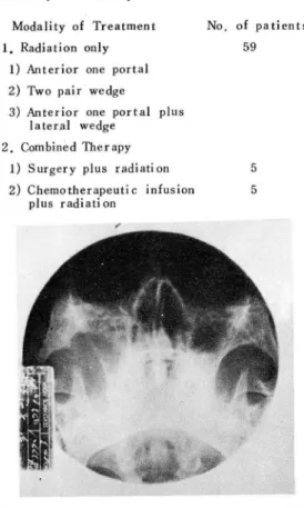
@ 전방일문수직조사(Anterior one portar)
영소에 충분한 종양선량을 조사할 수 있었고 반대 측 눈은 용이하게 방사선피폭으」로 부터 차폐할 수 있었다(Fig 1)
@ 파 o pair Wedge
45。쐐기형필타을 이용하여 2문직각조사하는 경우 (F ig 2 - A) 반매 측 안구에 방사선 피 폭선 량이 많아지 기 때문에 측연 조사시는 5 - 1 0。후땅으로 각도을 줌 으로서 t난대측 안구에 유해한 방사선을 줄일 수 있었
마{Fig 2 - 13)
(3) 전방-문수직조사와 측쐐 기필터 조사방법 이 경우 종양부위에 이교적 많은 선량분포를 보이며 반대측 안우에도 비교적 적은 양의 방사선이 조사됨을
볼 수 있 다 (Fig. 3).
조사량 : 원 말부위 에 6000 ~ 7400 rads 를 6~8 주 에 걸쳐 분할 조사하였으며 부작용이 싱할 경우 일정 한 휴식기간을 두고 재치료를 시행하였마. 엄파선 천 이 부위 에 는 4000 ~ 5000 r ads 를 Co-60 원격 치 료 장 치냐 8 Mev 천자선을 이용하여 4'" 5 주에 걸쳐 분할 조사하였다. 원발부위에 재발암의 경우 3명의 환자 에서 적극적인 방사선 치료를 시행하였으며 이중 l영
。1 2년동얀의 장기 생손을 보여주였다.
B.
영용요법총 10명이 병용요법을 맏았으멕 5영은 수솔과 방 사선치료를 5명은 약물요법과 방사선치료를 받았다.
- 256-
ANT
F<
T
7~ LT ANTRT
짧 6주ζ- LT샌첼
ongJe 60。
/
Fig. 1. Isodose distribution of anterior one pOTtal Fig. 3. Isod,ose distribution of anterior one portal irri dation by Co-60 teletherapy unit. and lateral wedge irridation by Co-60
( 550. 80) teletherapy uni t.
RT
ANT 5X5
...--까-딛 LT
(4S'W)
(4면/lj 'V
F
I•‘X
m싸 90' ‘ α
ANT
RT
7 X 9- iE"""' _ __
~,
(4S'W)293' α
..
~~
111
• κ -g .‘ “‘、、
LT
A. Portal combination with right angle B. Posterior tilting of lateral portal with 50degree.
Fig. 2 Isodose distribution of two pair wedge portal irridation by Co-60 teletherapy unit.
Table 5. Correlat.Ïon with stage anà tumor location according toOhngren'
,
line5tage Location Int rastructure Suprastructure Exten si\e Total
11 2 0 0 2
1깅 3 3 0 6
TJ 16 11 0 27
]‘4 12 2 10 24
1'0131 33 16 10 59
@ 수술과 수숭후 방사선치 료
시행된 수술방엽은 부분척 상악 절제 혹은 근치 상 악 절제술이었고 수술후 3 r~ 4 주 후에 방사선 치료를 시행하였다. 치료량은 5000 ~ 6000 r ads 를 5~6 주 에 걸쳐 분할 조사하였 다.
@ 약물요법과 방사선치료
이 경 우에 는 배 액 처 치 (Drainage Procedure ) 와 같 은 최 초의 수술적 인 가료후 5-Fu 250 m
g/
day 를 10~ 14 일간에 결쳐 동맥내 주업하였거 나 500 m
g/
day 로 수얼동얀 측두동맥 을 통하여 주입 하였 다.Modali ty of Treatmen t
No. of patients 59
5 Table 6
Modality of Treatment 1. Radiation on\y
1) Anterior one portal 2) Two pair wedge
3) Anterior one portal plus lateral wedge
2. Combined Therapy 1) S urgery plus r.adi ati on 2) Chemotherapeuti c infusion
plus radiati on
5
/
i織 f나
Fig. 4-B. Two months after operation film shows no maxillary hazines s and no further bony destruction on water' s proj ection Fig. 4-A. Pre • operative radiogram of maxillary
sinuses shows total haziness and bony destruction of inferior lateral wall of
Rt maxillary sinus.
Survival Year after Treatment Table 7.
Foll ow up
Survival months Status
1 2 3 4
5.
6.
7.
8.
9.
10.
11 12 13
Ex pir e live Expi re Exp i re Exp i re Exp i r e Expi re Ex pi re Ex p i re Ex pi re Expire live 6 yrs 7mon
6yrs 4mon.
3yrs 4mon.
1 yr 2mon.
5 yrs lmon 2yrs 4mon.
1 yr 6mon.
1 yr 5mon.
3 yr s 6yrs 7mon.
2 yr s 8mon.
5 yr s.
9mon Location
? I I
?·?·S
S S S Treatment
Radi ati On only Radiation only Radiation only Radiati on only Radiation plus Surgery Radi ati on only Radiati on on Iy Radiation only Radiati on only Radiati on only Radiati on only Radiation plus surgery Radiation plus surgery Stage
q니 끼4 끼J
qtu
T T T T
qJ
?i
jaT
T T T T4
꺼
NFN
R
?
””
Pathology
Squarnous cell Ca.
Epidermoid Ca.
Epidermoid Ca.
Epidermoid Ca.
Epidermoid Ca Epidermoid Ca Squamous ce 11 Ca. Reticulum celJ
sarcoma Epidermoid Ca.
Squamous cell Ca.
Squamous cell Ca Epidermoid Ca.
Squamous cell Ca.
Case
S
Ex p i r e
*
1 ’ Infrastructure? :
Undetermine258 -
S: Suprastructure
Fig. 5-A. Radiogram before irridation of. maxill- ary sinus shows total hariness with bony destruction on anterior inferior and lateral wall
Fig. 5-B. Film taken one month after radiation thcrapy shows more clear bony margin and air shadow.
l‘ iμ. 5-{‘ Film taken 4 months after radiation
therapy shows nearly normal findings .
- 259
•
화학요법제 주업시작 수일이내에 방사선 치료를 시 작하였 고 6000 ~ 7000 rads 를 6~7 주에 걸쳐 분할 조사하였다.
6. 증례
증례 1 : 40세 냥자 환자로 치 료당시 벙 기 는 T2NoM。
이고 우측 상악동암이었다. 종양선량 6600 radc를 조 사하고 8주후 근치수술을 받은 환자로 치료천 사진상 CFig 4-A) 에는 우측상악동내에 Total haziness와 하 외측백의 골파괴를 볼 수 있었다. 수술 2 개월 후 사 진상 (Fig. 4 - 13) 에는 상악동내의 haziness는 볼 수 없고 골파괴의 소견은 아직 남아있으나 치유펀 소 견을 보이고 있다.
중례 2 40 세 남자로 우측 상악동 악성임파종으로 병기는 T4NoMo 이었고 치료천 사진상 (Fig.5-A) 에
전, 하, 외측벽의 골칭윤을 올 수 있었고 이 학적으로 쿠강상악동누판이 판찰되였던 환자로 방사선 치료 l개 월 후 단순 x-선 사진상 (Fig. 5-13) 에서 골연이 좀 더 영확하게 나타났고 상악동에 공기가 보이기 시작하 였다 4 개월 후 사진상 (Fig . 5 -C)에서 브연 거의 정상 소견을 보여주었다.
증례 3 45 세 납자 환자로 뱅기는 T4NoMo 였으며 치료전 x-신 사진상 (Fig. 6-A) 에서 내 , 측벽의 골
*1_9 ... .Q.. ~ ζ느 。 1어 d
디 ι" λAμη
방사선단독치료를 받았고 치료 l개월 후 단순
x-
선 사잔 상 CFig. 6-13) 에 는 Haziness 가 현저 히 줄
Fig. 6-A. Pre- treatment film of maxillary sinus shows haziness and bony destruction of medial and lateral wall of Ht maxi- llary sinus.
;?~) -.f'-
3c
Fig. 6-B. Film taken one month after radiation therapy shows decrease in haziness and abscence of bony invasion.
7. 추적조사결과
1970 년 이후 치료를 말고 치료 완료일로부터 5 년 이상 경과한 환자 31 영 중- 추적조사가 가능했던 환 자는 13 예였마 (Table 7). 이들 중에 방사선 단독 치료를 받았던 환자는 10 명이었고 방사선 치료와 수 술의 영용치료를 받은 환자는 3 영이었다. 충분한 추 적조사는 아니지만 추정되는 5년생존율은( Table 8 ) 방사선 단독치 효가 φ/10 ( 30
%)
였 고 수울석 방뱀 파 방사선 명용치료에서는 2/3 (66.7 %) 였마. 또한 13 명 의 환자중 Infrastructure ‘ 가 5 명 으로 평균 생 촌월 수가 49 개월이었고 suprastructure 가 5 명으로 31.8 개월 이었다 (Table 9).Table 9. Survival Months accoTding to Location
LocatÍon No. of SUTvival Month
patlent
Infrastructure 5 49 Mon.
Suprastruct.ure 5 31.8 M"n.
Undetennined 5 51.3 Mon.
ill. 고 안
상악동암의 예후는 일반적으로 나쁜 종양중의 하나 이며 보고펀 5 년 생흔율은 10- 37 %이다 3, 4 , 5 , 6 ,7 ) 상악동암의 치료는 방사선 단독치료나 수술단독치료 혹은 수술과 방사선치료의 영용치료에 의존하고 있는 실정。l마 81
1950 년 이션의 치료 방법은 주로 땅사션 단독치료 블 시행하였으며 제한된 수술후에 라마웅을 상악동에 Fig. 6-C. Film taken 9 months after radiation 삼입 하고 종양신 량이 :3000 - 8000 Tad 5 가 되 깨 조사하 therapy shows marked bony destruction 였 마. 필요에 따라 추가로 200 - 250 Kvp 와 심부 x- of superior and lateral wall. 선 치 료장치 로 2000
•
000 Tads 을 2 - 4 주에 걸처 분할 조사하였다. 이 방법에 의한 5 년 생흔율은 Gibb6l 고 곰침윤 소견이 없어졌으냐 9개월 후 X~
선 사잔상 는
21%을 보고 하였고
Bac lesse
9J등은
20%을 보
(Fig. 6-{;) 에 서 는 상악동의 상연과 측벽 의 성 한 골 고 하였다i 보다 좋은 성적으로는 Dalley4J 등에 의 하 연 파괴 현상이 마시 판찰됨으혹서 국소부위의 재 딸로 사 40 %을 보고 하고있다.
료되었다 1950 년 이후에는 보마 적극적인 수술방법이 시행되
Tabl e 8. Modali ty of Trea tmen t and 5-year sur vi val
Modality of Treatment
Radiati 011 only
Radiation olus surgery
No. of 5 years survival Patients
10 ν/10 (30ro)
3 ι/3 (67%)
- 260
었고 수숭후 방사선치료의 병용치료가 시행되었으역 Zange 와 Schol tz1 0 J는 근치수술 단독치료로 5 년 생 존율을 37%로 보고하였고 FTazel15 J 등은 29 %을 보 고하였마. 영용치료 방법 인 수술과 수술후 방사선치료 에 대 한 보고로는 Zange 와 Scholtz 는 5 년 생존율을 30 %로 보고하였고 Hol sti 오1. Ri nne71 는 33% 을 보 고하였마. 그러 냐 이와같은 망사선 혹은 수술 단독 치
효냐 땅사선치료와 수숨의 병용치료의 정우 좋은 결과 을 얻기 위해서는 치료방법의 선택보냐는 환자야 선택
。iI 있마고 할 수 있다.
Jessell1등은 병용요법으호 수숭전 혹은 수술후 방 사션 조사가 에후어| 별마른 영향을 마치지 옷하고 비
슷한 결과플 보여 둔마고 보고 한바 있으나 Pearlm-
an2)등에 의하연 수술전 방사선 치료의 이점으로 수 술후 치료할 경우보다종양 부위에 혈판 분포가 잘 보 존되어서 산소의 공급이 보마많아 종양이 방사선 감수 성 을 높알 수 있으며 Tumor free Margin 이 넓 어 보마 성공적인 수숨을 할 수 있다고 한다
또한 근래 Nervi 12) 등에 의하연 동액내화학요법제의 주엽과 방사선 치료들 병용함으로서 보다 좋은 성적 을 얻고 있마고 하내 Goepfe r tJ3) 등에 의 하연 5 년 생 존율을 26 %로 보고하고 있으며 기존치료에 비해 그 성적이 마을 바 없다고 한다.
종양으| 치료방볍으로 적은 부위의 끌파괴 나 저l 한된 부위에 펴져있는 경우에는 수숭적인 방법이나 방사선 단독치료 어느 쪽이나 치료 성적이 비슷하마고 하역 좀 진행된 정우에는 방사선치료와 수술적인 치료을 영용하는 것。l 좋다고 한다 13 )
저자들의 69예중 환자의 성떨비쓴 남자에서 3 배가 -향 높은 발생빈도를 보였고 연령 분포는 40-69세 사 이가 가장 많은 82.6 %을 차지하여 대부분이 중장년 층에 많은 빌생빈도을 보여 다른 보고자으| 성적과 일
치 히였마 1 , 2 )
상악동암의 뱅리학적 소견은 펀펑상파 암이 가장높 은 빌생빈도을 보이역]) 그외에 선양냥암, 악성입파종, 형질세포좋, 흑색종이 발생한다고 한다. 저자의 경우에 도 펀평상피종이 76.8% 호 가장 높은 빈도들 보여 주 었다‘
임 상적 인 소견은 Sche chter 와 Ogur퍼 ) 에 으1'1 비 강증상이 32 %로 가장많고 이학적 소견은 비종괴가
78 %로 비강내 소견이 가장 많이 관찰되었다고 하1.-1 저자의 경우에 는 상악부종이 72.5 %로 가장 높은 빈 도을 보였다.
TNM 분류법에 의한 벙기분류는 어려운점이 있으예 4) 치료의 계획을 세우는 일파 결과의 분석을 위하여 필 요성이 요망되고 있마. 저자등은 SiSSOr.15J의 분류엽 을 이용하여 분류하였고 대부분이 진행펀 1'3
+
T4의명소로 1>3.7 %을 핀찰할 수 있었마 이는 PearJman
의 보고인 84.2 %와 비 슷한 소견을 보여 주고 있 다.
Ohngren씨 선에 의하여 상악동임의 원발병쇼을분 류해 관 결괴 Infrastructllre 각 2 매 많이 l살생 하였 는 데 다른 저 자도 이와짙은 결과을 보고하였다 16시7 )
1970년 이후 치료을 받고 치료완료일로부터 5년이
정과한 한자 31 영중 추적조사가 가능했던 환자는 13
멍이었고 이중 빙사신 단독치료자 10'성중 3명이 5 년 생존을 보여주었고 근치수술파 방사신 1녕용치료자 3 l갱숭 2명이 5년 생손풀을 보여주어 수숭과 방사선 병용 치료의 에에샤 마소 여)후가 좋지않냐 생각된마.
Schechter 외 Ogllra 등에 의 하연 재 딸암의 경 우 좀 너 적극적인 치료을 할 경우 생존에 도웅을 준마고 보 고한바 있으며 저자의 경우 재멜암 3이l 에서 적닥적인 근치 방사선 치료들 시행히여 。 1 중 최장 생손을 ]깅-인것은
2 년이 었마.
N.
결 론1970 년 l 월부터 1978년 3월까지 연세대학교 암 센다 치료싣에서 치료→쓸 맨은 69영의 환자을 대상~
로 잉상적 소견, 영피조직학적 소견, 액기분류, 치료외 치료후 추적조사의 결과는 다읍과 같다
1. 낭여 성별비는 남자가3 로 우월하였마.연행 운포는 40 ~ 60세 사이 가 선체 한자의 82.6 %을 차 지함으로서 풍장년층에 호발함을 볼 수 있었마.
2. 병리조직학적 분류을보면 펀펑상피엽이 53 예 (76.8%) 로 가장 높은 딴생빈도란 보여주었고 엄파선 종과 선양낭암이 각기 3 예 (4.3%) , 마분화셔1포암괴섬 유육종이 각기 2예 (2.9%), 유두선이성씨포암,골원성 육종, 형짚서|포암, 퇴행성 싱파세포염, 맥관육종이 각 기 l예였고 병랴학적으로 규명이 안된것이 l예였마.
3. TNM분류법응 S is son 에 의한 분류법을 사용하 였고 총 69영 의 환자중 49 영 。 1 분유지 기능하였 마. 이중 대부분이 1'3 + 1'4료서 49영숭 4,에(83.7 %) 을 차지하였마 Ohngren 씨 선에 의히여 싱악동암을 분류한 결 파 Infrastructure 가 33 예 suprastructure 가 16 예 로 lnf rast ru 야 ure 에 서 많이 발생 함을 볼 수 있었다.
4. 임 상적 인 소견으로쓴 싱 악부종이 72.5%(50/ 69 )호 가장 흔한 소견이었마.
5. 총 69 명 중 59 영 은 방 사선 단독치 료 을 받 았고 5 영은 수숭과 방사선병용요법을, 5 영은 호l 학요법제 의 옹액내주잉 치료와 빙사선치료의 영용요법을 받았 마.
6. 치료후 5 년이 정파한 31영의 환자중 13 명만 이 추적조사가 가능했우역 이좋 10 영은 방사신 단독 치료을 받고 3 '딩은 슈 술과 빙사선 병용요엽을 받았으 며 추정 5 년 생 존윤은 방사선 단독치료의 경우 :30%
0 / 10), 수송파 빙사산 뱅용 치료로 3 영중 2영。1 5 년이상 생존하여 망사선 단독치료보다 수숨과 방사선 l영용치료가 보다 。jl3휴가 좋지않나 생각된마 13 영의
환자중 5 명 이 Infra st ruct ure 에 생 긴 환자로 평 균 생존웰수는 49 개원이었고 5영의 Suprastructure 에생 긴 환자는 31.1; 개원 이 었다.
REFERE NCES
1.5chechter, G.L., and Ogura, J.H. : Maxillary sinus malignancy. Laryngoscope 82: 796-806
,
7972.2. Pearlman, A.O., and Abadir, R. Carcinoma of maxil!ary antrum : The role of preoperative irridation
,
Laryngoscope 84:400-409,
7974.3. Badir, A.O., Kurohar!i, 5.5., Webster, J.H. and 5hedd, D. P. Treatment of cancer of paranasal sinuses
,
Cancer 23:533-537,
1967.4. Dalley
,
V. M. Malignant disease of antrum,
Brit. J, Radiol. 32: 3 78-385,
7959.5. Frazell, E. L., and Lewis, J.5. Cancer of nasal cavity and accessory sinuses Report of management of 476 patients. Cancer 76:7293-7307
,
7963.6. Gibb
,
R. : Treatment of maxillary antrum and ethmoid by radium,
Proc. Roy. 50c. Med. 50:534-537,
7957.7. Holsti, L. R., and Rinne, R. Treatment of maxillary tumorsof paranasal sinuses
,
A cta Radiol. (Theraphy) 6’377-349,
7967.8. Kurohara, 5.5., Webster, J.H., and Ellis, F., Fitzgcrald,
J. P., 5hedd, D. P., and Badid, A.O. Role of radiation theraphy and surgery in the management of localized epidermoid carcinoma of the maxillary sinus
,
Am.;.Roentgenol. Radium Ther. Nuc!. Med. 774:35-42
,
7972.
S. Baclesse, F., Dechaume, M., and Calle, R. Les
ameloblastomes ( OLl adamantinomes) et les epithe- liomas adamantins (Les resultat obtenus a longue echeance par les in띠átions),
; .
Radiol. E1ectral. 46:77 3-7 22
,
7965.10. Zange, J., and 5choltz, H.J. 25 ;ahr Behandulung bosartiger bosartiger Geschwulster c!er Nase und Nebenholen in ; ena und ihr Ergebniss. Ztschr. Laryng.
,
Rhin.,
Otol. 42:674-640,
7963.11. Jesse, R.)-l., Gocpfert, H., and Lindberg, R.D.
5quamous cell carcinoma of maxillary and ethmoid sinus
,
Proc., Natl. Cancer Conf. 7: 793-79 끼 7973,12. Nervi C, Arcangeli G, Casale C, Cortese M, Guadagni A, Lepera V A reappraisal of intraarterial chemotheraphy
,
Cancer 26:577,
7970,13. Goepfert, H., J esse, R. H" and Lindberg, R. D. : Arterial infusion and radiation theraphy in the treatment of ac!vanced cancer of the nasal cavity and paranasal sinuses
,
Am.;. 5urg. 726:464-468,
7973.14. Fletcher, G. H., and Mac Comb, W.5. Cancer of Head and Nec/? Williams and Wilkins company
,
Baltimore,
Md.,
7966.15. 5isson, G.A., Johnson, N.E., and Amir, C.5. Cancer of the maxillary sinus : C!inical classiη'cation and management. Ann. Otol.
,
Rhinol and Laryngol.,
72;705α 7963
16. 5ato Y, Morita M, Takahashi 너, Watanabe N. Kirikac 1: Combined sugerι radi’otheraphy and regional chemo- theraphy in carcinoma of the paranasal sinuses
,
Cancer 25:577,
7970.17. Del Regato, J.A, 5piut, H.J. Cancer 2nd ed. Mosby Company 5t. Louis
,
7977: page 233.- 262-