Angiographic Results of Indirect and Combined Bypass Surgery for Adult Moyamoya Disease
In Jae Choi, MD, Sung Jin Cho, MD, PhD, Jae Chil Chang, MD, PhD, Sukh Que Park, MD, PhD, Hyung Ki Park, MD, PhD
Department of Neurosurgery, Soonchunhyang University Hospital, Seoul, Korea
Objective : The aim of this study was to compare the efficacy of indirect and combined bypass surgery for treatment of adult moyamoya disease (MMD). The definition of combined bypass surgery is a combination of superficial temporal artery-middle cerebral artery (STA-MCA) anastomosis and indirect anastomosis. Development of collateral circulation after surgery was investigated.
Methods : Forty three patients (58 hemispheres) with MMD were fol- lowed by cerebral angiography for at least six months after surgery, be- tween May 2002 and July 2011. Indirect and combined revascularization surgeries were performed in 33 and 25 cases, respectively. Good outcome was defined as more than group B, in accordance with the method sug- gested by Matsushima.
Results : Development of collateral circulation was not affected by sex (p
= 0.493), clinical features (p = 0.206), or Suzuki stage (p = 0.428). Based on postoperative cerebral angiography, the combined bypass surgery group showed a better angiographic outcome, than the encephaloduroarter- iomyosynangiosis (EDAMS) group (p = 0.100, odds ratio [OR] 4.107, 95%
confidence interval [CI] 0.700 – 24.096). The combined bypass group showed a better response than the encephaloduroarteriogaleosynangiosis (EDAGS) group (p = 0.088, OR 4.600, 95% CI 0.721 – 29.332). Similar re- sponses were observed for EDAGS and EDAMS (p = 0.886, OR 1.120, 95% CI 0.239 – 5.251). The combined bypass group showed a better re- sponse than the indirect group (p = 0.064, OR 4.313, 95% CI 0.840 – 22.130).
Conclusion : Results of this study demonstrate that combined bypass re- sults in better revascularization on angiographic evaluation in adult MMD.
Therefore, among surgical procedures, combined bypass is a choice that can be recommended.
J Cerebrovasc Endovasc Neurosurg.
2012 September;14(3):216~222 Received : 2 August 2012
Revised : 5 September 2012 Accepted : 19 September 2012 Correspondence to Sung Jin Cho, MD Department of Neurosurgery, Soonchunhyang University Hospital, 657 Hannam-dong, Yongsan-Gu, Seoul 140-743, Korea
Tel : (001) 82-2-709-9268 Fax : (001) 82-2-792-5976 E-mail : chosj@schmc.ac.kr
This is an Open Access article distributed under the terms of the Creative Commons Attribution Non- Commercial License (http://creativecommons.org/li- censes/by-nc/3.0) which permits unrestricted non- commercial use, distribution, and reproduction in any medium, provided the original work is properly cited.
Keywords Moyamoya disease, Cerebral revascularization, Indirect bypass surgery, Combined bypass surgery
INTRODUCTION
Moyamoya disease (MMD) is characterized by grad- ual stenosis or occlusion of the bilateral internal car- otid arteries; this occlusion induces development of collateral circulation, called moyamoya vessels in the
basal ganglia.19)24) Occurrence of this disease is more frequent among young teenagers and adults in their 30s to 40s, and represents the most common cause of ischemic stroke in children.5) MMD is generally char- acterized by cerebral ischemia and cerebral infarction in children and by hemorrhagic lesions in adults.
Various symptoms, including aphasia and hemiparesis, may appear, depending on the sites of cerebral ische- mia or hemorrhage.7)19)
Medical treatment for MMD is ineffective,19) how- ever, there are three representative techniques used in surgical treatment.15) The first is a direct anasto- mosis, such as a superficial temporal artery-middle cerebral artery (STA-MCA) procedure; the second is an indirect anastomosis such as encephaloduroarter- iogaleosynangiosis (EDAGS), encephalomyosynangiosis (EMS), encephaloduroarteriomyosynangiosis (EDAMS), encephalogaleosynangiosis (EGS) and multiple burr- hole surgery; and the third is a combined bypass surgery.1)4)6)10)12)14)18)20)22) The definition of combined bypass surgery is a combination of STA-MCA anas- tomosis and indirect anastomosis. The goal of these surgical treatments is to minimize and prevent symptoms of cerebral ischemia by effectively forming collateral circulation from the outside to the inside of the cranial cavity.15)
Currently, among these methods, there are no clear data indicating definite superiority. The indirect re- vascularization method is aimed at stimulating devel- opment of new vascular networks and is thought to lead to delayed collateralization, however, the extent of revascularization is considered unpredictable, where- as, with direct revascularization, selective perfusion of ischemic areas can be performed immediately, how- ever, use of this procedure could result in hyper- perfusion syndrome and hemorrhage as a complication.
In the current study, we evaluated the effectiveness of various surgical methods for treatment of MMD by analysis of cerebral angiographic results. In addition, factors affecting efficient collateral circulation were al- so evaluated, in terms of sex, clinical presentation, and Suzuki stage.
MATERIALS AND METHODS
Fifty eight hemispheres of 43 patients over the age of 15 years (mean 42.3 ± 15.8 years, 16-62 years) with confirmed or probable MMD underwent indirect or
combined bypass surgery between May 2002 and July 2011. Each patient was followed by cerebral angiog- raphy for at least six months. There were 21 male hemispheres (36.2%) and 37 female hemispheres (63.8%). The average follow-up period was 23 ± 20.0 months (range, 6-67 months). Indirect revasculariza- tion surgery was performed as EDAMS in 20 cases, EDAGS in 13 cases, and combined revascularization surgery was performed in 25 cases. The combined group included 20 cases of STA-MCA plus EDAGS, and five cases of STA-MCA plus EDAMS.
Surgery was performed under general anesthesia.
During surgery, patients were monitored continuously in order to maintain normal carbon dioxide tension (PCO2), blood pressure, and body temperature. Urine output was measured, and appropriate fluids were supplied in order to prevent dehydration.20)
For EDAGS, the scalp incision was made directly above the parietal branch of the STA and extended anteriorly in a blunt angle to the distal frontal branch of the STA. Both branches of the STA were exposed for as long as possible, and the galea and skin were widely divided in order to obtain sufficient galea at- tached and surround the STA. Then, two lines of in- cisions were placed in the galea two to three cm apart from each side of both branches of the STA to the main trunk of the STA in the shape of the letter Y, such that a pedicle of the galea was attached to the STA over its entire exposed length and separated them from the underlying temporal fascia or the peri- osteum in a bridge form. After the Y-shaped STA–ga- leal flap (STAGF) was retracted to either side, the ex- posed periosteum and the fascia were incised by means of an electric cautery in a straight line in order to expose the cranium underneath. One burr hole was then made for performance of craniotomy: one was placed just proximal to the main STA, then, the tem- poral base was removed piece by piece in order to preserve the middle meningeal artery (MMA) after craniotomy. The dura was incised into the pedi- cle-based bone window and rolled back on the brain surface. The larger of two branches of the STA was
cut at its distal side with the attached galea together in a flap. Then, the STAGF was inverted so as, through its richly vascularized outer surface, to con- tact the brain cortex exposed at the site of the dural defect. The arachnoid membrane was opened at many sites. The galeal edges of the STAGF were connected to the margins of the opening of the dura mater by suturing (using No. 3 silk thread) for completion of EDAGS. Consequently, the exposed brain surface was fully covered with STAGF as widely and closely as possible.
For EDAMS, the skin was incised beginning 1 cm above the root of zygoma of the temporal bone and continuing along the STA, while sufficiently preserv- ing the surrounding soft tissues, and curving toward the back from the intersection with the linea temporalis. After detaching the temporal muscle, an incision 5-6 cm in length and 6-7 cm in width was made in the skull. The dura was incised, while pre- serving the main branch of the MMA, and the dura was pushed inside between the dura and the arach- noid membrane so that the periosteal layer of the in- cised dura mater would contact the surface of the arachnoid membrane. The arachnoid membrane was opened at many sites; and the STA was sutured to the pia mater using 10-0 nylon sutures. Then, the tempo- ralis muscle was sutured to a dural edge, and the in- ner table of the bone flap was removed in order to re- duce the mass effect. If the mass effect was a concern due to the thickness of the temporalis muscle, the muscle layer was divided into two halves, and only the inner half was transplanted to the pia mater, with its edge sutured to the dura.20)
For the direct technique, the parietal or, less often, the frontal branch of the STA (donor vessels; over 1 mm in diameter) was first checked by doppler sonog- raphy and then dissected along its course via a linear incision (8 to 10 cm of dissection). This was followed by performance of a small craniotomy (5 to 6 cm in diameter) on Chater’s point, which corresponds to the end of the sylvian fissure. After finding a suitable branch of the MCA, direct anastomosis between the
STA branch and the cortical MCA branch was ach- ieved with 8 to 10 interrupted stitches of 10-0 or 11-0 suture. We used the thickest branch among the angular, posterior temporal, and posterior parietal arteries.20)
Cerebral angiography was performed for a mini- mum of six months after surgery, in order to evaluate development of collateral circulation. In accordance with the method suggested by Matsushima et al.,21) a good score (group A) indicated revascularization of more than two-thirds of the MCA distribution through the external carotid artery (ECA), a fair score (group B) indicated revascularization of one- to two- thirds of the MCA distribution, and a poor score (group C) in- dicated slight or no revascularization. Subjects in group A and B were considered as having a good an- giographic outcome.
SPSS version 12.0 (SPSS Inc., Chicago, IL) was used in performance of statistical analysis. Pearson’s Chi- square test was used for analysis of development of collateral circulation according to gender, surgical technique, clinical presentation and Suzuki classification.
RESULTS
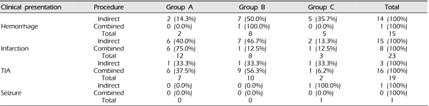
There were 21 male hemispheres (36.2%) and 37 fe- male hemispheres (63.8%). In the indirect group, cere- bral infarction was the most frequent clinical feature at the time of incidence (15 cases; 45.5%). In the com- bined group, transient ischemic attack (TIA) was the most frequent clinical feature at the time of incidence (16 cases; 64.0%). Six complications occurred. In the combined group, there were three hemorrhages and one infarction. In the indirect group, there was one hemorrhage and one infarction (Table 1).
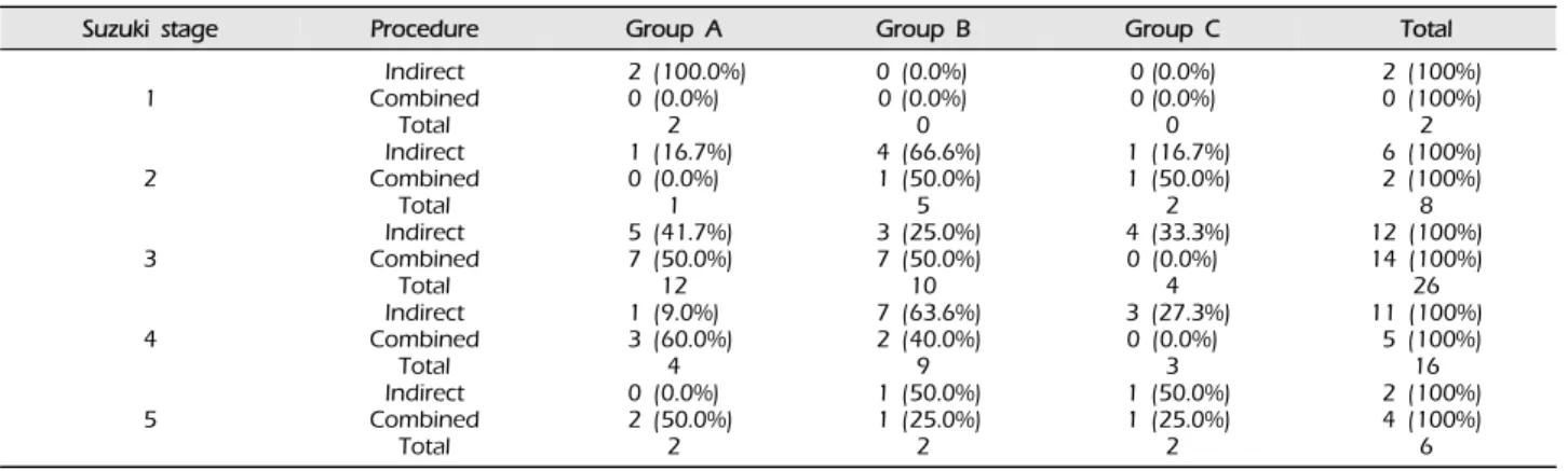
Angiographic outcome after surgery did not differ according to sex (p = 0.493, Table 2), clinical pre- sentation (p = 0.206, Table 3), or Suzuki stage (p = 0.428, Table 4).
As shown in Table 5, in the combined group, 23 cas- es (92.0%) showed a good outcome. In the EDAMS and EDAGS groups, positive outcomes were observed 15 (75.0%) and nine cases (69.2%), respectively. A bet-
Parameter EDAMS
(n = 9) EDAMS + BEGPS
(n = 11) EDAGS
(n = 11) EDAGS + BEGPS
(n = 2) Combined
(n = 23 ) Combined +
BEGPS (n = 2) Total, n = 58 (%) Gender
Males
Females 1
8 4
7 1
10 0
2 13
10 2
0 21 (36.2)
37 (63.8) Clinical presentation
Hemorrhage Infarct TIASeizure
44 10
54 20
5 5 0 1
02 00
1 8 14 0
00 20
15 (25.9) 23 (39.7) 19 (32.8) 1 (1.6) Suzuki stage
1 23 4 5
1 24 2 0
1 24 4 0
0 2 2 5 2
0 02 0 0
0 14 2 4 3
0 00 1 1
2 (3.4) 8 (13.8) 26 (44.8) 16 (27.6) 6 (10.4) Complications
Hemorrhage
Infarction 0
0 1
1 0
0 0
0 3
1 0
0 4 (6.9)
2 (3.4) EDAMS = encephaloduroarteriomyosynangiosis; EDAGS = encephaloduroarteriogaleosynangiosis; BEGPS = bifrontal encephalogaleoperiosteal synangiosis; Combined = direct + indirect; TIA = transient ischemic attack
Table 1. Clinical summary of the 58 hemispheres with moyamoya.
Sex Procedure Group A Group B Group C Total
Male
Female
Indirect Combined
Total Indirect Combined
Total
2 (33.3%) 7 (46.7%) 7 (25.9%)9 5 (50.0%)
12
2 (33.3%) 7 (46.7%) 13 (48.2%)9 4 (40.0%)
17
2 (33.3%) 1 (6.6%) 7 (25.9%)3 1 (10.0%)
8
6 (100%) 15 (100%) 27 (100%)21 10 (100%)
37 Analyzed using the chi-square test.
Group A: Revascularization area was more than two-thirds of the middle cerebral artery (MCA) distribution.
Group B: Revascularization area was between one-third and two-thirds of the MCA distribution.
Group C: Revascularization area was less than one-third of the MCA distribution.
Table 2. Surgical outcomes of bypass according to gender (p = 0.493).
Clinical presentation Procedure Group A Group B Group C Total
Hemorrhage
Infarction
TIA
Seizure
Indirect Combined
Total Indirect Combined
Total Indirect Combined
Total Indirect Combined
Total
2 (14.3%) 0 (0.0%) 6 (40.0%)2 6 (75.0%) 1 (33.3%)12 6 (37.5%)
7 0 (0.0%) 0 (0.0%)
0
7 (50.0%) 1 (100.0%)
7 (46.7%)8 1 (12.5%) 1 (33.3%)8 9 (56.3%)
10 0 (0.0%) 0 (0.0%)
0
5 (35.7%) 0 (0.0%)
5 2 (13.3%) 1 (12.5%) 1 (33.3%)3 1 (6.2%)
2 1 (100.0%)
0 (0.0%) 1
14 (100%) 1 (100%) 15 (100%)15 8 (100%) 3 (100%)23 16 (100%)
19 1 (100%) 0 (100%)
1 Analyzed using the chi-square test.
Group A: Revascularization area was more than two-thirds of the middle cerebral artery (MCA) distribution.
Group B: Revascularization area was between one-third and two-thirds of the MCA distribution.
Group C: Revascularization area was less than one-third of the MCA distribution.
Table 3. Surgical outcomes of bypass according to clinical presentation (p = 0.206).
Procedure Group A Group B Group C Total Combined
EDAMS EDAGS
12 (48.0%) 7 (35.0%) 2 (15.4%)
11 (44.0%) 8 (40.0%) 7 (53.8%)
2 (8%) 5 (25.0%) 4 (30.8%)
25 (100%) 20 (100%) 13 (100%) Group A: Revascularization area was more than two-thirds of the middle cerebral artery (MCA) distribution.
Group B: Revascularization area was between one-third and two-thirds of the MCA distribution.
Group C: Revascularization area was less than one-third of the MCA distribution.
EDAMS = encephaloduroarteriomyosynangiosis; EDAGS = encephaloduroarteriogaleosynangiosis; Combined = direct surgery + indirect surgery
Table 5. Surgical outcomes of bypass according to procedure.
Suzuki stage Procedure Group A Group B Group C Total
1
2
3
4
5
Indirect Combined
Total Indirect Combined
Total Indirect Combined
Total Indirect Combined
Total Indirect Combined
Total
2 (100.0%) 0 (0.0%)
2 1 (16.7%)
0 (0.0%) 5 (41.7%)1 7 (50.0%) 1 (9.0%)12 3 (60.0%)
0 (0.0%)4 2 (50.0%)
2
0 (0.0%) 0 (0.0%)
0 4 (66.6%) 1 (50.0%) 3 (25.0%)5 7 (50.0%) 7 (63.6%)10 2 (40.0%) 1 (50.0%)9 1 (25.0%)
2
0 (0.0%) 0 (0.0%)
0 1 (16.7%) 1 (50.0%) 4 (33.3%)2 0 (0.0%) 3 (27.3%)4
0 (0.0%) 1 (50.0%)3 1 (25.0%)
2
2 (100%) 0 (100%)
2 6 (100%) 2 (100%) 12 (100%)8 14 (100%) 11 (100%)26 5 (100%) 2 (100%)16 4 (100%)
6 Analyzed using the chi-square test.
Group A: Revascularization area was more than two-thirds of the middle cerebral artery (MCA) distribution.
Group B: Revascularization area was between one-third and two-thirds of the MCA distribution.
Group C: Revascularization area was less than one-third of the MCA distribution.
Table 4. Surgical outcomes for bypass according to Suzuki stage (p = 0.478).
Factor Estimated OR (95% CI) p value
Combined vs. EDAMS Combined vs. EDAGS EDAMS vs. EDAGS Combined vs. Indirect
4.107 (0.700-24.096) 4.600 (0.721-29.332) 1.120 (0.239-5.251) 4.313 (0.840-22.130)
0.100 0.088 0.886 0.064 Analyzed using the chi-square test.
Group A: Revascularization area was more than two-thirds of the middle cerebral artery (MCA) distribution.
Group B: Revascularization area was between one-third and two-thirds of the MCA distribution.
Group C: Revascularization area was less than one-third of the MCA distribution.
Table 6. Multiple logistic regression analysis in surgical outcomes.
ter response was observed in the Combined group, compared with the EDAMS group (p = 0.100, odds ra- tio [OR] 4.107, 95% confidence interval [CI] 0.700 - 24.096). Patients in the Combined group showed a better response than those in the EDAGS group (p = 0.088, OR 4.600, 95% CI 0.721 - 29.332). EDAGS and EDAMS showed a similar response (p = 0.886, OR 1.120, 95% CI 0.239 - 5.251). The Combined group showed a better response than the indirect group (p
= 0.064, OR 4.313, 95% CI 0.840 - 22.130, Table 6).
DISCUSSION
The aim of surgical treatment for patients with MMD has been creation of new collateral channels be- tween the external carotid system and cerebral cort- ical arteries.11) All surgical techniques are essentially based on the same surgical principle in which arteries or vascularized tissues (eg, the temporal muscle, the dura mater, the galea, and the omentum) are placed indirectly on the brain surface or direct anastomosis.
In this study, we found that combined bypass tended to result in better development of collateral circulation.
This result suggests that an indirect operation is some- times insufficient, and combined surgery induces not only an immediate but also a slow yet long-standing revascularization by use of all vessels capable of pro- viding the collateral circulation from the external car- otid artery to the brain cortex in MMD.
Therefore, we confirmed that the combination of STA-MCA anastomosis and other indirect surgery is an attempt to achieve a maximal increase in collateral circulation from the STA with surrounding wide ga- leal tissue by contact through its highly vascularized external surface with the ischemic brain cortex as widely and closely as possible.16) Many recent studies have reported good results using combined revascula- rization, rather than a single direct or indirect bypass operation.2)10)17)19)23)
However, our study has some limitations. First, we analyzed revascularization extent using a lateral an- giographic view, which excluded the anteroposterior view of the angiographic image. Thus, a true, accurate three-dimensional volume-concept analysis of re- vascularization extent was not performed. Second, the number of cases was relatively small (43 patients, 58 hemispheres). To solve these problems, we will con- duct a comparison of the degree of collateral circu- lation on accurate three-dimensional volume-concept analysis and study more patients with long-term fol- low-up. Finally, in this study, we used only angio- graphic results. Angiographic results do not always show correlation with clinical ones; therefore, in order to perform a more exact evaluation of the effect of various surgical methods, both results should be con- sidered in a future study.
CONCLUSION
Although the difference was not statistically significant, compared with indirect, combined bypass surgery re- sulted in better development of collateral circulation.
A similar response was observed for EDAMS and EDAGS. Therefore, among surgical procedures, com- bined bypass is a choice that can be recommended.
REFERENCES
1. Amine AR, Moody RA, Meeks W. Bilateral temporal-mid- dle cerebral artery anastomosis for Moyamoya syndrome.
Surg Neurol. 1977 Jul;8(1):3-6.
2. Czabanka M, Vajkoczy P, Schmiedek P, Horn P. Age-de- pendent revascularization patterns in the treatment of moyamoya disease in a European patient population.
Neurosurg Focus. 2009 Apr;26(4):E9.
3. Dauser RC, Tuite GF, McCluggage CW. Dural inversion procedure for moyamoya disease. Technical note. J Neurosurg. 1997 Apr;86(4):719-23.
4. Erickson DL, Koivukangas J. The treatment of Moyamoya disease by superficial temporal-middle cerebral artery (STA-MCA) anastomosis. Ann Clin Res. 1986;18 Suppl 47:21-4.
5. Goda M, Isono M, Ishii K, Kamida T, Abe T, Kobayashi H. Long-term effects of indirect bypass surgery on col- lateral vessel formation in pediatric moyamoya disease. J Neurosurg. 2004 Feb;100(2 Suppl Pediatrics):156-62.
6. Golby AJ, Marks MP, Thompson RC, Steinberg GK. Direct and combined revascularization in pediatric moyamoya disease. Neurosurgery. 1999 Jul;45(1):50-8;discussion 58-60.
7. Han DH, Nam DH, Oh CW. Moyamoya disease in adults:
characteristics of clinical presentation and outcome after encephalo-duro-arterio-synangiosis. Clin Neurol Neurosurg.
1997 Oct;99 Suppl 2:S151-5.
8. Houkin K, Kamiyama H, Takahashi A, Kuroda S, Abe H.
Combined revascularization surgery for childhood moya- moya disease: STA-MCA and encephalo-duro-arterio- myo-synangiosis. Childs Nerv Syst. 1997 Jan;13(1):24-9.
9. Ishii R. [Surgical treatment of moyamoya disease]. No Shinkei Geka. 1986 Aug;14(9):1059-68. Japanese.
10. Ishikawa T, Kamiyama H, Kuroda S, Yasuda H, Nakayama N, Takizawa K. Simultaneous superficial temporal artery to middle cerebral or anterior cerebral artery bypass with pan-synangiosis for Moyamoya disease covering both anterior and middle cerebral artery territories.
Neurol Med Chir (Tokyo). 2006 Sep;46(9):462-8.
11. Karasawa J, Kikuchi H, Furuse S, Kawamura J, Sakaki T. Treatment of moyamoya disease with STA-MCA anastomosis. J Neurosurg. 1978 Nov;49(5):679-88.
12. Karasawa J, Kikuchi H, Furuse S, Sakaki T, Yoshida Y.
A surgical treatment of moyamoya disease encepha- lo-myo synangiosis. Neurol Med Chir (Tokyo). 1977;17(1 Pt 1):29-37.
13. Kashiwagi S, Kato S, Yamashita K, Takasago T, Akimura T, Okamura S, et al. Revascularization with split duro-en- cephalo-synangiosis in the pediatric moyamoya disease- surgical result and clinical outcome. Clin Neurol Neurosurg.
1997 Oct;99 Suppl 2:S115-7.
14. Kawaguchi T, Fujita S, Hosoda K, Shose Y, Hamano S,
Iwakura M, et al. Multiple burr-hole operation for adult moyamoya disease. J Neurosurg. 1996 Mar;84(3):468-76.
15. Kim DS, Kang SG, Yoo DS, Huh PW, Cho KS, Park CK. Surgical results in pediatric moyamoya disease: an- giographic revascularization and the clinical results. Clin Neurol Neurosurg. 2007 Feb;109(2):125-31.
16. Kim DS, Yoo DS, Huh PW, Kang SG, Cho KS, Kim MC. Combined direct anastomosis and encephalodur- oarteriogaleosynangiosis using inverted superficial tem- poral artery-galeal flap and superficial temporal artery–
galeal pedicle in adult moyamoya disease. Surg Neurol.
2006 Oct;66(4):389-94;discussion 395.
17. Kim SH, Choi JU, Yang KH, Kim TG, Kim DS. Risk factors for postoperative ischemic complications in pa- tients with moyamoya disease. J Neurosurg. 2005 Nov;
103(5 Suppl):433-8.
18. Kinugasa K, Mandai S, Kamata I, Sugiu K, Ohmoto T.
Surgical treatment of moyamoya disease: operative tech- nique for encephalo-duro-arterio-myo-synangiosis, its fol- low-up, clinical results, and angiograms. Neurosurgery.
1993 Apr;32(4):527-31.
19. Kuroda S, Houkin K. Moyamoya disease: current con-
cepts and future perspectives. Lancet Neurol. 2008 Nov;
7(11):1056-66.
20. Lee JP, Cho SJ, Park HK, Park SQ, Chang JC, Choi SK.
Angiographic and Clinical Results of Indirect Bypass Surgery for Moyamoya Disease. Korean J Cerebrovasc Surg. 2010 Dec;12(4):250-8.
21. Matsushima T, Fukui M, Kitamura K, Hasuo K, Kuwabara Y, Kurokawa T. Encephalo-duro-arterio-synangiosis in children with moyamoya disease. Acta Neurochir (Wien).
1990;104(3-4):96-102.
22. Park JH, Yang SY, Chung YN, Kim JE, Kim SK, Han DH, et al. Modified encephaloduroarteriosynangiosis with bi- frontal encephalogaleoperiosteal synangiosis for the treat- ment of pediatric moyamoya disease. Technical note. J Neurosurg. 2007 Mar;106(3 Suppl):237-42.
23. Starke RM, Komotar RJ, Connolly ES. Optimal surgical treatment for moyamoya disease in adults: direct versus indirect bypass. Neurosurg Focus. 2009 Apr;26(4):E8.
24. Suzuki J, Takaku A. Cerebrovascular moyamoya disease.
Disease showing abnormal net-like vessels in base of brain. Arch Neurol. 1969 Mar;20(3):288-99.