55 I
Innttrroodduuccttiioonn
Sinus of Valsalva aneurysms (SVAs) are relatively uncommon and may be either congenital or acquired in origin.1) An unruptured SVA is usually asymptomatic and thus often remains undetected. Although the occurrence of rupture is commonly encountered complication related to SVA, unrup- tured SVAs are not infrequently detected recently, thanks to the wide performance of noninvasive echocardiography. An unruptured SVA rarely protrudes into the right ventricular outflow tract (RVOT) and in turn generates the RVOT obstruction.2)Here, I describe an asymptomatic patient who was diagnosed as having an unruptured SVA with the RVOT obstruction.
C Caassee
A 44-year-old female with an 8-year history of hyperten- sion visited our outpatient clinic for surgical treatment of varicose vein. She was referred to the cardiology department for perioperative risk stratification. The patient complained of mild dyspnea on exertion. Her blood pressure was 130/70 mmHg and her pulse rate, 66 beats per minute. On physical examination, an ejection systolic murmur of grade III/VI
was audible at the left second intercostal area. She was in sinus rhythm on electrocardiogram (Fig. 1). A chest X-ray showed mild cardiomegaly without abnormal finding in the lung parenchyma (Fig. 2). Transthoracic echocardiography (TTE) clearly showed an SVA involving the right coronary sinus (Fig.
3A and B), along with mild aortic regurgitation. The ascend- ing aorta was not dilated and the left ventricular systolic func- tion was normal without any demonstrable regional wall
pISSN 1975-4612/ eISSN 2005-9655 Copyright © 2009 Korean Society of Echocardiography www.kse-jcu.org DOI: 10.4250/jcu.2010.18.2.55
C
CAASSEE RREEPPOORRTT J Cardiovasc Ultrasound 2010;18(2):55-57
• Received: November 11, 2009 • Revised: April 5, 2010 �Accepted: May 18, 2010
• Address for Correspondence: Sung-Hee John, Division of Cardiology, Department of Internal Medicine, Presbyterian Medical Center, 68 Seowon-ro, Wansan-gu, Jeonju 560-750, Korea Tel: +82-63-230-8913, Fax: +82-63-230-8945, E-mail: [email protected]
A
A R Ra ar re e C Ca as se e o of f U Un nr ru up pt tu ur re ed d S Siin nu us s o of f V
Va al ls sa al lv va a A An ne eu ur ry ys sm m O Ob bs st tr ru uc ct tiin ng g t th he e R Riig gh ht t V
Ve en nt tr riic cu ul la ar r O Ou ut tf fl lo ow w T Tr ra ac ct t
S
Suunngg--HHeeee JJoohhnn,, MMDD
Division of Cardiology, Department of Internal Medicine, Presbyterian Medical Center, Jeonju, Korea
An unruptured sinus of Valsalva aneurysm is rare and is usually asymptomatic until a symptom associated with its complication develops. Hence, an unruptured sinus of Valsalva aneurysm is not infrequently missed unless echocardiogram is performed with other indications. An unruptured sinus of Valsalva aneurysm rarely protrudes into the right ventricular outflow tract, causing the right ventricular outflow tract obstruction. In this report, I describe a rare case of unruptured sinus of Valsalva aneurysm producing the right ventricular outflow tract obstruction, which was incidentally detected by echocardiography.
KEY WORDS:Sinus of Valsalva∙Aneurysm∙Ventricular outflow obstruction.
online ©ML Comm
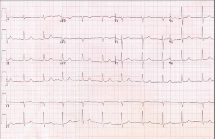
Fig. 1. Electrocardiogram shows normal sinus rhythm.
motion abnormalities. Despite no evidence of intracardiac abnormal shunt flow, abnormal systolic flow acceleration was noted at the RVOT adjacent to the right SVA, sugges- tive of the RVOT obstruction (Fig. 3C). For confirmation, transesophageal echocardiography (TEE) was performed. As a result, similar to TTE findings, a large right coronary SVA encroaching the RVOT and producing the RVOT obstruc- tion was clearly identified (Fig. 4). There was no shunt flow suggesting SVA rupture. The patient underwent elective surgery for SVA repair. Postoperative course was uneventful, and she was allowed to leave hospital.
D
Diissccuussssiioonn
SVAs are relatively uncommon with a reported incidence of 0.14-0.23%.1) Most SVAs are congenital in origin, but they may be seen after bacterial endocarditis, atherosclerosis or chest trauma.3)4)SVA rupture can also occur with ventri- cular septal defect.5) SVAs are thought to result from the absence of normal elastic and muscular tissue, leading to thinning of the wall of the aortic sinus and its dilation, finally resulting in SVA rupture, most commonly into the right ventricle or the right atrium. In a previous study rec- ruiting 332 patients, pathological rupture of a SVA occurred, in descending order, in the right (76.8%), the noncoronary (20.2%), and the left sinus of Valsalva (3%).6)
Unruptured SVA is rare and is usually asymptomatic, until a symptom of the accompanied complications develops. In this respect, an unruptured SVA is likely to be missed. Even more uncommonly, an unruptured SVA encroaches into the RVOT and can cause the RVOT obstruction with or without subjective symptoms.2)7)
Initially, invasive angiography was considered the gold standard for diagnosing this disease, TTE and/or TEE em- erged as preferred modalities of choice nowadays. In parti- cular, TTE and/or TEE is a quick and noninvasive method that can provide additional information on the size and location of aneurismal dilatations, the presence of fistulous
Journal of Cardiovascular Ultrasound 18 || June 2010
56
Fig. 2. Chest X-ray shows mild cardiomegaly.
Fig. 4. Transesophageal echocardiography. Midesophageal aortic valve long-axis view at 144。C rotation demonstrating the SVA originating from the right coronary sinus of Valsalva (arrow) (A). Short-axis view at 27。C rotation demonstrating the SVA protruding into the RVOT (arrowheads) (B). Color Doppler shows flow acceleration due to the RVOT obstruction without shunt flow suggesting SVA rupture (arrow) (C). SVA: sinus of Valsalva aneurysm, RVOT: right ventricular outflow tract, LV: left ventricle, LA: left atrium, Ao: aorta.
A B C
LV LA
AO
LA
RVOT Fig. 3. Transthoracic echocardiography. Parasternal long axis view (A) shows an aneurysm of the right sinus of Valsalva (arrow). Parasternal short axis views (B and C) show also the aneurysm (arrowheads) and systolic flow acceleration suggesting the RVOT obstruction (arrow). RVOT: right ventricular outflow tract, LV: left ventricle, LA: left atrium, Ao: aorta.
A B C
LV
LA AO
LA RVOT
LA RVOT
LA
RVOT
tract, the presence or absence of cardiac chamber involve- ment, the degree of aortic insufficiency, and identification of any associated anomaly or complication.
Optimal management of an asymptomatic, unruptured SVA is not established, since no precise natural history is currently available. Unruptured SVAs, although remaining silent, may expand, cause more severe symptoms, and require more extensive corrective procedures in the future. For this reason, some authors recommend early correction in these types of lesions, even without any subjective symptom.8) Furthermore, surgical treatment of SVAs can be performed with an acceptably low operative risk and, if successful, good long-term survival can be secured. Taken together, early sur- gical intervention is now recommended that is expected to prevent the development of more severe symptoms, and more extensive disease leading to more complicated and less satis- fying repairs.8)9)
In conclusion, I report here, for the first time in Korea, a rare case of an unruptured SVA obstructing the RVOT de- tected incidentally by echocardiography in an asymptomatic patient.
R
Reeffeerreenncceess
1. Goldberg N, Krasnow N. Sinus of Valsalva aneurysms. Clin Cardiol 1990;13:831-6.
2. Rosenberger P, Cohn LH, Fox JA, Locke A, Shernan SK. Sinus of Valsalva aneurysm obstructing the right ventricular outflow tract. Anesth Analg 2006;102:1660-1.
3. Freedom RM, Yoo SJ. Sinus of Valsalva aneurysm. In: Freedom RM, Yoo SJ, Mikailian H, William WG editors. The natural and modified history of congenital heart disease.1st Edition. New York: Blackwell Publishing;
2004. p.183-5.
4. Vereckei A, Vándor L, Halász J, Karádi I, Lengyel M. Infective endo- carditis resulting in rupture of sinus of Valsalva with a rupture site commu- nicating with both the right atrium and right ventricle. J Am Soc Echocar- diogr 2004;17:995-7.
5. Bansal RC, Wangsnes KM, Bailey L. Right aortic sinus of Valsalva-to- right ventricle fistula complicating bacterial endocarditis of membranous ventricular septal defect: evaluation by two-dimensional, color flow, and Doppler echocardiography. J Am Soc Echocardiogr 1993;6:308-11.
6. Chu SH, Hung CR, How SS, Chang H, Wang SS, Tsai CH, Liau CS, Tseng CD, Tseng YZ, Lee YT, et al. Ruptured aneurysms of sinus of Valsalva in Oriental patients. J Thoracic Cardiovasc Surg 1990; 99:288-98.
7. Regueiro Abel M, Penas Lado M, López Ciudad V, Castro Beiras A.
[Sinus of Valsalva aneurysm as a cause of acute myocardial infarction.] Rev Esp Cardiol 2002;55:77-9.
8. Takach TJ, Reul GJ, Duncan JM, Cooley DA, Livesay JJ, Ott DA, Frazier OH. Sinus of Valsalva aneurysm or fistula: management and out- come. Ann Thorac Surg 1999;68:1573-7.
9. Vural KM, Sener E, Tasdemir O, Bayazit K. Approach to sinus of Valsalva aneurysm: a review of 53cases. Eur J Cardiothorac Surg 2001;20: 71-6.
Unruptured Sinus of Valsalva Aneurysm|| Sung-Hee John
57