수궐음 심포경근의 해부학적 고찰
박 경 식1
1상지대학교 한의과대학 해부학교실
Study on the Anatomical Pericardium Meridian Muscle in Human
Kyoung-Sik Park
11
Dept. of Anatomy, College of Oriental Medicine, Sangji University
Abstract
Objectives : This study was carried to identify the component of the Pericardium Meridian Muscle
in human.Methods : The regional muscle group was divided into outer, middle, and inner layer. The
inner part of body surface were opened widely to demonstrate muscles, nerve, blood vessels and to expose the inner structure of the Pericardium Meridian Muscle in the order of layers.Results : We obtained the results as follows;
․The Pericardium Meridian Muscle composed of the muscles, nerves and blood vessels.
․In human anatomy, it is present the difference between terms (that is, nerves or blood vessels which control the muscle of the Pericardium Meridian Muscle and those which pass near by the
Pericardium Meridian Muscle).
․ The inner composition of the Pericardium Meridian Muscle in human is as follows ; 1) Muscle
P-1 : pectoralis major and minor muscles, intercostalis muscle(m.) P-2 : space between biceps brachialis m. heads.
P-3 : tendon of biceps brachialis and brachialis m.
P-4 : space between flexor carpi radialis m. and palmaris longus m. tendon(tend.), flexor digitorum superficialis m., flexor digitorum profundus m.
P-5 : space between flexor carpi radialis m. tend. and palmaris longus m. tend., flexor digitorum superficialis m., flexor digitorum profundus m. tend.
P-6 : space between flexor carpi radialis m. tend. and palmaris longus m. tend., flexor digitorum profundus m. tend., pronator quadratus m.
H-7 : palmar carpal ligament, flexor retinaculum, radiad of flexor digitorum superficialis m. tend., ulnad of flexor pollicis longus tend. radiad of flexor digitorum profundus m. tend.
H-8 : palmar carpal ligament, space between flexor digitorum superficialis m. tends., adductor pollicis m., palmar interosseous m.
▪교신저자 : 박경식, 강원도 원주시 우산동 660, 상지대학교 한의과대학 해부학교실, Tel. 033-730-0667, Fax. 033-730-0653, E-mail : [email protected]
∙본 연구는 2004년도 교내연구비 지원에 의하여 수행되었음.
·접수 : 2005/02/22 ·수정 : 2005/03/16 ·채택 : 2005/03/22
The K orean Journal of M eridian & Acupoint
Ⅰ. Introduction
The concept of Meridian Muscle shown in Ling Shu (Miraculous Pivot) of HUANDI
NEIJING (A Bible in traditional chinese
medicine for about two thousand years) is closely connected with the Twelve MainMeridian. Main Meridian or Meridian Muscle
is a general term of muscular system distributed in circulation of the Twelve Main Meridian, classified into 3Yin (The Negative)-3Yang (ThePositive) of upper & low limb
1) and composed of muscular tissue such as muscle (involvingtendon), fascia, ligament,2) which Chi (Gie : life energy) in the Twelve Main Meridian is collected for or concluded or translated into.3)
The Twelve Meridian Muscle is distributed in
the body surface of limb, trunk or head part, and in most case it's way is made in the opposite direction to the tip of limb.The term of Meridian Muscle has a deep meaning in myology, arthrology, rehabilitation, and the other clinics. Since anatomical, constituent elements of individual Meridian
Muscle are wrongly known to the academic
world of oriental medicine, it bring about a H-9 : radiad of extensor tend. insertion.2) Blood vessel
P-1 : lateral cutaneous branch of 4th. intercostal artery, pectoral br. of thoracoacromial art., 4th.
intercostal artery(art.)
P-3 : intermediate basilic vein(v.), brachial art.
P-4 : intermediate antebrachial v., anterior interosseous art.
P-5 : intermediate antebrachial v., anterior interosseous art.
P-6 : intermediate antebrachial v., anterior interosseous art.
P-7 : intermediate antebrachial v., palmar carpal br. of radial art., anterior interosseous art.
P-8 : superficial palmar arterial arch, palmar metacarpal art.
P-9 : dorsal br. of palmar digital art.
3) Nerve
P-1 : lateral cutaneous branch of 4th. intercostal nerve, medial pectoral nerve, 4th. intercostal nerve(n.)
P-2 : lateral antebrachial cutaneous n.
P-3 : medial antebrachial cutaneous n., median n. musculocutaneous n.
P-4 : medial antebrachial cutaneous n., anterior interosseous n. median n.
P-5 : median n., anterior interosseous n.
P-6 : median n., anterior interosseous n.
P-7 : palmar br. of median n., median n., anterior interosseous n.
P-8 : palmar br. of median n., palmar digital br. of median n., br. of median n., deep br. of ulnar n.
P-9 : dorsal br. of palmar digital branch of median n.
Conclusions : This study shows some differences from already established study on Meridian Muscle.
Key words : Meridian Muscle, meridian point(P1∼9), muscle, blood vessel, nerve
mistaken clinical application or a wrong diagno- sis.
This study was carried out in order to investigate correct elements of the Twelve
Meridian Muscle and to support the meridia-
nology or oriental clinics .At this time we report the Pericardium
Meridian Muscle in human following the Lung Meridian Muscle,
4)the Large Intestine Me- ridian Muscle,
5)the Spleen Meridian Muscle,
6)the Small Intestine Meridian Muscle,
7) and theHeart Meridian Muscle
8).Ⅱ. Materials and methods
1. Reagents and injection
1) The preparation of a preservative
Phenol weighing one kilogram was dissolved in one litre methylalcohol (The 1st solution).
The 500 ㎖ of glycerin was dissolved in 2 ℓ of methylalcohol and thereafter the additional 500 ㎖ of glycerin was dissolved in this solution (The 2nd solution). The 1st and 2nd solution were well mixed, and made warm (30min, 20℃). The 1 ℓ of methylalcohol was added to this mixed solution, and was stirred for 10minutes. For the last time 1.5 ℓ of formalin was added to this mixed solution.
2) Injection
The sheath of femoral artery & vein were exposed by vertical incision at the medial third of inguinal ligament, and femoral artery is carefully separated from femoral vein.
A preservative was injected into femoral artery at the speed of 150 ㎖ per minute.
After 6 ℓ of preservative was injected, A needle-inserted part was ligated, and subseque- ntly injector needle was inserted downwards for the preservation of the leg.
2. Embalmment of cadaver and experimental procedure
1) Cadaver was pending in the embalm- ment system for 40 hrs at 40℃.
2) Cadaver was exposed for 1hr at the normal temperature, and after that, was kept in refrigerated storage (3℃, 30%
humidity).
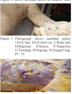
3) The Pericardium Meridian Muscle was labelled by latex at the surface of cadaver, subsequently photographed.
4) Pore was made by drill in the vertical direction at each meridian point.
5) Skin and superficial fascia were stripped off in the order and thereafter were labelled by latex at the exposed deep fascia surface, once more was photographed.
6) Deep fascia was also removed.
7) Subsequently muscle, tendon, nerve, blood vessels were investigated, photographed, and being divided into three layers (outer, middle, and inner layer).
Ⅲ. Results
The Pericardium Meridian Muscle was
marked at the surface of cadaver, and investigated. And also constituent elements was divided into three layers (outer, middle, inner layer)and identified as follows
1) A schema of the Pericardium Meridian
Muscle (Figure 1, and referred to Figure 8)
2) Muscle, blood vessels, nerve constitu- ting the Pericardium Meridian Muscle.
1. Ch'ōnji (P1)
As shown in figure 2, muscle group at outer layer is composed of pectoralis major muscle, that at middle layer is composed of pectoralis minor muscle, at deep layer intercos-
talis muscle.
Lateral cutaneous branch of 4th. interco- stal artery belongs to blood vessel at outer layer, and there is pectoral br. of thoracoacromial artery at middle layer, 4th. intercostal artery, at inner layer.
In case of nerves there are lateral cutane- ous branch of 4th. intercostal nerve, medial pectoral nerve in middle layer, and 4th. inter- costal nerve at inner layer.
2. Ch'ōnch'ōn (P2)
Muscle related to this meridian muscle is biceps brachialis muscle at outer layer, because Ch'ōnch'ōn corresponds to space between bra- chii biceps muscle heads (Figure 3).
Nerve are composed of lateral antebrachial cutaneous nerve.
3. Kokt'aek (P3)
As gathering from this study, muscle group constituting this meridian muscle are biceps brachialis muscle tendon at outer layer and brachialis muscle at inner layer (Figure 3, 4), as for blood vessel, intermediate basilic vein and brachial artery at outer layer.
Medial antebrachial cutaneous nerve and median nerve lie at outer layer, musculocutane- ous nerve at middle layer (Figure 3).
4. Kūgmun (P4)
Constituent elements of this meridian muscle are space between flexor carpi radialis muscle and palmaris longus muscle tendon Figure 1. Photograph shows meridian points
(A:Ch'ōnji, B:Ch'ōnch'ōn, C:Kokt'aek, D:Kūgmun, E:Kansa, F:Naegwan, G:Taenūng, H:Nogung, I:Chungch'ung, P1∼9)
Figure 1 shows meridian points in cadaver skin.
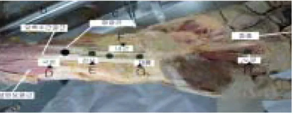
Figure 2. Photograph shows meridian points (Ch'ōnji:P1) and muscles (a:pectoralis minor muscle, b:5th.intercostalis muscle, c:4th.intercostalis muscle, d:3th.inter- costalis muscle)
(Figure 5), intermediate antebrachial vein, medial antebrachial cutaneous nerve at outer layer, flexor digitorum superficialis muscle at middle layer (Figure 6), and flexor digitorum profundus muscle, anterior interosseous artery, anterior interosseous nerve, median nerve at inner layer (Figure 7).
5. Kansa (P5)
Muscular elements of this meridian muscle are space between flexor carpi radialis muscle tendon and palmaris longus muscle tendon at outer layer (Figure 5), flexor digitorum superficialis muscle at middle layer (Figure 6),
flexor digitorum profundus muscle tendon at inner layer (Figure 7).
There are intermediate antebrachial vein at outer layer, anterior interosseous artery at Figure 3. Photograph in middle layer level
shows meridian points (b:Ch'ōnch'ōn, C:Kokt'aek), muscles (a:coracoid m., d:brachialis m., e:biceps brachialis m.), and musculocutaneous nerve (c)
Figure 4. Photograph in inner layer level shows meridian points (P2:Ch'ōnch'ōn, P3:
Kokt'aek), muscles (a:biceps brachialis m. insertion, b:brachialis m., c:biceps brachialis m. short head), vessels (e,f:brachial v.a), and nerves (d:mus- culocutaneous n., g:median n.)
Figure 5. Photograph at outer muscular level shows meridian points (D:Kūgmun, E:Kansa, F:Naegwan, G:Taenūng, H:Nogung) and muscles (a:brachiora- dialis m., b:flexor carpi radialis m., c:palmaris longus m. tend.)
Figure 6. Photograph at middle muscular level shows meridian points (D:Kūgmun, E:Kansa, F:Naegwan, G:Taenūng), muscles (a:flexor digitorum superficialis m., b:flexor pollicis longus m.) and median nerve.
Figure 7. Photograph at inner muscular level shows meridian points (D:Kūgmun, E:Kansa, F:Naegwan, G:Taenūng), muscles (a:flexor pollicis longus m. tend., flexor digitorum profundus m. tend., c:flexor digitorum superficialis m.)
middle layer.
Median nerve exist at outer layer, and also anterior interosseous nerve at middle layer.
6. Naegwan (P6)
Constituent elements of this meridian muscle are similar to above-mentioned Kūg- mun, Kansa. Space between flexor carpi radialis muscle tendon and palmaris longus muscle tendon constitute muscle group at outer layer (Figure 5), flexor digitorum profundus muscle tendon at middle layer, and pronator quadratus muscle at inner layer (Figure 7).
Outer layer are composed of intermediate antebrachial vein, median nerve, and inner layer, anterior interosseous artery & anterior interos- seous nerve.
7. Taenūng (P7)
Outer layer (Figure 5)are composed of palmar carpal ligament & flexor retinaculum, intermediate antebrachial vein, palmar br. of median nerve and middle layer, composed of space between flexor carpi radialis muscle tendon and palmaris longus muscle tendon, radiad of flexor digitorum superficialis muscle tendon (Figure 5, 6), median nerve. There are ulnad of flexor pollicis longus muscle tendon, radiad of flexor digitorum profundus muscle tendon, palmar carpal br. of radial artery, anterior interosseous artery, anterior interos- seous nerve at inner layer (Figure 7).
8. Nogung (P8)
At outer layer (Figure 5) palmar carpal
ligament (palmar aponeurosis), space between flexor digitorum superficialis muscle tendons, lumbrical muscle, superficial palmar arterial arch palmar br. of median n., palmar digital br.
of median nerve exist. There are adductor pollicis muscle, br. of median nerve at middle layer (Figure 6), palmar interosseous muscle, palmar metacarpal artery, deep br. of ulnar n.
at inner layer.
9. Chungch'ung (P9)
Constituent elements of outer layer are composed of radiad of extensor tend. insertion, dorsal br. of palmar digital artery, dorsal br. of palmar digital br. of median nerve (Refer to Figure 1).
Ⅳ. Discussion
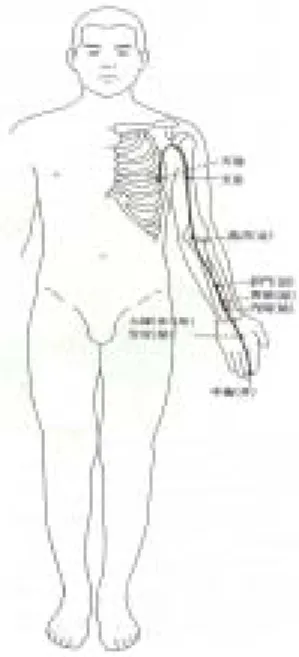
The Pericardium Meridian Muscle origi-
nates from inner chest, goes down piercing diaphragm, subsequently is connected with triple energizer meridian. Main branch reaches axillary region , especially Ch'ōnch'ōn meridian point. That passes by anterior & medial side of brachium and lies at space between the LungMeridian Muscle and the Heart Meridian Muscle, and subsequently arrives at Kokt'aek
situated at anterior condyle of humerus, passes by anterior of antebrachium, shortly it goes through anterior side of wrist joint, after that anterior surface of palm, finally reachesChungch'ung at the tip of middle finger.
1) (Figure 8)Meridian Muscle in oriental medicine
means a concept comprising soft tissue such as muscle, fascia ligament, and nerve on the out skirts of them.9) But we mean to comprice blood vessel additionally in this concept.
It is possible to know the mode of action of
Meridian Muscle
if we analyze the distribution of Meridian Muscle in connection with human anatomy.10,11,12) In the view of clinical application, Meridian Muscle plays an important role in the flexion & extention of muscle or joint or limb, since the abnormality of Meridian Muscle is expressed as the abnormalities of Meridian Muscle-piercing part, such as stretching, convulsion, relax, rigidity, displacement.13,14,15,16)Referring to the disability of Meridian Muscle, the chapter
Meridian Muscle of Ling Shu (Miraculous
Pivot) explains the following meaning "if Yang is over, the muscle extended and so long as Yinis over, then the muscle flexed. Cold brings about the muscle contraction, and hot, muscle slackness."17) This means the symptom of disease induced by abnormal meridian muscle subsquent to Yang or Yin over.
As mentioned above, the anatomical knowledge of muscle is essentially required for the clinical application of Meridian Muscle. And also at the same time such a knowledge must be exact. Such a knowledge guarantees the exact and effective application of Meridian
Muscle to clinics.
This study shows some differences from already established study1,17) on Meridian
Muscle ; that is, constituent elements of Meri- dian Muscle such as muscle, nerve, blood
vessels, ligament, fascia, and assay method.Above all the structure of each meridian point investigated in this study was divided into three layers according to depth from body surface but on the other hand we came across that it may be wide differences in opinion according to the disparity of real meridian point or the angle of acupunctuation.19)
References
1. A joint work of the allied department of acupuncture & moxibustion, college of ori- ental medicine. Acupuncture & Moxibustion (the 1st volume). 3Eds. Seoul : Gypmundang.
1991 : 565-79.
2. Song CH. Study on the 12 Meridian Muscle and the 12 Skin. The Journal of Korean Acupuncture & Moxibustion. 1989 ; (6)1 : 179-86.
Figure 8. The scheme of the Pericardium
Meridian in human.
3. 南京中醫學院. 鍼灸學. 上海科學技術出版社.
1984 : 7-8.
4. Lee JM, Park KS. Anatomy of the Lung
Meridian Muscle in Human. Kor J AM-
Meridian & Pointology. 2001 ; 18(2) : 19-25.5. Lee JM, Sim Y, Park KS. Anatomy of the
Large Intestine Meridian Muscle in Human.
Kor J Meridian & Acupoint. 2002 : 19(1) : 15-23.
6. Park KS. Anatomy of the Spleen Meridian
Muscle in Human. Kor J Meridian & Acupoint.
2003 : 20(4) : 65-76.
7. Park KS. Anatomical Study on the Small
Intestine Meridian Muscle in Human. J of
Kor Inst of Herbal Acupuncture. 2004 ; 7(2) : 57-64.8. Park KS. Anatomical Study on the Heart
Meridian Muscle in Human. J of Kor Orient
Med. 2005 ; 26(1) 45-55.9. 上海中醫學院. 傷科學. Hongkong : 商務印書 館. 1977 : 6-14.
10. PO Abrahams, RT Hutchings, SC Marks Jr.
Mc Minn's Color Atlas of Human Anatomy.
Wolfe Publishing. 1977 : 109-19.
11. H Ferner, J Stubesand. Sobotta's Atlas of Human Anatomy(Anatomy 1). 10th Eds.
Munich : Urban & Schwarzenberg. 1983 : 318-63.
12. Frank H Netter. Atlas of Human Anatomy (CIBA). 1987 : Plate 401-52.
13. P Butler. Imaging of the Nervous System.
Springer-Verlag. 1990 : 231-40.
14. RS Snell. Neuroanatomy. 3rd Ed. Little &
Brown. 1992 : 417-559.
15. C. Liebenson etc. Rehabilitation of the Spine.
Williams & Wilkins Inc. 1996 : 61-71.
16. Roentgen Interpretation, J. H. Juhl, 5th Ed., Harper & Row, 1981.
17. Jang MH. HUANDI NEIJING (A Korean version). Seoul : Sungbosa, 1975 : 145-54.
18. Ahan YK. Acupuncture & Moxibustion. Seoul : Sungbosa. 1991 : 259-79.
19. Choi HS. A Search on New Direction of Meridian Study. Kor J Meridian & Acupoint 2004 ; 21(3) : 1-20.