Anti-Oxidant Property and Inhibition of Melanin Synthesis of Eight Plant Extracts
Kim, Jae Young1, Jin Young Lee2, Wi Young Lee3, Yongsub Yi1,2*, and Yoongho Lim4
1Department of Biochemistry, Hoseo University, Asan, Korea
2Department of Herbal Cosmetics Science, Hoseo University, Asan, Korea
3Biotechnology Division, KFRI, Suwon 441-350, Kora
4Department of Bioscience and Biotechnology, Konkuk University, Seoul 143-701, Korea
Plants extracts are good resources to find functional compounds for human health. The following eight plants were collected and total phenolic contents were determined. Acer psedo-siebolianum showed the highest phe- nolic contents, 16.4 mg/g, whereas Cercidiphyllum japonica showed the lowest contents, 1.9 mg/g. The DPPH free radical scavenging capacities of the plant extracts showed high activity in following order : Acer ginnala (21.3 µg/mL) > Cornus walteri (23.9 µg/mL) > Distylum racemosum (29.2 µg/mL) > Castanopsis cuspidata var. Thunbergii (31.7 µg/mL) > Acer psedo-siebolianum (34.6 µg/mL) > Thuijopsis dolabrata cv. Aurea (53.1 µg/mL) > Cercidiphyllum Japonica (115.2 µg/mL). Also the mushroom tyrosinase inhibitory activities of total extracts were determined at different concentration. D. racemosum extract showed highest (49.1% at 1,000 mg) in inhibitory activity than other seven extracts. The ethanol fraction (IC50 value: 118.1 µg/mL) from D.
racemosum showed more inhibitory activity than ethyl acetate fraction (IC50 value: 203 µg/mL). The ethanol fraction on showed no significant cytotoxicity in B16/F1 cells line up to 60 µg/mL. Over 80 µg/mL of ethanol fraction showed cytotoxicity in B16/F1 cells. The melanin contents of cells were significantly attenuated by ethanol fraction in a dose-dependent manner. The IC50 value of ethanol fraction was 75.4 µg/mL.
Key words: Antioxidant activity, tyrosinase inhibition, plant extracts, depigmentation agent
Introduction
Plant extracts have been traditionally used as medicines for disease treatments, and widely used as topical applica- tions for wound-healing and anti-aging. Examples of these include ginkgo biloba, echinacea, ginseng, grape seed, green tea, lemon, lavender, rosemary, sarsaparilla, soy, prickly pear, sagebrush, jojoba, aloe vera, allantoin, feverwort, bloodroot, apache plume, and papaya. With the above reason, plant extracts were thought as good materials for human health care [14, 22].
Reactive oxygen species (ROS) are produced during several intracellular pathways, and they induce oxidative stress. Accumulation of ROS can cause oxidative alteration on cell constituents, and the alteration cause an irreversible oxidative damage over lifetime of a cell [11]. Oxidative
stress is generally related to generation of several diseases such as Alzheimer's diseases [18], Parkinson's disease [5], rheumatoid arthritis [25], the pathologies caused by dia- betes [7, 13], neurodegeneration in motor neurone disease [9], and cardiovascular disease [6, 23]. Antioxidants were used to represent the chemicals that prevented the con- sumption of oxygen. For this reason, antioxidants become necessary for the use as supplements to human health.
Although several strong synthetic antioxidants have already been reported and used, the antioxidants from natural sources are used as supplements to human health. A wide range of natural compounds including phenolic compounds, nitrogen compounds, and careteniods has good antioxida- tive properties [22].
Melanin is the major pigment secreted by melanocyte cells in basal layer of human skin, and overproduced by chronic sun exposure, or other hyperpigmentation diseases [4, 12]. Tyrosinase is a key enzyme that catalyzes melanin synthesis in melanocyte cells. Melanin biosynthesis can be inhibited by inhibition of melanocyte metabolism and
*Corresponding author
Tel: 82-41-540-5979, Fax: 82-41-541-5979 E-mail: [email protected]
proliferation, by inhibition of tyrosinase.
A number of tyrosinase inhibitors have been identified from both natural and synthetic sources and may be used for the treatment of some skin disorders associated with melanin hyperpigmentation. Also, they are important in cosmetics for skin whitening effects [15]. Thus, there is a need to identify the compounds that inhibit tyrosinase activity.
Materials and Methods
Plant materials and extraction
Eight plants, Distylum racemosum, Thuijopsis dolabrata cv. Aurea, Cercidiphyllum japonica, Acer psedo-siebolia- num, Acer geiseum, Castanopsis cuspidata var. thunbergii, Conus walteri, Acer ginnala were collected from various places in Korea in September, 2008, and all identified before extracted.
The dried plants were crushed with a pulverizer. The plant materials were extracted two times with a 70%
ethanol at room temperature for 24 hours and centrifuged to remove the debris. The extracts were evaporated to dryness using a rotary evaporator (Eyela, Japan). The extract powders were stored at -20oC until use.
Chemical and media
Folin-Ciocalteu reagent was purchased from Wako chemical Co. (Japan), and catechin, 1,1-diphenyl-2-picryl- hydrazyl, linoleic acid, muchroom tyrosinase, dihydroxy- phenylalanine (L-DOPA), 3-(4,5-Dimethylthiazol-2-yl)-2,5- diphenyltetrazoliumbromide (MTT), trichloroacetic acid (TCA), Dimethyl sulfoxide (DMSO) and all other chemical were purchased from Sigma (WI, USA). Organic solvents were purchased from J.T. baker (USA) at HPLC grade.
Dulbecco's Modified Eagle's Medium, fetal bovine serum, DPBS, trypsin, FBS, penicillin, and streptomycin were purchased from the Thermo Scientific Co. (USA).
Total polyphenolic content assay
Polyphenolic contents were determined using a protocol similar to the method described by Folin-Denis method [10]. The total extracts (1.0 mL) was quantitatively trans- ferred to a glass tube with a mixture of 95% ethanol (1.0 mL), distilled water (5.0 mL), and 50% Folin-Ciocalteu reagent (0.5 mL). The mixtures were allowed to react for 5 min and the 1mL NaCO3 was added. The mixture was
thoroughly mixed and placed in the dark for 1 hour. Absor- bance of each sample was measured by spectrophotometer (Bio-Rad, USA) at 735 nm, and catechin was used as a standard for the calibration of phenolic content.
Radical scavenging activity using 1,1-Diphenyl-2-Pi- crylhydrazyl (DPPH)
The free radical scavenging activity of plant extracts was measured by DPPH using the method of Blios [2]. The plants extract (0.1 mL) were transferred to a glass tube with a mixture of 95% ethanol (2.5 mL), and 0.2 mM DPPH solutions (0.5 mL) and mixed throughly, and allowed at room temperature in the dark, and in the absorbance was measured at 517 nm after 15 min.
All experiments were performed in triplicate. Linoleic acid was used as a standard. The IC50 (inhibitory concen- tration) is a concentration of plant extracts required to scavenge 50% DPPH radical.
DPPH radical scavenging activity was calculated as follows: Scavenging effect (%) = (A-B)/A × 100, where A
= absorbance at 517 nm without test sample, where B = absorbance at 517 nm with test sample.
Tyrosinase inhibition assay
The procedure developed by Yagi et al. [26] was adapted for tyrosinase inhibition assay. The test substance was dissolved in 60 mM sodium phosphate buffer (pH 6.8). The reaction mixture was prepared by adding different concen- tration of plants extract (0.5 mL) and was incubated at 25oC for 10 min. After incubation, mushroom tyrosinase (0.2 mL, 110 U/mL) and 10 mM L-DOPA (0.5 mL) were added. The reaction mixture was incubated for 2 min at 25oC. The amount of dopachrome formed was determined by measuring absorbance at 475 nm. The percent of inhi- bition of tyrosinase was calculated as follows: % inhibition
= (A-B)/A × 100, where A = absorbance at 475 nm without test sample, where B = absorbance at 475 nm with test sample. The IC50 (inhibitory concentration) is a concentra- tion of plant extracts required to inhibit 50% tyrosinase activity.
Cell cuture and cell viability assay
The B16/F1 melanoma cells line was obtained from Korean cell Line Bank (Seoul, Korea). Cells were cultured in DMEM containing FBS (10%), penicillin (100 U/mL), streptomycin (0.1 mg/mL) at 37oC in a humidified atmos-
phere of 5% CO2. Cells were harvested after incubation for 24 hours.
Viability of cultured cells was determined by MTT method [17]. Cells were added to each well in 96-well plates, and cultured for 24 hours. After samples treatment, MTT (5 mg/mL in PBS) was added 100 µL to each well.
Cells were incubated at 37oC for 30 min, and DMSO was added to dissolve the formazan crystals. The absorbance was measured at 560 nm with a spectrophotometer.
Melanin contents
The melanin content of the cultured B16/F1 cells was measured as described previously [27]. The cells were washed twice with PBS buffer, and lysed with 20 mM Tris- 0.1% Triton X-100 (pH 7.5). Cell lysates were precipitated with the same amount of 20% TCA. After washing twice with 10% TCA, the pellets were treated with a 3:1 mixture of ethyl alcohol and diethyl ether. Samples were air-dried, dissolved in 1mL of 0.85 M KOH, and boiled for 15 min.
After cooling, absorbance was measured with a spectro- photometer at 440 nm. The amount of cellular melanin was corrected according to the protein content of the samples.
The protein content was determined by the method of Bradford [3].
Results and Discussion
Determination of total polyphenolic content
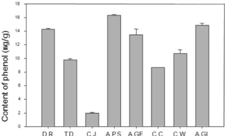
The polyphenolic contents of eight extracts varied sig- nificantly (Fig. 1). Acer psedo-siebolianum showed the highest phenolic content, 16.4 mg/g, whereas Cercidiphyllum japonica showed the lowest content, 1.9 mg/g. The other extracts were shown as follow : Acer ginnala (14.9 mg/g)
> Distylum racemosum (14.2 mg/g) > Acer geiseum (12.6 mg/g) > Conus walteri (10.3 mg/g) > Thuijopsis dolabrata cv. Aurea (9.8 mg/g) > Castanopsis cuspidata var. thun- bergii (8.7 mg/g).
Antioxidant activity of plant extracts
Antioxidant activity of the eight plant extracts was deter- mined by DPPH free radical scavenging activity (Table 1).
DPPH is a free radical donor widely used to evaluate the free radical scavenging effects of natural antioxidants [16].
The DPPH free radical scavenging capacities of the plant extracts showed high activity in following order : Acer ginnala (21.3 µg/mL) > Cornus walteri (23.9 µg/mL) >
Distylum racemosum (29.2 µg/mL) > Castanopsis cuspi- data var. Thunbergii (31.7 µg/mL) > Acer psedo-siebolia- num (34.6 µg/mL) > Thuijopsis dolabrata cv. Aurea (53.1 µg/mL) > Cercidiphyllum Japonica (115.2 µg/mL).
The antioxidant activity of phenolic compound depends on properties of their molecular structure, such as the availability of phenolic hydrogen and the possibility of stable phenoxyl radical formation. In addition, it has been reported that there is linear correlation between DPPH radical scavenging activity and concentration of phenolic compounds in various vegetable and fruits [20]. The above report showed the same results that there was a nearly linear correlation between DPPH radical scavenging activity and concentration of phenolic compounds.
Inhibitory effect of mushroom tyrosinase by total extracts
The mushroom tyrosinase inhibitory activities of total extracts were determined using L-DOPA as a substrate Fig. 1. Concentration of total polyphenolic compounds of eight plant extracts. D.R : Distylum racemosum, T.D: Thuijopsis dola- brata cv. Aurea, C.J: Cercidiphyllum japonica, A.P.S: Acer psedo- siebolianum, A.GE: Acer geiseum, C.C: Castanopsis cuspidata var. thunbergii, C.W: Conus walteri, A.GI: Acer ginnala.
Table 1. Antioxidative activity of total extracts by DPPH assay.
Plant extract Antioxidative activity (IC50µg/mL)
Acer geiseum 38.5
Acer ginnala 21.3
Acer psedo-siebolianum 34.6
Castanopsis cuspidata var. thunbergii 31.7
Cercidiphyllum japonica 115.2
Cornus walteri 23.9
Distylum racemosum 29.2
Thuijopsis dolabrata cv. Aurea 53.1
(Fig. 2). Total extracts were assayed at different concentra- tion. The Distylum racemosum extract showed the highest (49.1% at 1,000 mg) in tyrosinase inhibitory activity than other plant extracts, whereas the Thuijopsis dolabrata cv.
Aurea extract showed the lowest activity.
Tyrosinase inhibitory and antioxidant activity of Dis- tylum racemosum fractions
The tyrosinase inhibitory and antioxidant activity of fractions were determined using L-DOPA and DPPH as substrates (Table 2). The IC50 concentration of fractions was calculated after linear regression analysis. The ethanol fraction (IC50 value: 118.1 µg/mL) from D. racemosum showed more inhibitory effect than ethyl acetate fraction (IC50 value: 203 µg/mL) on tyrosinase inhibition. Also the ethanol fraction (0.9 µg/mL) showed the highest DPPH free redical scavenging capacities.
Effect of ethanol fraction on cytotoxicty
B16/F1 cells were treated with ethanol fraction to deter- mine whether it has a cytotoxicity effect. The results showed that ethanol fraction was not significant cytotoxicity to B16/F1 cells up to 60 µg/mL. Over 80 µg/mL of ethanol fraction showed cytotoxicity in B16/F1 cells (Fig. 3).
Effect of ethanol fraction on melanin content
Changes of the melanin contents in the B16/F1 cells treated with ethanol fraction were evaluated for depigment- ation activity. The melanin contents of cells were signifi- cantly attenuated by ethanol fraction in a dose-dependent manner (Fig. 4). The IC50 value of ethanol fraction was 75.4 µg/mL.
Fig. 2. Tyrosinase inhibition activity of eight plant extracts. : Distylum racemosum, : Cercidiphyllum japonica, : Acer psedo- siebolianum, : Acer geiseum, : Castanopsis cuspidata var.
thunbergii, : Cornus walteri, : Acer ginnala, : Thuijopsis dolabrata Cv. Aurea (not detection).
Table 2. DPPH radical scavenging activity and tyrosinase inhi- bition of fractions of Distylum racemosum.
treatment
Antioxidative activity (IC50µg/mL)
Tyrosinase inhibition activity
(IC50µg/mL)
Total extract 29.2 1010
Ethyl acetate fraction 2.6 203
Ethanol fraction 0.9 118
Fig. 3. Effects of ethanol fraction on cell viability were deter- mined by MTT assay. C: control.
Fig. 4. Effect of ethanol fraction on melanin content in B16/F1 melanoma cells incubated with various concentration of etha- nol fraction for 24 hours. C: control.
All living organisms have self defence mechanism against reactive oxygen species inside the cell. But sometimes there are the disturbances in normal metabolic process which disrupt the balances by increasing the formation of free radicals. As the results, these free radicals damage various components of the cell including protein, lipids and DNA. In human, oxidative stress is related to generation of several diseases such as Alzheimer's diseases [19], the pathologies caused by diabetes [7, 13], and cardio- vascular disease [23]. In addition to these, oxidative stress is also related to aging [1, 8]. However, it is still the matter of confusion whether the oxidants trigger the disease, or they were produced as a consequence of the disease and cause the disease [19]. Melanocytes contain a large number of specific membranous organelles, the melanosomes which hold the full capacity for biosynthesis and distribu- tion of the melanins [24]. Although melanins play a crucial role in the absorption of free radicals generated within the cytoplasm and protecting the host from UV light, the over- production and accumulation of melanin in skin could be a serious problem resulting in a large number of skin disorders such as melanoma, freckles, senile lentigines, and sites of actinic damage [21]. In cosmetics industry, melanin contents in human skin play a negative role due to reducing skin whitening effects. As the results, there is a need to find new compounds to reduce melanin contents in human skin, and the use of the inhibitors is primary in the cosmetic industry. In this study, we attempted to find new compo- nents to reduce melanin synthesis in cells, and found some possibility.
Acknowledgement
This work was supported by a grant (Code 20100301- 061-049) from Biogreen 21 Program, rural Development Administration, Republic of Korea.
REFERENCES
1. Aviram, M. 2000. Review of human studies on oxidative damage and antioxidant protection related to cardiovascular disease. Free radic. Res. 33: 85-97.
2. Blios, M. S. 1958. Antioxidant determination by the use of a stable free radical. Nature 26: 1190-1200.
3. Bradford, M. M. 1976. A rapid and sensitive method for the quantitation of microgram quantities of protein utilizing the principle of protein-dye binding. Anal. Biochem. 72: 248-
254.
4. Briganti, S., E. Camera, and M. Picardo. 2003. Chemical and instrumental approaches to treat hyperpigmentation. Pig- ment Cell Research 16: 101-110.
5. Christen, Y. 2000. Oxidative stress and Alzheimer disease.
Am. J. Clin. Nutr. 71: 621-629.
6. Cookson, M. and P. Shaw. 1999. Oxidative stress and motor neurone disease. Brain Pathol. 9: 165-186.
7. Davi, G., A. Falco, and C. Patrono. 2005. Lipid peroxidation in diabetes mellitus. Antioxid. Redox signal. 7: 256-268.
8. Finkel, T. and N. J. Holbrook. 2000. Oxidants, oxidative stress and the biology of aging. Trends Biochem. Sci. 25:
502-508.
9. Giugliano, D., A. Ceriello, and G. Paolisso. 1996. Oxidative stress and diabetic vascular complications. Diabetes Care 19:
257-267.
10. Gutfinger, T. 1981. Polyphenols in olive oils. J. Am. Oil Chem. Soc. 58: 966-968.
11. Halliwell, B., R. Aeschbach, J. Lolinger, and O. I. Aruoma.
1995. The characterization of antioxidants. Food Chem. Tox- icol. 33: 601-617.
12. Hearing, V. J. 2005. Biogenesis of pigment granules: a sensi- tive way to regulate melanocyte function. Journal of Derma- tological Science 37: 3-14.
13. Hitchon, C. and H. El-Gabaawy. 2004. Oxidation in rheuma- toid arthritis. Arthritis Res. Ther. 6: 265-278.
14. Hsu, S. 2005. Green tea and the skin. J. Am. Acad. Derma- tol. 52: 1049-1059.
15. Maeda, K. and M. Fukuda. 1996. Arbutin : mechanism of its depigmenting action in human melanocyte culture. J. Phar- macol. Exp. Ther. 276: 765-769.
16. Matusukawa, R., Z. Dubinsky, E. Kishimoto, K. Masak, Y.
Masuda, T. Takeuchi, M. Chihara, Y. Yamamoto, E. Niki, and I. Karube. 1997. A comparison of screening methods for antioxidant activity in seaweeds. Journal of Applied Physiol- ogy 9: 29-35.
17. Mosmann, T. 1983. Rapid colorimetric assay for cellular growth and survial: application to proliferation and cytotox- icity assays. J. Immunol. Methods 65: 55-63.
18. Nunomura, A., R. Castellani, X. Zhu, P. Moreira, G. Perry, and M. Smith. 2006. Involvement of oxidative stress in Alzheimer disease. J. Neuropathol. Exp. Nerrol. 65: 631-641.
19. Raha, S. and B. Robinson. 2000. Mitochondria, oxygen free radical, antioxidants and human disease: where are we now?
J. Lab. Clin. Med. 119: 598-620.
20. Rice-Evans, C., N. J. Miller, and G. Paganda. 1996. Struc- ture-antioxidant activity relationships of flavonoids and phe- nolic acid. Free Radical Biology and Medicine 20: 933-956.
21. Seiji, M., K. Shimao, M. S. C. Birbeck, and T. B. Fitz- patrick. 1963. Subcellular localization of melanin biosynthe- sis. Ann. N. Y. Acad. SCI. 100: 497-533.
22. Shimizu, K., R. Kondo, K. Sakai, N. Takeda, T. Nagahata, and T. Oniki. 2001. Novel vitamin E derivative with 4-sub- stituted resorcinol moiety has both antioxidant and tyrosi- nase inhibitory properties. Lipids 36: 1321-1326.
23. Van-Gaal, L., I. Mertens, and C. De-Block. 2006. Mecha- nisms linking obesity with cardiovascular disease. Nature 444: 875-880.
24. Velioglu, Y. S., G. Mazza, Y. L. Gao, and B. D. Oomah.
1998. Antioxidant activty and total phenolics in selected fruits, vegetables and grain products. J. Agric. Food Chem.
46: 4113-4117.
25. Wood-kaczmar, A., S. Gandhi, and N. Wood. 2006. Under- standing the molecular causes of Parkinson's disease. Trends Mol. Med. 12: 521-528.
26. Yagi, A., T. Kanabara, N. Morinobu. 1986. The effect of tyrosinase inhibition for aloe. Plantamedica 3981: 517-519.
27. Yang, J. Y., J. H. Koo, Y. G. Song, K. B. Kwon, J. H. Lee, H.
S. Sohn, B. H. Park, E. C. Rhee, and J. W. Park. 2006. Stim- ulation of melanogenesis by scoparone in B16 melanoma cells. Acta Pharmacol. Sin. 27: 1467-1473.
(Received Aug. 12, 2010/Accepted Nov. 16, 2010)
국문초록
수종의 식물수출물의 항산화 및 Melanin 합성 억제효과 김재영1·이진영2·이위영3·이용섭1,2*·임융호4
1호서대학교 생화학과, 2호서대학교 한방화장품과학과
3국립산림과학원, 4건국대학교 생명공학과
본 연구에서는 식물 추출물을 이용하여 항산화 활성 및 tyrosinase 활성 억제 효과를 측정하였다. 식물 추출물의 폴 리페놀물질의 총 함량은 Acer psedo-siebolianum의 추출물이 16.4 mg/g로 가장 높은 추출량을 나타내었다. 항산화 활성 측정에서는 Acer ginnala 에서 IC50값으로 21.3 µg/mL으로 가장 좋은 활성을 나타내었다. 반면에 L-DOPA를 기질로 하여 mushroom tyrosinase의 활성 억제측정에서는 Distylum racemosum 1,000 mg에서 49.1%로 다른 추출 물에 비하여 상대적으로 높은 활성을 나타내었다. Tyrosinase 활성 억제력이 가장 높은 D. racemosum의 추출물을 이 용하여 ethanol 분획과 ethyl acetate 분획으로 분리하여, 이중 D. racemosum의 ethanol 분획에서 항산화 활성 IC50
값은 0.9 µg/mL, tyrosinase 활성억제는 IC50값이 118.1 µg/mL로 ethyl actate 분획보다 높은 활성을 나타내었다. 또 한 ethanol 분획을 이용하여 B16/F1 melanoma cell에서는 60 µg/mL까지는 세포독성을 나타내지 않았으며 80 µg/
mL의 농도에서 약간의 세포독성을 나타내었다. 에탄올 분획을 이용한 세포내 melanin 색소의 생산억제 IC50값은 75.4
µg/mL로 분석되었다. 이러한 결과로 D. racemosum의 에탄올 추출물이 B16/F1 melanoma cell세포의 melanin색소합 성대사에 관여하여 색소합성을 저해하는 것으로 보인다.