CASE REPORT 1) F/26, Mild Chest Discomfort
A 26-year-old woman visited our hospital due to inter- mittent discomfort on left lower hemithorax. The past history and family history of the patient were not remarkable. Phy- sical and laboratory findings were within normal limits. A
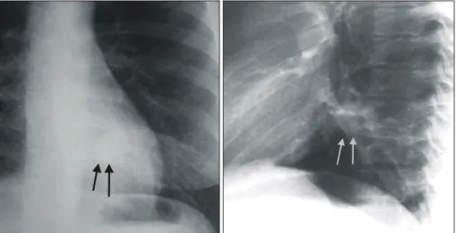
chest radiograph demonstrated about 2 cm sized well-defined nodular density at left retrocardiac area (Fig. 1). Chest CT with contrast enhancement showed a large anomalous artery arising from lower thoracic aorta supplying basal segments of left lower lobe. No evidence of bronchial abnormalities was noted in the lung (Fig. 2). Pulmonary arteriography showed absence of normal pulmonary artery and vein to basal seg- 김응수*․전의용**․이광우**․구동억***
Treatment of Systemic Arterial Supply to Lower Lobe of Left Lung (Operation vs. Embolotherapy): Comparison of Two Cases and Literature Review
Eung-Soo Kim, M.D.*, Eui-Yong Jeon, M.D.**, Gwang-Woo Rhee, M.D.**, Dong-Erk Goo, M.D.***
Systemic arterialization of lung with/without sequestration (Sequestration/Anomalous Origin of Left Pulmonary Artery, AOLPA) is a rare form of congenital anomalous systemic arterial supply to the lungs. In this anomaly, the arterial supply of one or more arteries of the basal segments of the lower lobe derives from an aberrant vessel arising from the aorta. We report two adult cases of systemic arterialization of normal basal segments of left lower lobe lung with/without sequestration. The one (AOLPA) was treated by left lower basal segmentectomy and the other (Sequestration) by therapeutic angiographic embolization. Based on the favorable follow-up result in our patients, al- though lobectomy (segmentectomy) is the basic treatment modality, embolotherapy could also be a mode of treat- ment that could be selectively applied to elderly, infirm patients or high risk patients with poor pulmonary function.
(Korean J Thorac Cardiovasc Surg 2006;39:230-235) ꠏꠏꠏꠏꠏꠏꠏꠏꠏꠏꠏꠏꠏꠏꠏꠏꠏꠏꠏꠏꠏꠏꠏꠏꠏꠏꠏꠏꠏꠏꠏꠏꠏꠏꠏꠏꠏꠏꠏꠏꠏꠏꠏꠏꠏꠏꠏꠏꠏꠏꠏꠏꠏꠏꠏꠏꠏꠏꠏꠏꠏꠏꠏꠏꠏꠏꠏꠏꠏꠏꠏꠏꠏꠏꠏꠏꠏꠏꠏꠏꠏꠏꠏꠏꠏꠏꠏꠏꠏꠏꠏꠏ Key words: 1. Lung, anomaly
2. Pulmonary arteries 3. Embolization
*한전의료재단 한일병원 흉부외과
Department of Thoracic & Cardiovascular Surgery, Hanil General Hospital, KEPCO Medical Foundation
**한전의료재단 한일병원 진단방사선과
Department of Radiology, Hanil General Hospital, KEPCO Medical Foundation
***순천향대학교병원 진단방사선과학교실
Department of Radiology, College of Medicine, Soonchunhyang University Hospital
†Presented at the Vascular 2004, Rotorua, New Zealand, September 3-8, 2004.
논문접수일:2005년 9월 23일, 심사통과일:2005년 12월 7일
책임저자:김응수 (132-703) 서울시 도봉구 쌍문 3동 388-1, 한전의료재단 한일병원 흉부외과 (Tel) 02-901-3090, (Fax) 02-901-3094, E-mail: [email protected]
본 논문의 저작권 및 전자매체의 지적소유권은 대한흉부외과학회에 있다.
ments of left lung. The pulmonary artery supplied entire right lung, left upper lobe and superior segments of left lower lobe and the pulmonary vein drained from concomittent parenchy-
me. Thoracic aortography showed anomalous systemic artery from left lateral margin of descending aorta, supplying left lower lobe basal segments of lung, a normal parenchymal
About 2 cm sized nodular density at left retrocardiac area (arrows) in normal lung parenchyme is noted on chest PA and left lateral views.
Fig. 2. Well-enhancing nodular lesion at left lower medial lung zone (A), increased vascularity of left lower lobe (B), and normal pulmonary parenchyma and bronchial structure are noted (C, D) in chest CT with enhancement.
A B
C D
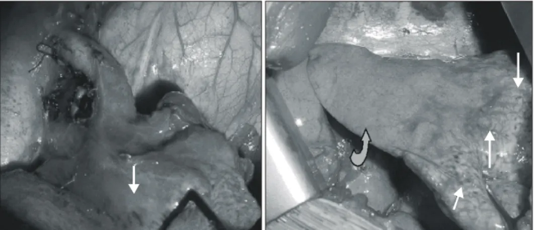
phase and a draining vein to normal left lower pulmonary vein (Fig. 3). On the diagnosis of anomalous systemic ar- terial supply to the basal segments of left lower lobe from descending aorta without sequestration, left lower basal segmentectomy with ligation of the abnormal artery was do- ne. Operative findings revealed similar findings to radiogra- phic results, and the involved lung surface showed multiple tiny telangiectases. The anomalous artery was about 1 cm in diameter in the left pulmonary ligament at its origin and be- came tortuous cranially to the proximal part of lower lobe like a swan neck-shaped curve, and then aneurysmally dilated up to 1.5 cm running along with normal pulmonary artery in the basal segments (Fig. 4). The pathologic examination showed a large anomalous artery of elastic type, normal bronchial communication, and multiple telangiectases in the visceral pleural surface. The patient did well after operation without evidence of complications.
2) M/16, Incidental Lung Nodule
A 16-year-old man admitted to the hospital with acute right chest pain due to pneumothorax. Closed thorocostomy was done. About 1 cm sized nodular density was found inci- dentally at left retrocardiac area on chest radiograph. Chest CT showed well-enhancing vascular structure at left lower medial lung zone (Fig. 5A), and the fissure density at left retrocardiac area. The findings suggested it to be pulmonary sequestration. The patient's past and family history were not remarkable. On the diagnosis of pulmonary sequestration, tho- racic aortography was done. It showed a curved shadow of left lower lung that suggested an accessary fissure artifact of the pulmonary sequestration, and an about 0.7 cm sized ano- malous systemic artery from the descending thoracic aorta supplying left lower lobe basal segments of lung. A draining vein to left lower pulmonary vein and no evidence of ab- Fig. 3. Thoracic aortography shows typical anomalous systemic artery with swan neck appearance and draining vein to left inferior pulmonary vein from left lower lobe basal segments of lung (A, B). Pulmonary arteriography shows absent segmental arterial and venous branches of left lower lobe basal segments (C∼F).
A B C
F E
D
normal fistula draining to the pulmonary vein was found (Fig.
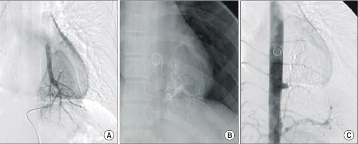
5B, C). Therapeutic embolization was done with metallic coils and gelfoam particles (Fig. 6). The patient was discharged in a week, and did well after embolotherapy for 2 year follow-up without evidence of lung abscess, pneumonia, or systemic thromboembolism.
DISCUSSION
Anomalous systemic arterial supply to the lungs has been described in bronchopulmonary sequestration, systemic arteria- lization of normal lung (Anomalous Origin of Left Pulmonary Artery, AOLPA) and the scimitar syndrome. This vessel al- ways supplies one of the lower lobes, frequently one of the
basal segments[1-6]. The absence of parenchymal abnorma- lities and a normal bronchial supply clearly distinguishes systemic arterial supply without sequestration from true se- questration[4]. In AOLPA, the feeding vessel in systemic ar- terial supply of normal lung without sequestration arises from the distal part of the thoracic descending aorta, the proximal abdominal aorta or the celiac axis, and it is an elastic type artery[2-6]. The systemic artery can measure up to 1 cm in diameter, is smaller at its origin in the pulmonary ligament, but getting dilated just beyond the site entering the lobe, especially in the left-sided cases, as in our case 1. In most cases atresia of the corresponding pulmonary artery was present[2,4]. There were some cases with normal branching of the pulmonary artery in the involved segment[7,8]. The lower lobe superior segment of left lower lobe of lung (curved arrow).
Fig. 5. Anormalous artery is noted at left lower medial lung zone on en- hanced CT (A). Aortography shows anomalous artery from left side of lower thoracic aorta (arrow) (B). Se- lective arteriography shows localized parenchymal lung staining without evi- dence of fistulous lesion (C).
A
C B
venous return was always via normal inferior pulmonary vein, causing a left-to-left shunt[2-8].
Symptoms of AOLPA were cough, sputum, fever, and dyspnea, but were usually asymptomatic in most patients. The- refore most of these were diagnosed as an incidental finding on the chest roentgenogram. However, left ventricular failure from left-to-left shunt, or massive hemoptysis may result[2,5,7].
We treated two patients with systemic arterialization of normal basal segments of left lower lobe by basal segmentectomy in the one (AOLPA) and therapeutic embolization in the other (Se- questration) without significant complications in 2 year fol- low-up. Based on the favourable follow-up result in our patients, lobectomy (segmentectomy) should be performed as the basic treatment modality. However, embolotherapy may be considered selectively an alternative to thoracotomy in patient with systemic arterialization of lower lung to high risk patients of old age or with debilitation, or with poor pulmonary function.
REFERENCES
1. Pryce DM, Sellors TH, Blair LG. Intralobar sequestration of
lung associated with an abnormal pulmonary artery. Br J Surg 1947;35:18.
2. Painter RL, Billig DM, Epstein I. Anomalous systemic ar- terialization of the lung without sequestration. N Engl J Med 1968;279:866-7.
3. Pernot C, Simon P, Hoeffel JC, Worms AM, Marcon F, Pre- vot J. Systemic artery-pulmonary vein fistula without sequest- ration. Pediatr Radiol 1991;21:158-9.
4. Kirks DR, Kane PE, Free EA, Taybi H. Systemic arterial sup- ply to normal basilar segments of the left lower lobe. Am J Radiol 1976;126:817-21.
5. Flisak ME, Chandrasekar AJ, Marsan RE, Ali MM. Systemic arterialization of lung without sequestration. Am J Radiol 1982;138:751-3.
6. Yamanaka A, Hirai T, Fujimoto T, Hase M, Noguchi M, Konishi F. Anomalous systemic arterial supply to normal basal segments of the left lower lobe. Ann Thorac Surg 1999;68:332-8.
7. Campbell DC, Murney JA, Dominy DE. Systemic arterial blood supply to a normal lung. J Am Med Assoc 1962;182:497.
8. Curriano G, Willis K, Miller W. Congenital fistula between an aberrant systemic artery and a pulmonary vein without sequestration. J Pediatr 1975;87:554.
Fig. 6. Venous phase of selective arteriography shows draining vein to normal left inferior pulmonary vein (A). After embolization with microcoils and gelfoam particles, stasis of contrast and embolic material are noted (B). Postembolization aortography shows complete occlusion of anomalous artery with short residual stump at its origin (C).
A B C
중심 단어:1. 폐, 기형 2. 폐동맥 3. 색전술