DOI : 10.3341/jkos.2009.50.7.984
= 증례보고 = 접수번호 : 50-07-10-04
실리콘관의 돌출 및 누소관 열창 발생을 감소시키기 위한 변형된 관 삽입술
이태수⋅장민욱 고려대학교 의과대학 안과학교실
목적: 선천 비루관 폐쇄의 치료인 실리콘관 삽입술의 주된 합병증을 줄이기 위하여 개선한 방법을 소개하고 그 결과를 알아보고자 하였다.
대상과 방법: 선천 비루관 폐쇄를 진단받고 탐침법을 시행받았으나 호전을 보이지 않는 13개월 이상의 연령인 환자 46명을 대상으로 변형된 실리콘관 삽입술을 시행하여 수술 성공률 및 합병증 발생 정도를 조사하였다. 변형된 방법은 관의 길이를 조정하는 방법을 개선한 것으로 관 삽입 후 안구 내안각 쪽에서 튜브를 당긴 후 놓아 탄성에 의해 복원되는 정도를 통해 밴드의 위치를 조정하여 상⋅하 누점에 장력이 최소화되게 하고 탐침 끝에 실리콘 스폰지(5×5 mm)를 끼워 고정한 뒤 고정하는 것이다. 수술 성공의 정의는 유루증상이 없고 형광 색소 저류 검사상 저류가 없는 것으로 하였다.
결과: 수술 시 평균 연령은 32.8개월이었으며 남아가 22명, 여아가 24명이었다. 탐침법 시행 횟수는 평균 1.5회, 평균 추적관찰 기간은 12.6개월, 관 제거는 평균 5.4개월 후에 제거하였다. 수술 성공률은 88%였으며 관의 돌출은 3안, 눈물점 열창은 2안에서 발생하였다.
다른 특별한 합병증은 발견되지 않았다.
결론: 선천 비루관 폐쇄 환자의 치료로 소개한 변형된 방법은 수술 후 흔하게 발생하는 누소관 열창 및 관의 돌출을 줄이는 유용한 방법으로 생각된다.
<대한안과학회지 2009:50(7):984-988>
■ 접 수 일: 2008년 10월 10일 ■ 심사통과일: 2009년 4월 14일
■ 책 임 저 자: 이 태 수
서울시 구로구 구로동길 97번지 고려대학교 구로병원 안과 152-703 Tel: 02-2626-1260, Fax: 02-857-8580 E-mail : tsoooo@hanmail.net
* 본 논문의 요지는 2008년 대한안과학회 제99회 춘계학술대회에서 구연으로 발표되었음.
선천 비루관 폐쇄는 영유아의 눈물기관 이상 중 가장 흔한 질환으로 발생빈도는 저자에 따라 다르나 약 1.75~6% 정 도로 보고되고 있고, 우리나라에서의 발생빈도는 6.25% 정 도로 보고되고 있다.1-5이에 대한 치료로 점안약과 마사지 등의 보존적 치료를 우선적으로 시행하며 효과가 없을 경 우 1세 이하에서는 주로 탐침법을 시행한다. 탐침법의 보고 된 성공률은 약 70~93% 정도로 높으나6-14수회의 탐침법 으로도 호전이 없을 경우에는 수술적 치료를 고려하며 일반 적으로 실리콘관 삽입술을 시행한다. 소아의 경우 성공률은 보고에 따라 약 80~97%로 높으나15-19누점의 미란, 누소 관 열창, 관의 돌출, 관의 이탈, 결막 또는 각막 자극 등 합 병증이 발생할 수 있다.15-20특히 Anderson and Edwards21 은 실리콘관 삽입술 후 초기 합병증으로 관의 이탈 및 빠짐 현상이 가장 흔하게 발생한다고 보고한 바 있다. 이러한 합 병증을 줄이기 위한 방법으로 Hong and Chang은 실리콘
관의 탈출을 막기 위해 6~0 nylon으로 관을 묶었으나 효과 가 적었다고 보고하였다.22저자들은 선천 비루관폐쇄의 치 료로 시행되는 실리콘관 삽입술의 합병증인 관의 이탈 및 누 소관 열창의 발생률을 감소시키기 위하여 개선한 방법을 소 개하고 그 결과를 알아보고자 하였다.
대상과 방법
2000년 12월부터 2007년 12월까지 고려대 구로 병원 안과에 내원하여 선천 비루관 폐쇄로 진단된 환자 중 최소 1회 이상 탐침법을 받았으나 유루 증상이 호전되지 않은 환 자 46명 48안을 대상으로 하였다. 수술 시 연령이 13개월 이상인 경우로 포함하였으며 12개월 이내에 다른 보존적 치료를 받았거나 최소 1회 이상의 탐침법을 시행받은 환자 만을 대상으로 하였다. 그리고 누점이나 누소관협착이 있는 경우, 출혈 성향이 있는 경우, 스테로이드 치료에 금기가 되는 경우, 외상에 의한 경우, 누기에 종양이나 결석이 의심되는 경우, 급성 또는 만성 누낭염이 있는 경우 등은 대상에서 제외하였다.
선천성 비루관 폐쇄는 병력상 출생 후 수개월 내에 눈꼽 과 유루 증상이 있고, 다른 원인을 찾을 수 없는 경우로 진 단하였다. 수술 전후에 있어 추적기간 동안 형광 색소 저류 검사와 Munk’s scale을 평가하여 최종 관찰 시 형광 색소
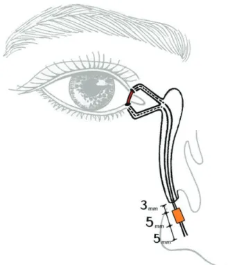
Figure 1. Bicanalicular silicone tube is in place and silicone sponge is placed 2~3 mm below the inferior meatus.
Figure 2. Schematic diagram shows that the ideal position of the silicone tube intubated in modified surgery is in place without tension and the silicone sponge is placed 2~3 mm below the inferior meatus.
Table 1. Characteristics of the patients
Mean±SD Range
Age at surgery (months) 32.8±18.9 13~96 Numbers of previous probing 1.5±1.3 1~3 Follow up period (months) 12.6±14.2 1~54 Timing of tube removal (months) 5.4±1.3 0.25~6
Table 2. Postoperative complications
No of patients (%)
Protrusions of tube 3 (6.2)
Slitting of canaliculus 2 (4.2)
Granuloma formations 3 (6.2)
Canaliculitis 1 (2.1)
Nasal bleeding 1 (2.1)
저류 grade 0~1, Munk’s scale grade 0~1인 경우를 성공 으로 정의하였다.
변형된 실리콘관 삽입술은 기존의 삽입술과 근본적으로 동일한 방법이지만 관의 길이를 조정하는 방법을 새롭게 고안한 것이었다. 우선 전신마취하에 하비갑개 밑으로 1:
100,000으로 희석된 에피네프린용액(보스민Ⓡ액, 제일약 품, Korea)으로 적신 5 cm×5 cm의 면거즈로 비강 점막을 수축시킨 다음, 누점을 확대하였고, 그 후 #00 Bowman 탐 침으로 탐침법을 시행하여 하비갑개 밑으로 탐침이 나옴을 확인한 후, BIKAⓇ(Bicanalicular intubation set, S1.1000, FCI, Fracne)을 이용하여 삽입술을 시행하였다. 삽입된 실 리콘관이 하비도의 정확한 위치에서 나오는가를 비강 내시 경을 이용하여 확인하였다. 관 삽입 후 비강 내에서 탐침을 통하여 양실리콘관에 5×5 mm 크기의 실리콘 스폰지를 끼 워 넣고 안구쪽 내안각에서 사시구(Jameson muscle hook) 를 이용하여 튜브를 당긴 후 놓아 그 탄성에 의해 복원되는 정도를 보았다. 이를 수차례 반복하여 튜브의 장력이 상, 하 누점에 영향을 주지 않는 편안한 상태가 되면 이에 맞게 실
리콘 스폰지의 비강 내 위치를 조정한 뒤 5~0 prolene으로 실리콘 스폰지에 두 가닥의 실리콘관을 고정하였다. 이상적 실리콘 스폰지의 위치는 하비갑개에서 약 2~3 mm 정도 하 방으로 하였다(Fig. 1, 2). 이 후 안구쪽에서 수차례 튜브를 당겨 더 이상 누소관 열창을 일으킬 수 있는 장력이 존재하 지 않음을 확인한 뒤 수술을 마쳤다. 수술 후에는 점안 항 생제 및 점안 스테로이드 제제를 1개월에서 수개월간 사용 하였으며 수술 후 외래에서 1개월 마다 정기적으로 추적 관 찰하였다. 그리고 관제거는 전신마취하에 수술실에서 시행 하였다.
결 과
선천 비루관 폐쇄 환자는 총 46명 48안이었으며 남자가 22명 여자가 24명이었다. 수술 시 평균 연령은 평균 32.8±
18.9개월이었으며, 수술 이전에 시행받은 탐침 횟수는 1.5±
1.3회였다. 수술 후 평균 추적 관찰 기간은 12.6±14.2개월 이었으며, 실리콘관 제거는 수술 후 평균 5.4±1.3개월 이었 다(Table 1). 수술 성공률은 형광 색소 저류 검사와 Munk’s scale을 평가하여 최종 관찰 시 42안에서 성공하여 약 88%
의 수술 성공률 소견을 보였다. 합병증이 발생한 경우는 총 10안으로 관의 이탈이 3안(6.3%), 누소관 열창이 2안
(4.2%), 육아종 생성이 3안(6.2%), 비출혈이 1안(2.1%), 누소관 감염이 1안에서 발생하였다(Table 2). 이 중 누소관 열창은 수술 후 경과 관찰 중 열창이 점차 진행하여 4 mm 이상되었을 때로 정의하였고, 그 이하의 진행하지 않는 작은 크기의 열창은 포함하지 않았다. 위의 합병증 발생안 중 누 소관 열창 2안, 누소관 감염 1안에서 수술 후 1개월 이내에 관을 조기 제거하였다. 수술 실패는 총 6안에서 발생하였는 데, 관의 이탈이 2안, 누소관 열창이 2안, 누소관 협착이 1안, 그리고 합병증은 발생하지 않았으나 증상의 호전이 없던 경우가 1안이었다.
고 찰
실리콘관 삽입술의 초기 합병증으로 관의 이탈 및 누소 관 열창이 비교적 흔히 발생하며 이로 인해 수술 성공률에 영향을 미칠 수 있다. 국내외 보고에 따르면 선천 비루관 폐쇄 환자에게 실리콘관 삽입술을 시행한 이후 관의 이탈 및 누소관 열창의 발생 정도가 저자에 따라 다르나 20안 이 상을 보고한 연구에서는 약 25~36% 정도의 발생률을 보 였다.23-28Yoon et al23 20안 중 9안(45%)에서 발생하였다 고 보고하였으며, Cho et al24은 31안 중 11안(34%), 그리 고 Mun and Jung25은 55안 중 20안(36%)에서 관이 이탈 및 누소관 열창이 발생하였다고 보고하였다. 국외의 보고에 서 Lim et al27은 122안 중 31안(25%)에서 발생하였다고 보고하였으며, Ratliff and Meyer26은 30안 중 7안(23%)에 서 발생하였다고 보고하였다. 흔하게 발생하는 관의 탈출을 막기 위한 연구도 다양하게 보고되었는데, Jordan and Anderson29은 40명의 환자를 대상으로 수술 시 4~0 silk로 관을 묶음으로써 수술 후 관의 돌출이 발생하지 않았다고 보고하였으나 이는 추적관찰 기간이 3개월로 짧다는 한계 가 있었다. Yazici et al30은 50안을 대상으로 두 가닥의 실 리콘 관을 서로 묶은 뒤 5~0 또는 6~0 polyglactin으로 실 리콘 관과 비강 점막을 봉합하여 비강 내 고정하는 방법을 사용하였으나 50안 중 18안(36%)에서 관의 이탈이 발생 하였다고 보고하였으며, 국내에서도 Hong and Chang22은 관의 이탈을 막기 위해 6~0 nylon을 사용하여 관을 묶었으나 68예 중 9예(13.2%)에서 관의 이탈 및 탈출이 발생하였다.
그러나 본 연구에서는 관의 이탈이 3안(6.2%), 누소관 열창이 2안(4.1%)에서 발생하여 다른 보고들에 비하여 월 등히 합병증 발생률이 낮았다. 물론 환자의 수술 전 상태나 나이, 수술 성공의 정의 등의 차이로 발생률을 직접적으로 비교하는 것은 어려우나 본 연구에서 관의 이탈 및 누소관 열창의 발생 정도가 매우 낮았다.
수술 초기에 발생하는 관의 이탈 및 누소관 열창은 수술
성공 및 예후에 영향을 줄 수 있다. Welsh and Katowitz28 수술 후 3개월 이전에 관의 조기 제거 시 수술 성공률이 55.6%로 6개월 이상 유지하였을 때의 95.2%에 비하여 현 저하게 낮다고 보고한 바 있다. 즉 수술 초기에 관의 이탈 및 누소관 열창의 발생으로 인한 관의 조기 제거는 수술 예 후에 영향을 미칠 수 있다. 본 연구에서도 총 6안에서 수술 이 성공하지 못 하였는데 이 중 관의 이탈 및 누소관 열창 에 의한 실패가 4안으로 전체 실패의 67%를 차지하였다.
물론 본 연구에서의 수술 성공률은 약 88%로 다른 저자들 의 80~ 90% 정도의 성공률과 비교할 때16-20 월등히 높은 것은 아니지만 관의 조기 제거 원인이 될 수 있는 합병증을 줄임으로써 성공률을 높일 수 있다고 생각한다.
단, 이 수술방법의 한계점은 수술 및 관 제거 시에 모두 전신마취를 해야 한다는 것인데, 이에 대한 대안이 추후 연 구에서 필요할 것으로 생각된다.
결론적으로 저자들이 고안한 변형된 실리콘관 삽입술은 수술 및 관 제거 시에 모두 전신마취를 해야하는 단점이 있 으나, 관의 위치 이탈 및 누소관 열창을 최소화할 수 있는 좋은 방법으로 생각된다.
참고문헌
1) Cassady JV. Dacryocystitis in infancy. Am J Ophthalmol 1948;
31:773-80.
2) Guerry D III, Kendig EL Jr. Congenital impotency in the naso- lacrimal duct. Arch Ophthalmol 1948;39:193-204.
3) Chaabouni M, Zayani A, Chebini S, et al. Congenital obstruction of lacrimal ducts in 578 children. Arch Er Pediatr 1993;50:107-9.
4) Paul TO, Shepherd R. Congenital nasolacrimal duct obstruction:
natural history and the timing of optimal intervention. J Pediatr Ophthalmol Strabismus 1994;31:362-7.
5) Lee SY, Chung HS, Kim HB, et al. The incidence of congenital nasolacrimal duct obstruction on Korean neonates. J Korean Ophthalmol Soc 1989;30:5-8.
6) Ffooks OO. Dacryocystitis in infancy. Br J Ophthalmol 1962;
46:422-34.
7) Baker JD. Treatment of congenital nasolacrimal system obstruc- tion. J Pediatr Ophthalmol Strabismus 1985;22:34-6.
8) Kassoff J, Meyer DR. Early office-based vs late hospital-based nasolacrimal duct probing: a clinical decision analysis. Arch Ophthalmol 1995;113:1168-71.
9) El-Mansoury J, Calhoun JH, Nelson LB, Harley RD. Results of late probing for congenital nasolacrimal duct obstruction. Oph- thalmology 1986;93:1052-4.
10) Honavar SG, Prakash VE, Rao GN. Outcome of probing for congenital nasolacrimal duct obstruction in order children. Am J Ophthalmol 2000;130:42-8.
11) Katowitz JA, Welsh MG. Timing of initial probing and irrigation in congenital nasolacrimal duct obstruction. Ophthalmology 1987;
94:698-705.
12) Sturrock SM, MacEwan CJ, Young JD. Long term results after
probing for congenital nasolacrimal duct obstruction. Br J Ophthalmol 1994;78:892-4.
13) Ahn DH, Helen Lew, Kim HY, Lee SY. The effect of probing for congenital nasolacrimal duct obstruction. J Korean Ophthalmol Soc 1998;39:836-40.
14) Moon JS, Choi WC. Office probing of congenital nasolacrimal duct obstruction. J Korean Ophthalmol Soc 1999;40:2357-61.
15) Dortzbach RK, France TD, Kushner BJ, Gonnering RS. Silicone intubation for obstruction of the nasolacrimal duct in children.
Am J Ophthalmol 1982;94:585-90.
16) Leone CR, Van Gemert JV. The success rate of silicone intubation in congenital nasolacrimal duct obstruction. Ophthalmic Surg 1990;21:90-2.
17) Kraft SP, Crawford JS. Silicone tube intubation in disorders of the lacrimal system in children. Am J Ophthalmol 1982;94:290-9.
18) Beigi B, O’Keefe M. Results of Crawford tube intubation in children. Acta Ophthalmol 1993;71:405-7.
19) Veloudios A, Harvey JT, Philippon M. Long-term placement of silastic nasolacrimal tubes. Ophthalmic Surg 1991;22:225-7.
20) Kim KS, Park TK, Choi WC. Intranasal endoscopic diagnosis and treatment in congenital nasolacrimal duct obstruction. J Korean Ophthalmol Soc 2001;42:7-12.
21) Anderson, RL, Edwards, JJ. Indications, complications, and results with silicone stents. Ophthalmology 1979;86:1474-87.
22) Hong SW, Chang HK. The complications of silicone tube ntuba- tion after lacrimal surgery. J Korean Ophthalmol Soc 1998;39:
2469-76.
23) Yoon TJ, Na KS, Yoon WJ. The effect of silicone tube intubation in pediatric nasolacrimal duct obstruction. J Korean Ophthalmol Soc 2002;43:155-9.
24) Cho KW, Lee SY, Kim SJ. Treatment of congenital nasolacrimal duct obstruction using silicone intubation set. J Korean Ophthal- mol Soc 1995;36:553-8.
25) Mun HJ, Jung WS. Surgical efficacy of probing with silicone intubation for lacrimal apparatus obstruction in children. J Korean Ophthalmol Soc 2002;43:2375-81.
26) Ratliff CD, Meyer DR. Silicone intubation without intranasal fixation for treatment of congenital nasolacrimal duct obstruction.
Am J Ophthalmol 1994;118:781-5.
27) Lim CS, Martin F, Beckenham T, Cumming RG. Nasolacrimal duct obstruction in children: outcome of intubation. J AAPOS 2004;8:466-72.
28) Welsh MG, Katowitz JA. Timing of silastic tubing removal after intubation for congenital nasolacrimal duct obstruction. Ophthal Plast Reconstr Surg 1989;5:43-8.
29) Jordan DR, Anderson RL. Prevention of prolapsed silicone stents in dacryocystorhinostomy surgery. Arch Ophthalmol 1987;105:
455.
30) Yazici B, Akarsu C, Salkaya M. Silicone intubation with the Ritleng method in children with congenital nasolacrimal duct obstruction. J AAPOS 2006;10:328-32.
=ABSTRACT=
A Modified Technique of Bicanalicular Silicone Tube Intubation in Congenital Nasolacrimal Duct Obstruction
Tae Soo Lee, MD, PhD, Minwook Chang, MD
Department of Ophthalmology, Korea University College of Medicine, Seoul, Korea
Purpose: To introduce a modified technique of bicanalicular silicone tube intubation, which can reduce slitting of the canaliculus and protrusion of the tube.
Methods: This study included 46 patients who underwent modified surgery for Congenital Nasolacrimal Duct (CNLD) obstruction.
To be included in this study, patients were older than 13 months and had a history of failed probing. Using our modified technique, tube lengths can be appropriately adjusted by either pulling or releasing the tube at the medial canthus until a tube loop is in place without any tension to the upper and lower canaliculi. Two threads of silicone tube were tied together using 5-0 nylon over a silicone sponge (5×5 mm) and left within the nasal cavity for several months without fixation to the nasal mucosa. A successful surgery was clinically defined as no epiphora and no dye retention in the conjunctival sac.
Results: The mean age of patients at the time of surgery was 32.8 (±18.9) months. There were 22 males and 24 females. Prior to intubation, patients had been probed an average of 1.5 (±1.3) times, and the mean follow-up period was 12.6 (±14.2) months.
The tube was removed at 5.4 (±1.3) months postoperatively on average. The success rates were 88%. Tube protrusions occurred in three eyes, and canalicular splittings were recorded in two eyes. No other serious complications were encountered.
Conclusions: This new technique might enable us to remarkably reduce both protrusion and slitting of the canaliculus in bicanalicular silicone intubation for congenital nasolacrimal duct obstruction.
J Korean Ophthalmol Soc 2009;50(7):984-988
Key Words: Canalicular laceration, Congenital NLD obstruction, Silicone tube intubation, Tube protrusion
Address reprint requests to Tae Soo Lee, MD, PhD
Department of Ophthalmology, Guro Hospital, Korea University College of Medicine
#97 Gurodong-gil, Guro-gu, Seoul 152-703, Korea
Tel: 82-2-2626-1277, Fax: 82-2-857-8580, E-mail: tsoooo@hanmail.net