CASE REPORT
상피하종양 형태로 발현된 호산구성 대장염 1예
윤정희, 박상규, 박호준, 임원, 이태영, 송철수
좋은삼선병원 소화기내과
Eosinophilic Colitis that Presented with Subepithelial Tumor-like Lesions
Jeonghui Yun, Sanggyu Park, Hojun Park, Won Lim, Taeyeong Lee and Chulsoo Song Division of Gastroenterology, Department of Internal Medicine, Good Samsun Hospital, Busan, Korea
Eosinophilic colitis is a rare disease that is characterized by eosinophilic infiltration in the colon wall in symptomatic patients. Thus far, the epidemiology and pathophysiology of eosinophilic colitis have not been well defined, but the hypersensitivity response is likely to play a role in its pathogenesis. The clinical presentation of eosinophilic colitis is usually nonspecific and depends on the layer of the intestinal wall affected by the eosinophilic infiltrate. Eosinophilic colitis is diagnosed generally by exclusion, i.e., after all other causes of eosinophilic infiltration have been excluded. Although there is no consensus over its diagnostic criteria, the laboratory results and radiology and endoscopy findings can provide important diagnostic evidence. This paper reports a case of eosinophilic colitis presenting as subepithelial tumor-like lesions in a 41-year-old man with the chief complaints of abdominal pain and loose stools.
The patient had no diseases and no food or drug allergies in his medical history. In general, the endoscopic findings of eosinophilic colitis can vary from a normal mucosa to frank ulcerations. In this case, however, endoscopy revealed subepithelial tumor-like lesions.
The colon biopsy showed eosinophilic infiltration in the lamina propria. The patient was treated with steroids, and his symptoms re- gressed with no signs of relapse. (Korean J Gastroenterol 2021;77:300-304)
Key Words: Abdominal pain; Endoscopy; Eosinophilia; Colitis
Received January 6, 2021. Revised March 12, 2021. Accepted March 15, 2021.
CC This is an open access article distributed under the terms of the Creative Commons Attribution Non-Commercial License (http://creativecommons.org/licenses/
by-nc/4.0) which permits unrestricted non-commercial use, distribution, and reproduction in any medium, provided the original work is properly cited.
Copyright © 2021. Korean Society of Gastroenterology.
교신저자: 박상규, 47007, 부산시 사상구 가야대로 326, 좋은삼선병원 소화기내과
Correspondence to: Sanggyu Park, Division of Gastroenterology, Department of Internal Medicine, Good Samsun Hospital, 326 Gaya-daero, Sasang-gu, Busan 47007, Korea. Tel: +82-51-322-0900, Fax: +82-51-323-3308, E-mail: psg4864@hanmail.net, ORCID: https://orcid.org/0000-0002-3688-734X
Financial support: None. Conflict of interest: None.
INTRODUCTION
Eosinophilic colitis (EC) is a rare clinical condition included in the group of eosinophilic gastrointestinal disorders (EGIDs) characterized by abnormal infiltration of the gastrointestinal mucosa by eosinophils in the absence of secondary causes.1,2 EC is the least frequent manifestation of EGIDs with a bimodal age distribution. The condition appears to have a first peak in infants and a second peak in young adults. EC is a relatively common pathology in infants but rare in adults.3,4 Furthermore, the epidemiologic features and pathophysiologic mechanisms
have not been well defined.
Because the symptoms are similar to other gastrointestinal disorders, only a few case reports in which EC has been defini- tively diagnosed are available. Colitis dominated by eosino- philic infiltration may also occur secondarily in association with parasitic infections; inflammatory bowel disease; auto- immune diseases, such as systemic lupus erythematosus;
drug reaction; and hypereosinophilic syndrome. All these sec- ondary causes must be excluded before making a diagnosis of primary EC.1,5,6 Although the standardized criteria have not been established, a diagnosis of EC is normally made by com-
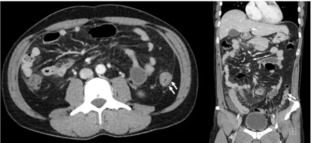
Fig. 1. Computed tomography findings were nonspecific. Mild wall thickening with mucosal enhancement in the descending colon.
A B
C D
Fig. 2. Colonoscopy findings. Multiple whitish subepithelial tumor-like lesions with nonspecific inflammatory changes, such as erythema and mucosal edema, were observed. (A) Cecum. (B) Transverse colon. (C) Descending colon. (D) Sigmoid colon.
bining the clinical, endoscopy and histology findings. This pa- per presents a patient with uncommon endoscopic findings diagnosed with EC who was treated successfully with steroids.
CASE REPORT
A 41-year-old man was admitted to hospital with complaints of diffuse abdominal pain and loose stools for 5 days. The frequency of loose stools increased to more than 5 times
A B
Fig. 3. Pathology findings. Numerous eosinophils in the lamina propria of the colonic mucosa. (A) H&E staining, ×200. (B) H&E staining, ×400.
A B
Fig. 4. Follow up endoscopy findings. The subepithelial tumor-like lesions were improved compared to the previous colonoscopy. (A) Descending colon. (B) Sigmoid colon.
a day, and the characteristics of the stools were mushy, like fluffy pieces with ragged edges. The abdominal pain had be- come more aggravating in both lower quadrants the day before he was admitted. The patient had no medical history of severe disease and allergic reactions, such as rhinitis, asthma, sinus- itis, dermatitis, food or drug allergies, or atopic conditions.
The physical examination was unremarkable, except for lower abdominal mild tenderness. The patient showed no signs of end-organ damage beyond the gastrointestinal tract, such as the heart and skin. The laboratory findings were as follows:
white blood cell count 15,100/µL (neutrophil 37%, lymphocyte 14%, monocyte 2%, eosinophil 47%), hemoglobin 14.9 g/dL, and platelet count 254,000/µL. The eosinophil count was ele- vated to 7,097/µL (reference 0-800/µL). The erythrocyte sed- imentation rate (ESR) was elevated at 30 mm/hours, and the CRP was modestly elevated at 1.40 mg/dL. The antibodies
against Clonorchis sinensis and Paragonimus westermani were negative. The anti-nuclear cytoplasmic antibody and an- ti-Saccharomyces cerevisiae antibody were negative. The serum IgE levels were normal, and the fecal parasite test results were negative. A CT scan of the abdomen showed only non- specific colitis with mild wall thickening of the descending colon and small fluid collections in the lower abdominopelvic cavity (Fig. 1). The CT scan did not reveal any other subepithelial tumor-like lesions except for mild enterocolitis. Nevertheless, the colonoscopy showed multiple whitish subepithelial tu- mor-like lesions from the cecum to the rectum with erythema and mucosal edema (Fig. 2). Multiple biopsies were taken from each segment of the colon and rectum. The histopathology examination revealed eosinophilic inflammatory infiltrates in the lamina propria from all segments of the biopsies (Fig. 3). There were >160 eosinophils per high power field.
The examination corresponded to mucosal involvement of the intestinal layers, and his clinical features were the most com- mon forms of EC without severe complications. Prednisolone 30 mg/day was started; the calculated dose was 0.5 mg/kg/day because his body weight was 60 kg. His symptoms resolved within a week. Prednisolone was tapered by 5 mg weekly until a 5 mg/day dose was reached in the last week. A follow-up colonoscopy was performed 2 weeks after he was given the last dose, and the findings were normal (Fig. 4). The patient was followed up as an outpatient for 6 months with no re- currence of his symptoms.
DISCUSSION
EC is a rare inflammatory disease that infrequently affects the colonic mucosa macroscopically and can be underdiagnosed.7 The histology findings will lead to a definitive diagnosis if other causes can be excluded. Secondary conditions that lead to the eosinophilic infiltration of the colonic mucosa need to be excluded to establish a correct diagnosis.1,5,6
Although the etiology and pathophysiology of this disease are not entirely understood, an interaction between genetic and environmental factors has been implicated. EC is asso- ciated with a wide spectrum of allergic diseases, such as rhini- tis, asthma, sinusitis, dermatitis, eczema, and urticaria.3,8 In most cases, primary EC is related to an allergic reaction, ei- ther an IgE-mediated anaphylactic-type food allergy or a food enteropathy not mediated by IgE. In infants, EC appears to be an IgE-associated disorder with mast cell accumulation and degranulation in the colonic tissue. On the other hand, in adults, EC is more commonly a non-IgE-mediated allergic reaction associated with a delayed CD4(+) Th2 lymphocytes response.1,9
The clinical features depend on the intestinal layers af- fected by the eosinophilic infiltration. Mucosal involvement is the most common form and presents with diarrhea, mal- absorption, and protein-losing enteropathy.10 Transmural dis- ease has a more severe presentation with wall thickening or strictures that can lead to intestinal obstruction, volvulus, and perforation.11 Subserosal disease, a very rare form, presents with eosinophilic ascites and is associated with a good prognosis.12 Endoscopic biopsies may be non-diagnostic in cases of muscular or subserosal involvement. Because endo- scopic biopsies do not sample the submucosa, muscularis
propria, or serosa, it is difficult to distinguish between the various forms.4
The laboratory findings might be indicative of EC, but they are typically not sufficient for a diagnosis. The blood eosino- phil count can be normal in up to 20% of patients. The radio- logical findings are often nonspecific and only present in 60-70% of cases in adults. CT imaging may show nodularity of the bowel wall, colonic wall thickening, mucosal fold thick- ening, and ascites in some cases.13,14 Imaging studies might help exclude inflammatory and infectious colitis.
The endoscopic findings are variable and usually non- specific, such as edematous mucosa with a loss of the normal vascular pattern, patchy erythematous changes, erosions, or aphthous ulcerations.15,16 Although endoscopic studies are frequently associated with normal macroscopic findings, mi- croscopically, EC shows diffuse patchy involvement. Therefore, multiple endoscopic biopsies are required if EC is suspected despite the visualization of a normal mucosa. EC is diagnosed by the appearance of sheets or clusters of eosinophils in the lamina propria. On the other hand, there are no guidelines that define an excessive versus normal number of eosinophils in the colonic mucosa.7
EC tends to be more aggressive in adolescents and adults, whereas it is relatively benign in infants and usu- ally resolves within days after removing the triggering food.
Adolescents and adults often require more aggressive medical management.1 Several treatment options are avail- able, even though the evidence for most is limited to a few case studies.
Corticosteroids are used as first-line pharmacological ther- apy if a dietary therapy fails to achieve an adequate clinical response. Oral prednisone at doses of 20-40 mg per day for 2 weeks has been shown to induce clinical remission in most patients.17 On the other hand, the main problem with corticosteroids is the high relapse rate. Maintenance treat- ment might be required in patients whose symptoms relapse during or after drug tapering with low-dose prednisone or budesonide.10
In severe or recurrent cases, long courses of steroids or immunomodulatory agents are needed. Mesalazine has been reported to be effective in some cases.2 Azathioprine and an- ti-tumor necrosis factor agents, such as infliximab and adali- mumab, have been attempted in the severe, steroid-re- fractory, or steroid-dependent EC.18,19 Other steroid-sparing
agents can be an option, including antihistamines, mast-cell stabilizers, and leukotriene receptor antagonists, even though their effectiveness in EC has yet to be evaluated.1,20
In summary, this paper presented a case of EC with the endoscopic appearance of subepithelial tumor-like lesions, which were proven to be eosinophilic infiltrates by multiple biopsies. In the absence of clear diagnostic guidelines, EC is diagnosed primarily by exclusion. Therefore, the most im- portant factor in making a correct diagnosis is having a high degree of clinical suspicion.
REFERENCES
1. Alfadda AA, Storr MA, Shaffer EA. Eosinophilic colitis: epidemiol- ogy, clinical features, and current management. Therap Adv Gastroenterol 2011;4:301-309.
2. Alfadda AA, Shaffer EA, Urbanski SJ, Storr MA. Eosinophilic colitis is a sporadic self-limited disease of middle-aged people: a pop- ulation-based study. Colorectal Dis 2014;16:123-129.
3. Jensen ET, Martin CF, Kappelman MD, Dellon ES. Prevalence of eosinophilic gastritis, gastroenteritis, and colitis: estimates from a national administrative database. J Pediatr Gastroenterol Nutr 2016;62:36-42.
4. Díaz Del Arco C, Taxonera C, Olivares D, Fernández Aceñero MJ.
Eosinophilic colitis: case series and literature review. Pathol Res Pract 2018;214:100-104.
5. Uzunismail H, Hatemi I, Doğusoy G, Akin O. Dense eosinophilic infiltration of the mucosa preceding ulcerative colitis and mimick- ing eosinophilic colitis: report of two cases. Turk J Gastroenterol 2006;17:53-57.
6. Ahmad M, Soetikno RM, Ahmed A. The differential diagnosis of eosinophilic esophagitis. J Clin Gastroenterol 2000;30:242-244.
7. Yan BM, Shaffer EA. Primary eosinophilic disorders of the gastro-
intestinal tract. Gut 2009;58:721-732.
8. Mansoor E, Saleh MA, Cooper GS. Prevalence of eosinophilic gastroenteritis and colitis in a population-based study, from 2012 to 2017. Clin Gastroenterol Hepatol 2017;15:1733-1741.
9. Guajardo JR, Plotnick LM, Fende JM, Collins MH, Putnam PE, Rothenberg ME. Eosinophil-associated gastrointestinal dis- orders: a world-wide-web based registry. J Pediatr 2002;141:
576-581.
10. Klein NC, Hargrove RL, Sleisenger MH, Jeffries GH. Eosinophilic gastroenteritis. Medicine (Baltimore) 1970;49:299-319.
11. Velchuru VR, Khan MA, Hellquist HB, Studley JG. Eosinophilic colitis. J Gastrointest Surg 2007;11:1373-1375.
12. Pineton de Chambrun G, Gonzalez F, Canva JY, et al. Natural his- tory of eosinophilic gastroenteritis. Clin Gastroenterol Hepatol 2011;9:950-956.e1.
13. Zhang M, Li Y. Eosinophilic gastroenteritis: a state-of-the-art review. J Gastroenterol Hepatol 2017;32:64-72.
14. Anuradha C, Mittal R, Yacob M, Manipadam MT, Kurian S, Eapen A. Eosinophilic disorders of the gastrointestinal tract: imaging features. Diagn Interv Radiol 2012;18:183-188.
15. Turner KO, Sinkre RA, Neumann WL, Genta RM. Primary colonic eosinophilia and eosinophilic colitis in adults. Am J Surg Pathol 2017;41:225-233.
16. Okpara N, Aswad B, Baffy G. Eosinophilic colitis. World J Gastroenterol 2009;15:2975-2979.
17. Lee CM, Changchien CS, Chen PC, et al. Eosinophilic gastro- enteritis: 10 years experience. Am J Gastroenterol 1993;88:
70-74.
18. Lampinen M, Rönnblom A, Amin K, et al. Eosinophil granulocytes are activated during the remission phase of ulcerative colitis. Gut 2005;54:1714-1720.
19. Al-Haddad S, Riddell RH. The role of eosinophils in inflammatory bowel disease. Gut 2005;54:1674-1675.
20. Gupta N, Aggarwal A, Gupta R, Sule S, Wolf DC. The management of eosinophilic gastroenteritis. Scand J Gastroenterol 2015;
50:1309-1314.