Volume 14, Number 1, June 2002
급성 슬개골 탈구의 수술적 치료
가천의과대학 부속 길병원 정형외과학교실
이범구・조현철・엄기석・김신웅
= 국문 초록 =
목적 : 급성 슬개골 탈구 환자에서 탈구와 연관된 슬관절 병변의 평가와 함께 조기에 수술적 방법으로 치 료 후 임상적 결과에 대하여 분석하여 보고자 한다.
대상 및 방법 : 1997년 2월부터 2 0 0 0년 1월까지 급성 슬개골 탈구로 조기에 수술적 치료한 1 1례를 대상 으로 하였으며, 슬개골 탈구의 과거력이 있거나 관절경 소견상 슬관절내 출혈이 없어 재발성 탈구가 의심 되 는 환자는 대상에서 제외 하였다. 평균 추시 기간은 2 3개월 이었다. 술 전 단순방사선 전후면 및 3 0도 굴곡 측면사진 과 Merchant 촬영을 하여 슬개골의 이상고위, 대퇴 활차의 이형성(crossing sign), sulcus a n g l e을 측정하였으며, 이학적 검사에서 Q - a n g l e을 측정하여 탈구 유발인자를 평가하였으며, 수술 전 후에 congruence angle을 측정하였다. MRI 검사, 관절경 소견, 및 수술 소견에서 내측지대 손상 유무와 손상 위치 골연골 손상 여부 및 슬관절내 동반 병변을 관찰하였다. 전 례에서 진단적 관절경을 시행 후, 내측지대 봉합술( 5례), 내측지대 봉합술과 외측이완술( 3례), 내측지대 보강 및 중첩술 즉, 변형된 Madigan 술식( 3 례)을 시행하여 최소 1 2개월 이상 추시 관찰하였다. 수술 후 임상적 결과는 Kujala score를 사용하여, 방사 선학적 결과는 술 후 congruence angle을 측정하여 평가하였다.
결과 : 슬개골의 이상고위 7례(63.6%), 대퇴활차 이형성 5례(45.5%), 증가된 sulcus angle은 5례 ( 4 5 . 5 % )에서 관찰되었으며, 20도 이상으로 증가된 Q - a n g l e은 없었다. 전체 1 1례중 9례(81.8 %)에서 한 가지 이상의 슬개골 탈구 유발인자를 관찰할 수 있었다. 내측 슬개대퇴 인대손상은 대퇴부착부(내전근 결절) 에서 5례, 슬개골에 근접한 중간부위에서 4례, 이완된 경우가 2례이었다. 임상적 결과 평균 Kujala score는 9 1점 이었다. 술 후 방사선 검사결과 congruence angle은 1 0례에서 정상범위로 회복되었고, 1례에서는 슬 개골 아탈구 소견을 보였으며, 추시기간중 재탈구는 없었다.
결론 : 급성 슬개골 탈구는 슬개골 이상고위, 대퇴활차 이형성, 증가된 sulcus angle이 동반된 경우와 연 관성이 있고, 급성 슬개골 탈구의 조기 수술적 치료로 2 3개월의 추시에서 우수한 임상결과를 얻을 수 있었다.
색인 단어 : 슬개골, 탈구, 수술적 치료
Corresponding Author : Beom Koo Lee, M.D.
Department of Orthopaedic Surgery, Gil Medical Center, Gacheon Medical Collage, 1198, Kuwol-dong, Namdon-gu, Incheon, Korea
Tel : 032-460-3384, Fax: 032-468-5437, E-mail : [email protected]
서 론
급성 슬개골 탈구는 발생의 병태 생리가 명확하지 않 고 저자에 따라 치료방법에 이견이 있는 슬관절 손상 중의 하나이다. 급성 탈구 시 일반적으로 보존적 치
료가 선호되어 왔으나, 슬개골의 재발성 탈구 및 슬 개골 부정정열의 빈도가 높고 만성적인 슬개-대퇴부 증상이 남는 경우가 많기 때문에 슬개골 탈구와 연관 된 병변을 교정하고자 다양한 수술적 방법이 연구되 어 시행되고 있다7,14,23,25,26). 이에 저자들은 수술적 방법 으로 치료한 급성 슬개골 탈구 환자에서 탈구와 연관
된 슬관절 병변의 평가와 함께 수술 후 임상적 결과 에 대하여 분석하여 보고자 한다.
재료 및 방법 1. 대상
1 9 9 7년 2월부터 2 0 0 0년 1월까지 급성 슬개골 탈 구로 수상 2주 이내 수술적 치료를 받은 후 1년 이상 추시 가능하였던 1 1례를 대상으로 하였다. 슬개골 탈구의 과거력이 있거나 관절경 소견상 슬관절내 혈 종이 없어 슬개골 재발성 탈구가 의심 되는 환자는 대상에서 제외 하였다. 남자가 2명, 여자가 9명 이였 고 평균 연령은 2 5 . 3세( 1 5 ~ 4 0세)였으며, 발생 부위 는 우측이 4례, 좌측이 7례였고 수상기전은 간접 손 상이 8례, 직접 타박에 의한 접촉성 손상이 3례였으 며 평균 추시 기간은 2 3개월이었다.
2. 평가방법
방사선학적 검사로 단순방사선 전 후면 및 3 0도 굴 곡 측면사진 과 Merchant 촬영을 하여, 측면사진에 서 슬개골의 이상고위(patella alta), 대퇴 활차의 이형성(crossing sign)7 )을, Merchant 사진에서 sulcus angle을 측정하였고, 이학적 검사에서 Q - a n g l e을 측정하여 탈구 호발인자를 평가하였으며, 수술 전 후에 congruence angle을 측정하였다. 수 술 전 8례에서는 MRI 촬영을 시행하였다. MRI 검 사, 관절경 검사소견 및 수술소견에서 내측지대 손상 유무와 손상위치, 골연골 손상 여부 및 슬관절내 동 반 병변을 관찰하였다. 술 후 임상적 결과는 재발성 탈구 및 아탈구, 동통, 활동제한 등 환자의 주관적 판단에 근거한 Kujala acore1 5 )로 평가하였으며, 방 사선학적 결과는 술 후 congruence angle을 측정 하여 평가하였다.
3. 수술방법
전 례에서 진단적 관절경을 시행 후, 내측지대 손 상부위가 슬개골에 근접한 중간 지점인 경우 관절경 적 봉합이 가능하였던 1례에서는 관절경을 사용하여 경피적 봉합을 시행하였고, 내측지대의 손상부위가 내전근 결절부착부인 경우와 같이 관절경적 방법으로 봉합이 불가능 하였던 4례의 경우 슬관절 내측에 내
전근 결절을 중심으로 사선의 피부 절개를 시행하여 손상된 내광배근 기시부와 내측 슬개대퇴인대가 내전 근결절에서 기시하는 부위를 내전근 결절에 남아 있 는 연부조직에 비흡수사를 사용하여 관혈적 방법으로 봉합하였다. 봉합시행 후 관절경 소견상 슬개골 이상 정렬이 남아 있었던 3례의 경우 관절경적 외측이완 술을 추가하였고, 내측지대 봉합 및 외측 이완술에도 불구하고 아탈구가 있었던 3례에서는 슬관절 전면에 종축으로 피부절개를 시행 후 내측지대의 이완 또는 손상부위에서 내광배근과 대퇴직근건 사이를 절개하 고 원위부로 절개를 진행하여 관절절개술을 시행한 후 내측지대와 내광배근위 원위부를 외측으로 이동시 켜 슬개골의 전면에 중첩되게 보강봉합하는 변형된 Madigan 술식을 사용하여 근위부 재배열을 시행하 였다. 슬개골 관절면으로부터 발생된 관절내 유리체 는 골연골 골편이 크고 정복이 가능한 2례의 경우 K -강선으로 골터널을 뚫고 비흡수 봉합사로 봉합하 여 고정하였고, 크기가 작고 고정이 불가능한 4례의 경우에는 제거하였다.
4. 술 후 재활
5례에서는 cylinder cast 나 splint 고정을 3주간 시행 후 슬관절 굴곡운동 및 체중 부하를 허용하였 고, 6례에서는 술 후 부종 과 통증이 감소할 때까지 cylinder splint 고정 후 장하지 보조기 착용 후 슬 관절 굴곡운동 및 물리치료를 시행하였다.
결 과
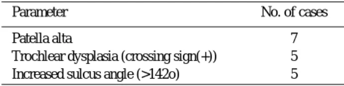
단순방사선 소견상 슬개골의 이상고위 7례 (63.6%), 대퇴활차 이형성(crossing sign)은 5례 (45.5%), 증가된 sulcus angle은 5례( 4 5 . 5 % )에서 볼 수 있었으며(Table 1), 관절내 유리체가 3례에서 관찰 되었다. 20도 이상으로 증가된 Q - a n g l e은 없 었으나, 15~20도 범위가 2례 있었다. 전체 1 1례중 9례(81.8 %)에서 한가지 이상의 슬개골 탈구 호발 인자를 관찰할 수.있었다. 관절경 및 수술소견에서 내측지대의 손상은 대퇴부착부(내전근결절)에서 내측
Table 1. Radiologic findings
Parameter No. of cases
Patella alta 7
Trochlear dysplasia (crossing sign(+)) 5 Increased sulcus angle (>142o) 5
슬개대퇴인대와 내광근 기시부의 손상이 동반된 경우 5례, 슬개골에 근접한 중간부위에서 손상된 경우 4 례, 이완된 경우가 2례였고(Table 2), 관절경 시행 중 관절내 유리체가 6례에서 있었고, 골연골 손상은 슬개골 내측 관절면에 발생한 경우가 5례, 대퇴골 외 과에 발생한 경우가 3례 있었고, 술 전 MRI 촬영을 시행한 8례 중 7례에서 대퇴골 외과의 골좌상 소견이 있었고, 내측 측부인대 손상은 1례 있었다(Table 3).
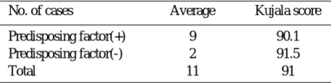
임상적 결과 전체 평균 Kujala score는 9 1점이었고 탈구 유발인자가 동반된 9례에서는 9 0 . 1점, 탈구 유 발 인자가 동반되지 않은 2례에서는 9 1 . 5점 이었다 (Table 4). 술 후 방사선 검사결과 c o n g r u e n c e a n g l e은 1 0례에서 정상범위로 회복되었고, 1례에서 는 슬개골 아탈구 소견을 보였으며, 재탈구는 없었 다. 합병증으로 관절강직이 1례에서 있었으나 관절경 적 연부조직 유리술 시행 후 물리치료로 정상 관절운 동 범위를 회복하였고, 복재신경 손상이 1례 있었다 (Table 5).
고 찰
급성 슬개골 탈구는 활동이 왕성한 젊은 연령에서 주로 발생하고, 대부분 외측으로 탈구되며, 탈구 시 내측지대의 파열과 골연골 골절이 흔하게 동반된다.
이는 정상 슬관절에서도 발생할 수 있지만, 이보다는 탈구를 쉽게 유발하는 해부학적 인자들을 가진 슬관 절에서 흔히 발생한다. 탈구를 쉽게 유발하는 해부학 적 비정상 소견에 대해 여러 저자들이 슬개골 이상 고위, 외반슬, Q-각의 증가, 하지의 회전 이상정열, 대퇴골 전염각의 증가 및 하퇴의 외회전 변형, 장경 대의 구축, 대퇴골 외과의 결손, 얕은 대퇴구, 내광 배근 발달 저하, 슬개골의 과운동성 , 선천성 인대이 완, 슬개골 경사 등을 언급하고 있다1 , 4 , 9 , 1 3 )
. 본 연구 에서도 2례를 제외한 9례(81.8 %) 에서 한가지 이 상의 탈구 유발인자가 관찰되었으며, 탈구 유발인자 동반 유무에 따른 임상적 결과에 유의한 차이는 없었 다(Table 4). 내측 슬개 대퇴인대는 슬개골의 외측 아탈구에 대하여 수동적 억제력으로 작용하며, 급성 슬개골 탈구시 손상되는 주요 구조물로 생각되고 있 다8 ). O’Donoghue 등1 8 )은 대부분의 예에서 내측 관절낭이 슬개골 부착 부위에서 견열된다고 생각하였 고, Sally 등1 9 )은 90% 이상에서 내측 슬개대퇴인대 의 대퇴기시부에서 손상이 있었음을 보고하였다.
Burks 등3 )도 사체 실험 모델에서 내측 슬개대퇴인 대손상은 대퇴기시부에서 주로 발생됨을 보고하였다.
내광배근은 슬개골을 동적으로 안정화시키는 구조물 로서 이 또한 급성 슬개골 탈구 시 흔히 손상된다1 ). Hunter 등1 2 ) 은 내광배근의 손상이 슬개골 부정 정 렬을 초래할 수 있다고 하였다. 본례의 경우 내측 슬 개대퇴인대 손상부위가 대퇴기시부(대퇴 내전근결절) 인 경우가 5례(45.5 %), 슬개골에 근접한 중간부위 인 경우가 4례(36.3 %)로 Sallay 등의 보고와 정도 의 차이는 있으나 대퇴기시부에서 손상이 주로 발생 하는 것으로 나타났고, 내광배근의 원위부 손상은 5 례(45.5 %)에서 관찰되어 Sally 등의 연구에서보다 는 낮은 결과를 얻었다. 급성 슬개골 탈구의 비수술 Table 2. The site of medial retinacular tear
Tear site No. of cases
Parapatellar tear 4
Adductor tubercle 5
with VMO* injury
No tear (lax retinaculum) 2
*VMO : vastus medialis obliquus muscle
Table 3. Pathologic findings in MRI
Pathology No. of cases
MPFL* injury
tear from adductor tubercle 4
parapatellar tear 3
sprain without tear 1
VMO†injury 3
Bone bruise
lateral femoral condyle 7
Loose bodies 4
MCL‡injury 1
* MPFL : medial patellofemoral ligament
†VMO : vastus medialis obliliquus muscle
‡MCL : medial collateral ligament
Table 4. Clinical results acording to the predisposing factor
No. of cases Average Kujala score
Predisposing factor(+) 9 90.1
Predisposing factor(-) 2 91.5
Total 11 91
Table 5. Complications
Complications No. of cases
Joint stiffness 1
Saphenous neuralgia 1
Patellar subluxation 1
A
B C
D
E
F
a b
c d e
적 치료 결과에 대하여 C o f i e l d와 B r y a n6 )은 약 4 4
% 의 재발성 탈구율을 보고하였고, Hawkins 등1 1 ) 은 4 0 ~ 7 0 %에서 만성적 슬개골 불안정으로 인한 만 성적 슬개대퇴 동통이 발생된다고 하였다. Maen- p a a와 L e h t o1 6 )도 1 3년 추시 결과, 44 % 의 재발성 슬개골 탈구가 발생하였으며, 재발성 탈구가 발생하 지 않은 경우에도 1 9 %에서 슬개대퇴 동통 또는 슬 개골 아탈구가 발생하여 평균 Kujala score 82점의 결과를 얻었다. 급성 슬개골 탈구의 수술적 치료로 여러 가지 방법이 고안되어 시행되어 왔다. Sher- man 등2 0 )은 4 5례의 환자에서 관절경적 외측이완술 을 시행한 결과 2 5 %에서 재발성 슬개골 탈구 등의 만족스럽지 못한 결과를 보고하였다. Harilainen 과 M y l l y n e n1 0 )은 관혈적 외측 이완술을 내측 관절 낭 축범술 (medial capsular reefing)과 병행하여 1 2개월 추시결과 평균 Lysholm score 94점의 결과 를 보고하였다. Small 등2 1 ) 은 관절경적 내측 중첩 술을 외측 이완술과 병행하여 1 2개월 추시결과 평균 Lysholm score 92점의 결과를 보고하였다. Sally
등1 9 )은 내측 슬개대퇴인대의 대퇴내전근 결절의 부착
부를 복원하는 술식을 시행하여 평균 L y s h o l m score 84점의 결과를 보고하였다. Christopher 등5 ) 은 내측 슬개대퇴인대의 대퇴내전근 결절의 부착부와 내광배근의 원위부 손상을 복원을 시행하여 평균 Kujala score 91점의 결과를 보고하였다. 본 연구
에서 내측지대 봉합 후 관절경적 외측이완술의 시행 여부에 따른 임상적 결과에서 유의한 차이는 없었으 며, 양군 모두에서 재발 없이 임상적으로 우수한 결 과를 얻었다. 관절경적 내측지대 봉합술을 시행한 2 례에서 낮은 Kujala score를 보였는데, 1례는 복재 신경손상으로 인한 슬관절 동통을 호소하였고, 다른 1례는 추적 검사 시 슬개골 아탈구와 슬관절 동통을 호소하였다. 관절경적 내측지대 봉합술은 비침습적인 장점이 있는 반면 동적 안정화 구조물인 내광배근과 정적 안정화 구조물인 내측 슬개대퇴인대의 내전 근 결절 부착부에 대한 평가와 이 구조물의 손상 시 해 부학적 복원이 어려운 점이 단점으로 생각된다. 이에 비해 슬관절 내측 절개 후 시행하는 관혈적 내측지대 봉합술은 내측지대의 손상부위를 육안으로 직접 확인 할 수 있고, 내전근 결절부에 손상이 있는 경우 손상 된 내측 슬개대퇴인대와 내광근 기시부의 해부학적 복원이 가능하다는 장점이 있는 것으로 생각되며, 저 자들의 경험으로도 관절경적 내측지대 봉합 시 술 후 슬개골 아탈구의 합병증이 발생한 예가 있었다. 저자 들의 경우 내측지대의 손상위치에 따라 수술방법을 달리하여 수술을 시행한 결과 비 수술적 치료를 시행 한 문헌상의 결과6 , 1 1 , 1 6 )
보다 재발 및 만성적 슬개대퇴 동통의 발생 없이 좋은 임상적 결과를 얻었으나 적은 수의 증례 및 짧은 추시 기간이어서 추후 더 많은 증 례와 추시 관찰이 요할 것으로 사료된다.
FIGURE 1. 22 year old woman suffered a patellar dislocation in the right knee and posterolateral injury in the left knee 25 months later.
A : A pre op. roentgenogram of right knee shows tilting & dislocation of the patella on a merchant view.
B : A follow up roentgenogram one year after operation shows well-seated patella on the Merchant view.
C : A lateral view of right knee shows trochlear dysplasia (=crossing sign). The line of the condyle are superimposed at the same point in the proximal part of the trochlaea(arrow). the proximal part of the trochlea is flat.
D : T2-weighted image of the axial MRI scan
a) in the right knee after patellar dislocation demonstrates increased T2-wieghted signal anterior to the adductor tuber- cle, a MPFL tear(large arrow), medially reflected synovial layer(small arrow)
b) in the left knee with the posterolateral injury, the MPFL is well visualized deep to the VMO muscle(medium arrow) and that the distal belly of the VMO muscle is normally present at this level.
E : T2 weighted image of the sagittal MRI scan
a) in the right knee after patellar dislocation shows a large triangle of T2-weighted signal anterior to the adductor tuber- cle (large arrow) where the VMO muscle has been peeled off the adductor tendon.
b) the left knee with the posterolateral injury, shows the normal relationship of the VMO muscle to the adductor tuber- cle(small arrow).
F : Arthroscopic findings
a) The torn medial retinaculum.
b) The dislocated patella before medial retinaculum repair.
c) The patella tiling and subluxation are still present even after medial retinaculr repair.
d) Arthroscopic lateral release was performed.
e) The well reduced patella after arthroscopic lateral release.
결 론
급성 슬개골 탈구는 슬개골 이상고위, 대퇴활차 이 형성, 증가된 sulcus angle이 동반된 경우와 연관성 이 있고, 급성 슬개골 탈구의 조기 수술적 치료로 2 3 개월의 추시에서 우수한 임상결과를 얻었다.
REFERENCES
11) Bassett FR : Acute dislocation of the patella, osteo- chondral fractures, and extensor mechnism of the knee.: instructional Course lectures, 25:40-49, 1976.
12) Boring TH and O’Donoghue DH : Acute patellar dislocation; Results of immediate surgical repair.
Clin Orthop, 136;182-185, 1978.
13) Burks Rt, Desio SM and Bachus KN : Biomechni- cal evaluation of lateral patellar dislocations. Am J Knee Surg, 11:24-31, 1998.
14) Cash JD and Hughston JC : Trearment of acute patellar dislocation. Am J Sports Med, 1 6 : 2 4 4 - 2 4 9 , 1988.
15) Christopher SA, Beth ES, Stein DM and Jack HH : Imediate surgical repair of the medial patellar sta- bilizers for acute patellar dislocation: A review of eight cases ; Am J sports Med, 28:804-810, 2000.
16) Cofield RH and Bryan RS : Acute dislocation of the patells; Results of conservative treatment. J Truma, 17:526-531, 1977.
17) Dejour H, Walch G, Neyret P and Adeleine P : La dysplastic delatrochlee femorale. Rev chir Orthop, 76;45, 1990.
18) Desio SM, Burks RT and Bachus KN : Soft tissue restraints to lateral patellar translation in the human knee. Am J Sports Med, 26:59-65, 1998.
19) FreemanIII BC : Recurrent Dislocations. In Camp- bell’s Operatibe Orthopaedic 8th ed. 1391-1405, St.
Louis, 1992.
10) Harilinen A and Myllunen P : Operative treatment in acute patellar dislocation: Radiological predispos- ing factors, diagnosis, and results. Am J knee Surg 1:
178-185, 1988.
11) Hawkins U, Bell RH and Anisette G : Acute patel- lar dislocation. The natural history. Am J Sports Med, 14:117-120, 1986.
12) Hunter SC, Marascalco R and Hughston JC : Disruption of the vastus medialis obliquus with medial knee ligament injuries. Am J Sports Med, 11:
427-431.
13) Insall J., Goldberg, V and Salvati E. : Recurrent dislocation and the highriding patella. Clin. Orthop.
88;67-69, 1972.
14) Jenson CM and Roosen JU : Acute trumatic dislo- cations of the patella. J Truma 23:160-162, 1985 15) Kujala UM, Osterman K and Kormano M : Scor-
ing of patellofemoral disorders. Arthroscopy 9:159- 163, 1993
16) Maenpaa H and Lehto MU : Patellar dislocation:
The long-term results of non-operative management in 100 patients. Am J Sports Med, 25:213-217,1997.
17) McManus F, Rang M and Heslin DJ : Acute dis- loction of the patells in children : The natural histo- ry. Clin Orthop, 13:88-91, 1979.
18) O’Donoghue DH : Treatment of Injuries to Ath- letes. Third edition. Philadelphia, WB Saunders Co, pp 600-617, 1976
19) Sally PI, Poggi J and Speer KP : Acute dislocation of the patella: A correlative pathoanatomic study.
Am J Sports Med, 24:52-60, 1996.
20) Sherman OH, Fox JM and Sperling H : Patellar instability: Treatment by arthroscopic electrosurgical lateral release. Arthroscopy, 9:63-67, 1993.
21) Small NC, Glogau Al and Berezin MA : Arthro- scopically assisted proximal extensor mechanism realignment of the knee. A r t h r o s c o p y , 9 : 6 3 - 6 7 , 1993.
─ Abstract ─
Operative Treatment in Acute Patellar Dislocation
Beom Koo Lee, M.D., Hyun Chul Jo, M.D., Gi Serk Eom, M.D., Shin Woong Kim, M.D.
Department of orthopaedic surgery, Gill Medical Center Gacheon Medical collage, Incheon, Korea
Purpose : To analyze factors related with patellar dislocation and evaluate clinical results after early surgical treatment in acute patellar dislocation.
Materials and Methods : The study was done in 11 patients who underwent surgery due to acute patel- lar dislocation from February 1997 to January 2000. patients who had history of previous patella disloca- tion were excluded in this study. The average follow up period was 23 months. Radiographs were antero- posterior, lateral at 30 degree of flexion, and Merchant view. We looked for of patella alta, trochlear dys- plasia, intraarticular loose body and measured sulcus angle, congruence angle, Q-angle. We checked the location of injured medial retinaculum and ostochondral lesions, and associated intraarticular lesions from MRI, arthrosopic and operative findings. We performed the 3 operative methods: Upon the completion of the arthroscopic evaluation, medial retinacular repair (5 cases), medial retinacular repair and lateral release(3 cases). augmented repair and plication of medial retinaculum ie, modified Madigan technique(3 cases).
R e s u l t s : patella alta was present in 7 cases(63.6%), trochlear dysplasia(crossing sign) in 5 cases(45.5%), and increased sulcus angle in 5 cases(45.5%). one or more predisposing factors contribut- ing to patella dislocation were seen in 9 of 11 cases(81.8 %). Medial retinacular tear was present in the adductor tubercle in 5 cases and midportion(parapatellar tear) in 4 cases, and relaxed without tear in 2 cases. The clinical results showed that the average Kujala score was 91 points. The congruence angle became normal in 10 cases and patella subluxation was seen in 1 case postoperatively.
Conclusions : Acute patellar dislocation was related in those cases with trochlear dysplasia and patella alta and increased sulcus angle . During the 23 months follow up preiods, Excellent clinical results were obtained with early surgical treatment.
Key Words : Patella, Dislocation, Surgical treatment