Vol. 46, No. 2, pp. 252 - 259, 2005
Pain is a major symptom in cancer patients, and most cancer patients with advanced or terminal cancers suffer from chronic pain related to treatment failure and/or tumor progression. In the present study, we examined the development of cancer pain in mice. Murine hepatocarcinoma cells, HCa-1, were inoculated unilaterally into the thigh or the dorsum of the foot of male C3H/HeJ mice. Four weeks after inoculation, behavioral signs were observed for mechanical allodynia, cold allodynia, and hyperalgesia using a von Frey filament, acetone, and radiant heat, respectively. Bone invasion by the tumor commenced from 7 days after inoculation of tumor cells and was evident from 14 days after inoculation. Cold allodynia but neither mechanical allodynia nor hyperalgesia was observed in mice that received an inoculation into the thigh. On the contrary, mechanical allodynia and cold allodynia, but not hyperalgesia, were developed in mice with an inoculation into the foot. Sometimes, mirror-image pain was developed in these animals. These results suggest that carcinoma cells injected into the foot of mice may develop severe chronic pain related to cancer. This animal model of pain would be useful to elucidate the mechanisms of cancer pain in humans. Key Words: Cancer pain, allodynia, hyperalgesia, behavior, mouse
INTRODUCTION
Chronic pain is a major symptom in cancer patients with advanced or terminal cancers. These
patients suffer from chronic pain related to treatment failure and/or tumor progression. Cancer pain might be related to invasion of either peripheral nerves or bones, which accounts for 75% of all chronic cancer pain.1
Clinical literature regarding cancer pain assess-ment and manageassess-ment is extensive. However, the understanding of the mechanisms that under-lie the production of pain associated with malignancy is meager at best. An understanding of these basic mechanisms is essential for the development of better therapeutic approaches to cancer pain treatment, but efforts to gain such in-formation are hampered by the lack of adequate animal tumor models. While investigations have been accumulated on neuropathic pain using ani-mal models,2-4 cancer pain by bone invasion hasn't been extensively studied. Recently, Schwei et al.5 described a femur model of bone cancer pain that allowed characterization of neuro-chemical changes in the spinal cord associated with development of tumor-induced nociception. Similarly, Medhurst et al.6 reported that rats receiving intra-tibial injections of MRMT-1 cells displayed the gradual development of mechani-cal allodynia and mechanimechani-cal hyperalgesia and reduced weight bearing on the affected limb.
Although the femur or tibial bone tumor model provides a valuable archetype to assess CNS changes, it is difficult to examine the func-tion of the hind paw which has been studied widely. This study was conducted to develop a new mouse model of cancer pain by comparing cancer pain-related behaviors produced by tu-mors located in different sites of the hind limbs of mice.
Behavioral Characteristics of a Mouse Model of Cancer Pain
Bae Hwan Lee1, Jinsil Seong2, Un Jeng Kim1, Ran Won1, and Jiyoung Kim2
1Medical Research Center, Department of Physiology, Brain Research Institute, Yonsei University College of Medicine, Seoul, Korea;
2Department of Radiation Oncology, Brain Korea 21 Project for Medical Science, Yonsei University College of Medicine, Seoul, Korea.
Received September 9, 2004 Accepted October 15, 2004
This work was supported by a grant from the Korea Health 21 R D Project, Ministry of Health Welfare, Republic of Korea (01-PJ8-PG1-01CN01-0005).
Reprint address: requests to Dr. Jinsil Seong, Department of Radiation Oncology, Yonsei University College of Medicine, 134 Shinchon-dong Seodaemun-gu, Seoul 120-752, Korea. Tel: 82-2-2228-8112, Fax: 82-2-3463-7441, E-mail: jsseong@yumc.yonsei. ac.kr
MATERIALS AND METHODS
Subjects
A total of 105 male C3H/HeJ mice (8-10 weeks old, 22-26 g) were used. Mice were bred and maintained at five animals per cage. The tem-perature and humidity were constantly controlled. Water and diet were supplied ad libitum. Ex-periments were carried out in accordance with the guidelines laid down by the NIH in the US re-garding the care and use of animals for ex-perimental procedures. All procedures were ap-proved by the Committee for Animal Experiments in Yonsei University College of Medicine.
Inoculation of carcinoma cells
A murine hepatocarcinoma syngeneic to C3H/ HeJ, murine hepatocarcinoma (HCa-1), was used in the present study. Suspensions of 106 tumor cells were prepared following Milas et al.7 Cells were injected unilaterally into the thigh or the dorsum of the foot of male C3H/HeJ mice. Mice were regularly sacrificed and bone invasion by the tumor was histologically examined.
Behavioral tests for cancer pain
Behavioral signs of different components of cancer pain were observed in all of the mice before the inoculation and then on days 1, 3, 5, 7, 14, 21, and 28 after inoculation. The behavioral testing procedures followed those reported by Lee et al.8 with some modifications. To measure mechanical allodynia, mice were placed on a metal mesh floor under a transparent plastic dome (8×5×4.5 cm), and allowed to habituate for at least 30 min. Mechanical allodynia of the plantar surface of the hind paw was determined by measuring the paw withdrawal threshold in response to probing with von Frey monofila-ments. The most sensitive area was first deter-mined by poking various areas of the paw with a von Frey hair. Testing for mechanical allodynia was then conducted by gently poking the most sensitive spot with the filament. Withdrawal threshold was determined by increasing and decreasing stimulus intensity between 0.2 and 12
g equivalents of force using a von Frey filament, and estimated when the force produced a 50% response rate after 10 applications to each hind paw. To quantify cold allodynia, mice were placed on a metal mesh floor under a trans-parent plastic dome and brisk paw withdrawal in response to acetone application was measured. An acetone bubble was applied 5 times (once every 5 min) to each paw. The frequency of paw withdrawal was expressed as a percentage re-sponse rate to the application of acetone 10 times to each hind paw. To quantify heat hyperalgesia, the latency of paw withdrawal to noxious heat stimuli was measured. Each mouse was placed on a wire mesh floor under which a light box was located. A radiant heat stimulus was applied by aiming a beam of light through a hole (10 mm) in the light box to the thigh or foot through the wire mesh. The paw withdrawal latency was defined as the time from the start of the light beam to the lifting of the paw from the floor. Fifteen seconds were allowed between stimula-tions and 5 measurements were made on each side of the hindlimb of each mouse on each day. The 5 measurements were averaged and re-corded as the paw withdrawal latency on a given day. Behavioral assessments of mechanical allodynia, cold allodynia, and heat hyperalgesia were made of both the ipsilateral and contra-lateral sides to the tumor.
Histology
Mice were sacrificed at regular intervals after the injection of tumor cells. Tumor-bearing hind paws were fixed in 4% zinc-buffered formalin in 0.1 M PBS at 4 overnight, decalcified in 10% EDTA (pH 7.4) for 2 weeks, and embedded in paraffin. Paraffin blocks were sectioned 7 m-μ thick, stained with Hematoxylin and Eosin, and examined under the light microscope.
Statistical analysis
Data are expressed as means ± SE. Differences in pain behaviors following inoculation of HCa-1 cells or vehicle (control medium) were tested using the Student's t-test at each time point. Statistical significance was set at p < 0.05.
RESULTS
Behavioral assessments
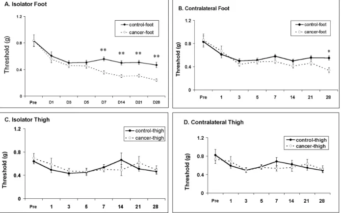
The response threshold of foot withdrawals to repeated mechanical stimulation was plotted for each group (the control group vs the cancer-treated group) against time (Fig. 1). It should be noted that mice showed a high response threshold to stimulation of the paw before tumor inocu-lation, and that vehicle treated mice maintained high threshold responsiveness to mechanical stimulation in all groups. The most vigorous mechanical allodynia was developed in the animals with a tumor in the hind paw (Fig. 1A). In the figures, asterisks indicate significant dif-ferences between HCa-1 inoculation and vehicle groups. Mice with HCa-1 inoculation in the foot showed remarkable mechanical allodynia from 7
days after inoculation (Fig. 1A). In addition, unexpectedly, these mice showed mechanical al-lodynia at 28 days after inoculation on the con-tralateral side, indicating mirror image pain (Fig. 1B). In contrast, mice with HCa-1 inoculation in the thigh did not show any remarkable mechani-cal allodynia either on the ipsilateral or contra-lateral side (Fig. 1C and 1D).
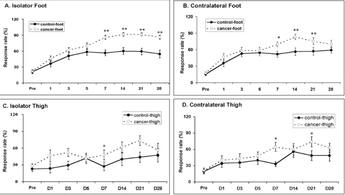
The frequencies of foot withdrawals to repeated cold stimulation (with acetone bubble) were plot-ted for each group against time (Fig. 2). The animals with a tumor in the hind paw showed the most vigorous cold allodynia (Fig. 2A). In the figures, asterisks indicate significant differences between HCa-1 inoculation and vehicle groups. Mice with HCa-1 inoculation in the foot showed remarkable cold allodynia from 7 days after inoculation (Fig. 2A). In addition, these mice also showed cold allodynia from 7 days after
inocula-Fig. 1. Development of mechanical allodynia after inoculation of hepatocarcinoma cells (HCa-1) into the thigh or the dorsum of the foot in mice. A: Mechanical allodynia on the ipsilateral foot, B: Mechanical allodynia on the contralateral foot, C: Mechanical allodynia on the ipsilateral thigh, D: Mechanical allodynia on the contralateral thigh. Response thresholds to von Frey filaments with different bending forces were used as an index of mechanical allodynia. Data were expressed as means ± SE. Abscissa was marked as Pre for pre-inoculation control and D for post-inoculation days. Asterisks (*) indicate significant differences between HCa-1 and vehicle groups at each time point by Student's t-test (p
tion on the contralateral side, indicating mirror image pain (Fig. 2B). In contrast, mice with HCa-1 inoculation in the thigh showed low levels of cold allodynia both on the ipsilateral and contralateral side (Fig. 2C and 2D).
The latencies of hind paw withdrawal to noxious radiant heat stimulus were plotted for each group against time (Fig. 3). As shown in the figure, heat hyperalgesia was not observed in any animals with a tumor in the foot or the thigh (Fig. 3A and C). In addition, these animals did not show sensitivity to heat on the contralateral side (Fig. 3B and D).
Histological examination
Bone invasion of cancer cells in the ipsilateral hind paw was detected from 7 days and was evident from 14 days after inoculation. Fig. 4 shows the histopathological examinations of ipsilateral hind paw at 7 and 14 days after injec-tion of HCa-1 cells. At day 7, bone invasion
com-menced and became obvious at day 14 with bone destruction.
DISCUSSION
In the present study, behavioral signs of pain gradually developed after the inoculation of HCa-1 cells into the foot but not the thigh of mice. Moreover, normally innocuous mechanical and thermal stimuli applied to the receptive field ipsilateral to the tumor produced a pain experi-ence, so called mechanical allodynia and cold allodynia, respectively. However, hyperalgesia (painful stimuli applied to the receptive field producing more increased pain) was not observed in our study. In addition, signs of spontaneous pain, such as lifting of the paw, were not observed (data not shown).
We also observed increased sensitivity (mirror image pain) on the contralateral side of the tumor. It is well established that the threshold of the
Fig. 2. Development of cold allodynia after inoculation of hepatocarcinoma cells (HCa-1) into the thigh or the dorsum of the foot in mice. A: Cold allodynia on the ipsilateral foot, B: Cold allodynia on the contralateral foot, C: Cold allodynia on the ipsilateral thigh, D: Cold allodynia on the contralateral thigh. Response rates to acetone application were used as an index of cold allodynia. Data were expressed as means ± SE. Abscissa was marked as Pre for pre-inoculation control and D for post-inoculation days. Asterisks (*) indicate significant differences between HCa-1 and vehicle groups at each time point by Student's t-test (p < 0.05).
reflexive withdrawal response of the ipsilateral paw to external stimuli decreases after various kinds of injury to the unilateral hind limbs of rats. This phenomenon indicates one of the typical neuropathic pain behaviors known as hyperal-gesia or allodynia. Similarly, the contralateral hind paw can be sensitive to stimuli applied to the same contralateral paw, a phenomenon termed “mirror image pain” behavior.
Sometimes, painful neuropathic symptoms spread to the side opposite the nerve injury in humans.9-12 For example, Livingston10 and Mitchell11reported about the later development of neuropathic signs on the side contralateral to the original painful injured side. Similar clinical observations that painful neuropathic symptoms may spread to the side opposite the nerve injury have been reported.9,12The condition whereby the signs and symptoms of a long-standing pain syndrome are reflected in the contralateral
ex-tremity is called “mirror image pain”.10
Mirror image pain has been reported in several experimental animal models of neuropathic pain13-17 and different experimental paradigms in the absence of lesions of the nervous system.18,19 All these observations suggest that otherwise insensitive body areas contralateral to an injured side can become sensitive following unilateral injury, and that originally painful states on one side may manifest into secondary pain on the other side known as “mirror image pain”. In the present study, unexpectedly the mice with a tumor developed mirror image pain.
Pain is the most frequently presented symptom in cancer. The character of pain from cancer can differ depending on the tissue or organ involved and the tumor type.20For example, Wacnik et al.21 observed hyperalgesia produced by implantation of fibrosarcoma cells into the C3H/HeJ mouse calcaneus bone but not by melanoma tumors. In Fig. 3.Development of heat hyperalgesia after inoculation of hepatocarcinoma cells (HCa-1) into the thigh or the dorsum of the foot in mice. A: Heat hyperalgesia on the ipsilateral foot, B: Heat hyperalgesia on the contralateral foot, C: Heat hyperalgesia on the ipsilateral thigh, D: Heat hyperalgesia on the contralateral thigh. Response latency to radiant heat was used as an index of heat hyperalgesia. Data were expressed as means ± SE. Abscissa was marked as Pre for pre-inoculation control and D for post-inoculation days. Asterisks (*) indicate significant differences between HCa-1 and vehicle groups at each time point by Student's t-test (p < 0.05).
the present study, we observed severe bone inva-sion after the inoculation of HCa-1 cells. Patholo-gical changes in the bone itself or surrounding tissues may contribute to pain in chronic states of cancer.
At present, the mechanisms of cancer pain remain to be determined, at least in part, due to a lack of animal models of cancer pain. Recently, however, several animal models of cancer pain were reported. Schwei et al.5 have reported the first animal model of bone cancer pain, Subse-quently, other animal models of cancer pain have been developed by implantation of tumor cells within bone or overlying tissues adjacent to bones in rats6and mice.21,22-24 According to these animal models of cancer pain, different models display different behavioral signs of cancer pain. Our model of cancer pain appears to show somewhat different aspects of cancer pain compared to other animal models of cancer pain.
The severity of cancer pain does not depend on the size, location, or type of tumor. Thus, different patients with the same type of cancer located in the same peripheral tissue may experience dif-ferent type and severity of cancer pain.25 These clinical evidences may explain behavioral dif-ferences in animal models of cancer pain.
In order to try to elucidate the mechanisms of cancer pain, Honore et al.23compared the neuro-chemical changes in the spinal cord and sensory neurons using three different murine pain models. According to their study, substance P, calcitonin gene-related peptide (CGRP), and protein kinase Cγ were up-regulated in the spinal cord in the inflammatory pain model and down-regulated in the neuropathic pain model. In contrast, there were no significant changes in either of these neurotransmitters in the cancer pain model. Galanin and neuropeptide Y were dramatically up-regulated in dorsal root ganglion neurons in Fig. 4. Morphological changes in the hind paw after inoculation of hepatocarcinoma cells. Gross photos (A) and histopathological examinations (B, ×100, H & E staining) of ipsilateral hind paw, 7 and 14 days after injection of HCa-1 cells. At day 7, bone invasion commences and becomes obvious at day 14 with bone destruction, as indicated by arrows.
A B
Day 7
neuropathy, whereas no change in these neur-opeptides was observed in the cancer pain model. However, the up-regulation of glial fibrillary acidic protein (GFAP) in the spinal cord was observed in the cancer pain model. Similarly, Medhurst et al.6 observed significant enhancement of GFAP staining in the corresponding segments of the ipsilateral spinal cord, suggesting the possible involvement of astrocytes. These results indicate that cancer pain is not merely a form of pain due to inflammation and/or neuropathy but rather is a distinct pain state.23
Even now, detailed mechanisms of cancer pain still remain unclear. For example, the neural circuits and putative transmitters related to cancer pain have not been determined. Despite the lack of information of detailed mechanisms, cancer pain appears to reflect central changes involving various morphological, physiological, and bioche-mical processes. Thus, special efforts should be oriented to reveal the mechanisms and modula-tions of cancer pain using animal models of cancer pain.
REFERENCES
1. Portenoy RK, Lesage P. Management of cancer pain. Lancet 1999;353:1695-700.
2. Won R, Lee BH, Park S, Kim SH, Park YG, Chung SS. Role of different peripheral components in the expres-sion of neuropathic pain syndrome. Yonsei Med J 2000; 41:354-61.
3. Lee WT, Sohn MK, Ahn SK, Lee JE, Park KA. Studies on the changes of c-fos protein in spinal cord and neurotransmitter in dorsal root ganglion of the rat with an experimental peripheral neuropathy. Yonsei Med J 2001;42:30-40.
4. Lee YW, Park KA, Lee WT. Effects of MK-801 and morphine on spinal C-Fos expression during the devel-opment of neuropathic pain. Yonsei Med J 2002;43: 370-6.
5. Schwei MJ, Honore P, Rogers SD, Lalak-Johnson JL, Finke MP, Ramnaraine ML, et al. Neurochemical and cellular reorganization of the spinal cord in a murine model of bone cancer pain. J Neurosci 1999;19:10886-97. 6. Medhurst SJ, Walder K, Bowes M, Kidd BL, Glatt M, Muller M, et al. A rat model of bone cancer pain. Pain 2002;96:129-40.
7. Milas L, Hunter N, Mason K, Withers HR. Immu-nologic resistance to pulmonary metastases in C3Hf/Bu mice bearing syngeneic fibrosarcoma of different sizes. Cancer Res 1974;34:61-71.
8. Lee BH, Won R, Baik EJ, Lee SH, Moon CH. An animal model of neuropathic pain employing injury to the sciatic nerve branches. Neuroreport 2000;11:657-61. 9. Bonica JJ. Causalgia and other reflex sympathetic
dystrophies. In: Bonica JJ, Liebeskind JC, Albe-Fessard DG, editors. Advances in Pain Research and Therapy. New York: Raven; 1979. p.141-66.
10. Livingston WK. The mirror image. In: Fields HL, editor. Pain and Suffering. Seattle: IASP Press; 1998. p.79-85.
11. Mitchell SW. Injuries of Nerves and Their Consequ-ences. Philadelphia: Lippincott; 1872.
12. Thomas PK. Clinical features and differential diagnosis of peripheral neuropathy. In: Dyck PJ, Thomas PK, Lambert EH, and Bunge R, editors. Peripheral Neu-ropathy. Philadelphia: Saunders; 1984. p.1169-90. 13. Attal N, Jazat F, Kayser V, Guilbaud G. Further
evi-dence for pain-related behaviours in a model of uni-lateral peripheral mononeuropathy. Pain 1990;41:235-51.
14. Kayser V, Basbaum AI, Guilbaud G. Deafferentation in the rat increase mechanical nociceptive threshold in the innervated limbs. Brain Res 1990;508:329-32.
15. Seltzer Z, Dubner R, Shir Y. A novel behavioral model of neuropathic pain disorders produced in rats by partial sciatic nerve injury. Pain 1990;43:205-18. 16. Sinnott CJ, Garfield JM, Strichartz GR. Differential
efficacy of intravenous lidocaine in alleviating ipsila-teral versus contralaipsila-teral neuropathic pain in the rat. Pain 1999;80:521-31.
17. Vos BP, Strassman A, Maciewicz RJ. Behavioral evi-dence of trigeminal neuropathic pain following chronic constriction injury to the rat's infraorbital nerve. J Neurosci 1994;14:2708-23.
18. Aloisi AM, Porro CA, Cavazzuti M, Baraldi P, Carli G. ‘Mirror pain’ in the formalin test: behavioral and 2-deoxyglucose studies. Pain 1993;55:267-73.
19. Grubb B, Stiller RU, Schaible HG. Dynamic changes in the receptive field properties of spinal cord neurons with ankle input in rats with chronic unilateral inflam-mation in the ankle region. Exp Brain Res 1993;92:441-52.
20. Caraceni A, Portenoy RK. An international survey of cancer pain characteristics and syndromes. Pain 1999; 82:263-74.
21. Wacnik PW, Eikmeier LJ, Ruggles TR, Ramnaraine ML, Walcheck BK, Beitz AJ, et al. Functional interactions between tumor and peripheral nerve: morphology, algogen identification, and behavioral characterization of a new murine model of cancer pain. J Neurosci 2001;21:9355-66.
22. Cain DM, Wacnik PW, Turner M, Wendelschafer-Crabb G, Kennedy WR, Wilcox GL, et al. Functional interac-tions between tumor and peripheral nerve: changes in excitability and morphology of primary afferent fibers in a murine model of cancer pain. J Neurosci 2001; 21:9367-76.
Luger NM, Sabino MC, et al. Murine models of inflam-matory, neuropathic and cancer pain each generates a unique set of neurochemical changes in the spinal cord and sensory neurons. Neuroscience 2000;98:585-98. 24. Luger NM, Honore P, Sabino MAC, Schwei MJ, Rogers
SD, Mach DB, et al. Osteoprotegerin diminishes ad-vanced bone cancer pain. Cancer Res 2001;61:4038-47.
25. Sabino MAC, Luger NM, Mach DB, Rogers SD, Schwei MJ, Mantyh PW. Different tumors in bone each give rise to a distinct pattern of skeletal destruction, bone cancer-related pain behaviors and neurochemical changes in the central nervous system. Int J Cancer 2003;104:550-8.