A Mild Form of COG5 Defect Showing Early-Childhood-Onset Friedreich’s-Ataxia-Like Phenotypes with Isolated Cerebellar Atrophy
Progressive cerebellar ataxias are rare diseases during childhood, especially under 6 years of age. In a single family, three affected siblings exhibited Friedreich’s-ataxia-like phenotypes before 2 years of age. They had progressive cerebellar atrophy, intellectual disability, and scoliosis. Although their phenotypes were similar to those observed in patients with autosomal recessive cerebellar ataxias, other phenotypes (e.g., seizure, movement disorders, ophthalmologic disturbance, cardiomyopathy, and cutaneous disorders) were not noted in this family. Whole-exome sequencing of the family members revealed one potential heterozygous mutation (c.1209delG, NM_181733.2; p.Met403IlefsX3, NP_859422.2) of the gene encoding conserved oligomeric Golgi complex subunit 5 (COG5). The heterozygous deletion at the fifth base in exon 12 of COG5 caused a frameshift and premature stop. Western blotting of COG5 proteins in the skin tissues from an affected proband showed a significantly decreased level of full length COG5 and smaller, aberrant COG5 proteins. We reported a milder form of COG5 defect showing Friedreich’s-ataxia-like phenotypes without hypotonia, microcephaly, and short stature that were observed in most patients with COG5 defect.
Keywords: Cerebellar Ataxia; Cerebellar Atrophy; COG5 Protein; Intellectual Disability;
Scoliosis; Child Young Ok Kim,1 Misun Yun,2
Jae-Ho Jeong,2 Seong Min Choi,3 Seul Kee Kim,4 Woong Yoon,4 Chungoo Park,5 Yeongjin Hong,2 and Young Jong Woo1
1Department of Pediatrics, Chonnam National University Medical School, Gwangju, Korea;
2Department of Microbiology, Chonnam National University Medical School, Gwangju, Korea;
3Department of Neurology, Chonnam National University Medical School, Gwangju, Korea;
4Department of Radiology, Chonnam National University Medical School, Gwangju, Korea;
5School of Biological Sciences and Technology, Chonnam National University, Gwangju, Korea Received: 23 March 2016
Accepted: 28 June 2016 Address for Correspondence:
Young Ok Kim, MD, PhD
Department of Pediatrics, Chonnam National University Medical School, 42 Jebong-ro, Dong-gu, Gwangju 61469, Republic of Korea
E-mail: [email protected]
Funding: This research was supported by Basic Science Research Program through the National Research Foundation of Korea (NRF) funded by the Ministry of Education, Republic of Korea (NRF-2014R1A1A4A01006920).
https://doi.org/10.3346/jkms.2017.32.11.1885 • J Korean Med Sci 2017; 32: 1885-1890
INTRODUCTION
Hereditary cerebellar ataxias comprise a heterogeneous group of neurodegenerative, metabolic, and genetic disorders (1-4).
They are classified based on their pattern of inheritance, and about 50 genes have been identified (1-4). Autosomal dominant cerebellar ataxias are typically observed in patients aged 20–50 years (1-3). During childhood, most of the hereditary cerebellar ataxias are autosomal recessive, X-linked, or mitochondrial (1- 4). Autosomal recessive cerebellar ataxias are categorized into three subclasses according to age at onset and coexistence of cerebellar atrophy: ‘Friedreich’s-ataxia-like,’ ‘Friedreich’s-atax- ia-like with cerebellar atrophy,’ and ‘early-onset ataxia with cer- ebellar atrophy (1,2).’ However, assessment of hereditary cere- bellar ataxias is confusing, as there are many chemical studies and genes to consider (1-4).
The conserved oligomeric Golgi (COG) complex, which con- sists of 8 subunits, is an important membrane protein for main-
taining the Golgi structure (5,6). It plays a critical role in retro- grade vesicular trafficking in the Golgi apparatus (5,6). COG deficiency that is one of the subtypes of congenital disorders of glycosylation (CDG)-II, causes glycosylation defects and other posttranslational modifications of glycoproteins (5,6). In hu- mans, mutations of the genes encoding the COG1 and COG4–
COG8 subunits have been reported (5-15). Since the first report in 2009, five mutations of the gene encoding COG5 have been reported in seven patients (11-13). Most of these patients had hypotonia, microcephaly, and developmental delay with or without short stature (11-13). Combined cerebellar atrophy was noted in two unrelated patients (11-13).
The present study identified a novel heterozygous deletion of COG5 in a family with three affected siblings presenting with early-childhood-onset Friedreich’s-ataxia-like phenotypes, iso- lated cerebellar atrophy, intellectual disability, and scoliosis. How- ever, the patients exhibited normal growth and muscle tone dif- ferently from the previous severe cases with homozygous or com- CASE REPORT
Pediatrics
1 / 1 CROSSMARK_logo_3_Test
2017-03-16 https://crossmark-cdn.crossref.org/widget/v2.0/logos/CROSSMARK_Color_square.svg
pound heterozygous mutations of COG5. This finding suggests that variations of COG5 need to be considered in patients with similar early-onset Friedreich’s-ataxia-like phenotypes and iso- lated cerebellar atrophy.
CASE DESCRIPTION
In January 2013, a girl was admitted to the hospital due to ataxic gait, scoliosis and intellectual disability. In her family, three fe- male siblings including her (proband) showed Friedreich’s-atax- ia-like phenotypes with isolated cerebellar atrophy (Fig. 1A).
They were born at full term to unrelated healthy parents and their perinatal history was uneventful. Their ataxia was first de-
tected below 2 years of age in all patients. Dysmetria, dysdiado- chokinesia, and dysarthria were observed with decreased deep tendon reflex. Scoliosis was detected between 1 and 4 years of age, and developmental slowing was recognized after the first year. All affected patients had mild-to-moderate intellectual disability but had no regression. The height and head growths were normal. No hypotonia, seizures, abnormal movements, ophthalmologic problems, neuronal hearing loss, facial dysmor- phism, skin lesions, or abnormalities in the internal organs were observed (Table 1). Brain magnetic resonance imaging (MRI) demonstrated isolated diffuse cerebellar atrophy with enlarged interfolial spaces in a normal-sized cerebellum, although the supratentorial structures of the brain appeared to be normal in
Fig. 1. Family pedigree and brain MRI. (A) Family pedigree of the family with early-childhood-onset Friedreich's-ataxia-like phenotype and isolated cerebellar atrophy. (B) Mid- sagittal T1-weighted brain MRI in patients III-3 and III-4 (proband) showing cerebellar atrophy with enlarged interfolial spaces in the cerebellum. No abnormalities in other parts of brain parenchyma were noted.
MRI = magnetic resonance imaging.
III-3 III-4
1 2 3 4
4 5 6 7 8 9 10
1 2 3 4
P
B B B
B 3
2 1
I
II
IIl
Early-childhood-onset Freidrich ataxia-like phenotypes with isolated cerebellar atrophy, intellectual disability and scoliosis B Blood available for study
P Proband A
B
Kim YO, et al. • Friedreich’s-Ataxia-Like Phenotypes by COG5 Defect
all patients (Fig. 1B). Extensive chemical, metabolic, and mo- lecular genetic studies were performed, including a test for the gene encoding frataxin. All other tests except for whole-exome sequencing (WES) failed to establish the causes in this family.
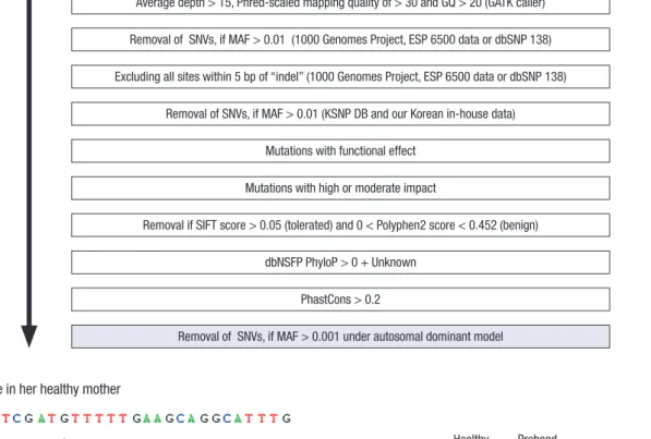
WES was performed for the three affected siblings and their mother using a TruSeq Exome Kit (Illumina Inc., San Diego, CA, USA) on a HiSeq2000 platform (Illumina Inc.). The blood sample from their father was not available. The obtained sequence reads were aligned to the human genome (hg19) using Bowtie 2. Allele frequencies of the known variants were confirmed from multiple databases, including the 1000 Genomes Project, the National Heart, Lung, and Blood Institute Exome Sequencing Project, the Single Nucleotide Polymorphism Database, and the Korean Single Nucleotide Polymorphism Database (400 Korean controls; http://nih.go.kr/NIH_NEW/main.jsp). To prioritize the variants, we established and tested our bioinformatics work- flow (Fig. 2A). No potential variant was found under autosomal recessive or compound heterozygous models. Under autoso- mal dominant model, only one potential variant was left: the bioinformatics pipeline detected a novel heterozygous deletion in exon 12 of COG5 causing a frameshift and premature stop (c.1209delG, NM_181733.2; p.Met403IlefsX3, NP_859422.2). San- ger sequencing reverified the presence of the COG5 variation only in the patients (Fig. 2B).
To assess the expression of COG5, Western blotting of the COG5 protein was performed. The skin tissues from the affect- ed proband and a healthy control were disrupted in liquid ni- trogen with a mortar and pestle, and the proteins were extract- ed. Western blotting of COG5 proteins was performed using an anti-COG5 antibody (ab90301, Abcam, USA). Although a single band with a COG5 protein of about 90 kDa was detected by West- ern blotting of normal skin tissue as reported previously (11), two bands — one of the same size (about 90 kDa) and one small-
er (about 40 kDa) — were detected in the affected proband. The intensity of the 90-kDa band in the affected proband appeared to be significantly decreased compared to that in the normal healthy control (Fig. 2C).
As there is no fundamental or curative treatment for COG5 defect, the patients were supportively treated for their scoliosis by an orthopedic surgeon.
Ethics statement
This study was approved by the Human Research Ethics Com- mittee of Chonnam National University Hospital (IRB No. CNUH- 2014-179). Informed consent to participate was obtained from the mother of the affected siblings. The biospecimens were pro- vided by the Chonnam National University Hospital Biomedi- cal Research Institute Biobank with informed consent, under Institutional Review Board-approved protocols.
DISCUSSION
Using the traditional classification, CDG can be subdivided into two types: CDG-I and CDG-II (5,6). CDG-I affects the addition of glycans to proteins and CDG-II affects processing of the pro- tein-bound N-glycans (5,6). CDG-I results only in an N-glyco- sylation defect, while CDG-II causes a combined N- and O-gly- cosylation defect (5,6). CDG can be screened with isoelectric focusing of N- or O-glycosylated proteins such as serum trans- ferrin or apolipoprotein CIII, respectively (5,6). However, iso- electric focusing is available only in some specialized clinical centers, usually takes a long time and its sensitivity and speci- ficity are unreliable (6). Moreover, transferrin glycan analysis reveals a nonspecific pattern in most patients with the CDG-II, in contrast to those with the CDG-I (6). Due to this limitation of screening tests for CDG-II, Western blotting or genetic analysis Table 1. Clinical features and radiologic findings of the patients
Features and findings III-2 III-3 III-4 (proband)
Gender Female Female Female
Age, yr 25 18 14
Progressive ataxia: age at detection, yr < 2 < 2 1–2
Other neurologic signs Data not available Dysarthria, dysmetria, dysdiadochokinesia,
decreased deep tendon reflex Dysarthria, dysmetria, dysdiadochokinesia, decreased deep tendon reflex
Scoliosis: age at detection, yr 2 4 1
Involvement of other organs No No No
Echocardiography Data not available Normal (at 10 yr) Normal (at 13 yr)
Developmental slowing: age at onset, yr < 1 < 1 < 1
Intellectual disability Moderate Mild Mild
Brain MRI Normal (at 2 yr) → diffuse isolated cerebellar atrophy (at 18 yr)
Diffuse isolated cerebellar atrophy (at 10 yr)
Diffuse isolated cerebellar atrophy (at 12 yr)
Spine MRI Data not available Data not available No abnormality in spinal cord (at 12 yr)
Array CGH* Data not available Data not available 46, XX, inv(9)(p12q13), arr[hg18] (1-22, X)x2†
Test for FXN mutations Data not available No abnormalities Data not available
MRI = magnetic resonance imaging, CGH = comparative genome hybridization, FXN = the gene encoding frataxin.
*Array CGH was performed with the Roche NimbleGen CGX-3 135K whole-genome array. †Pericentric inversions of chromosome 9 have been reported in some normal populations.
Kim YO, et al. • Friedreich’s-Ataxia-Like Phenotypes by COG5 Defect
Fig. 2. Mutation analysis of the gene encoding COG5. (A) Bioinformatic prioritization for variants in WES. (B) Electropherograms showing the wild-type reference sequence of COG5 in the healthy mother, and a heterozygous deletion (arrow) at the fifth base in exon 12 of COG5 causing a frameshift and premature stop (c.1209delG, NM_181733.2;
p.Met403IlefsX3, NP_859422.2) in the affected proband. (C) Western blotting of COG5 skin proteins from the affected proband and a healthy control with an anti-COG5 anti- body. The proband had two bands at about 90 and 40 kDa, representing the normal and aberrant COG5 proteins, respectively. The level of full-length COG5 at about 90 kDa in the proband appears to be significantly decreased compared to that in the normal healthy control showing a single meaningful band.
COG5 = conserved oligomeric Golgi complex subunit 5, WES = whole-exome sequencing, GQ = genotype quality score, GATK = Genome Analysis Toolkit, SNVs = single-nu- cleotide variants, MAF = minor allele frequency, ESP = Exome Sequencing Project, dbSNP = Single Nucleotide Polymorphism Database, KSNP DB = Korean Single Nucleotide Polymorphism Database, SIFT = Sorting Intolerant From Tolerant, Polyphen2 = Polymorphism Phenotyping v2, dbNSFP = Database for Nonsynonymous SNPs Functional Pre- dictions, PhyloP = phylogenetic P value, MW = molecular weight.
Average depth > 15, Phred-scaled mapping quality of > 30 and GQ > 20 (GATK caller)
Removal of SNVs, if MAF > 0.01 (1000 Genomes Project, ESP 6500 data or dbSNP 138)
Excluding all sites within 5 bp of “indel” (1000 Genomes Project, ESP 6500 data or dbSNP 138)
Removal of SNVs, if MAF > 0.01 (KSNP DB and our Korean in-house data)
Mutations with functional effect
Mutations with high or moderate impact
Removal if SIFT score > 0.05 (tolerated) and 0 < Polyphen2 score < 0.452 (benign)
dbNSFP PhyloP > 0 + Unknown
PhastCons > 0.2
Removal of SNVs, if MAF > 0.001 under autosomal dominant model
Wild type in her healthy mother
Mutant type in the affected proband
c.1209delG
Removal of SNVs, if MAF 0.01 (1000 Genomes Project, ESP 6500 data or dbSNP 138) Excluding all sites within 5bp of "indel” (1000 Genomes Project, ESP 6500 data or dbSNP 138)
Removal of SNVs, if MAF >0.01 (KSNP DB and our Korean in-house data) Mutations with functional effect
Mutations with high or moderate impact
Removal if SIFT score >0.05 (tolerated) and 0< Polyphen2 score <0.452 (benign) dbNSFP PhyloP >0 + Unknown
PhastCons >0.2
Removal of SNVs, if MAF 0.001 under autosomal dominant model
Mutant type in the affected proband
c.1209delG
Wild type in her healthy mother
A
B C
Healthy
control Proband (III-4)
90
40 (kDa)MW
Normal COG5
Aberrant COG5
Healthy
control Proband (III-4)
MW (kDa)
90
Normal COG5
Aberrant 40 COG5
A
B C
of COG subunits in suspected patients with COG deficiency is recommended (5,6,8-10). WES can be an alternative useful di- agnostic method in CDG-II or COG deficiency, as it can screen for diverse variants simultaneously and rapidly with only small
amounts of blood.
Deficiencies in COG subunits have been reported in patients with developmental delay, intellectual disability, growth failure, hypotonia, dysmorphic features, cerebral or cerebellar abnor-
Kim YO, et al. • Friedreich’s-Ataxia-Like Phenotypes by COG5 Defect
malities, and feeding problems (5-15). The most commonly re- ported defective subunit in humans is COG7, followed by COG5 (5-8,11-13). Although the clinical features are not easy to delin- eate in small numbers of patients, the phenotypes appear to differ between the different defective subunits (5-15). Most pa- tients with COG7 defects died below 1 year of age, and had se- vere intellectual disability, growth retardation, facial dysmor- phisms, hyperthermia, and congenital defects of multiple inter- nal organs (5-8). However, the patients with the other COG sub- unit defects showed lower mortality rates (5,6,9-15).
All seven previously reported patients with COG5 mutations had hypotonia, developmental delay and intellectual disability (11-13). Six of them had moderate-to-severe intellectual dis- ability; all six had microcephaly and five had short stature (11- 13). Only one patient with mild intellectual disability exhibited normal head and height growth (13). Two patients presented with deafness and blindness and one of them had convulsions (13). Brain MRI findings were abnormal in two patients: the one patient had global cerebral and cerebellar atrophy (13), while the other patient had diffuse cerebellar and brainstem atrophy (11). Neurogenic bladder was present in two patients (13). Con- tracture was noted in one patient (12). Most of the patients with COG5 mutations were from consanguineous parents and had homozygous or compound heterozygous mutations (11-13).
Based on the previous diagnostic algorithm for childhood ataxia, recessive cerebellar ataxias were initially suspected in the family described herein, such as ataxia telangiectasia, in- fantile-onset spinocerebellar ataxia, or Friedreich’s ataxia (1-4).
Prior to the WES study in this family, COG5 deficiency was dif- ficult to suspect initially because the patients had no hypotonia, microcephaly, or short stature (5-15). The mild phenotypes in the present family might be due to the heterozygous deletion involving only one allele, although a novel heterozygous dele- tion of COG5 (c.1209delG) resulted in a premature stop (p.Met- 403IlefsX3) causing a smaller, aberrant COG5 protein and a re- duction in normal COG5 expression. Isolated cerebellar atro- phy in this report is also rare radiologic finding in either of COG5 deficiency or early-childhood-onset cerebellar atrophy (4,11,13).
Associated cerebral abnormalities or abnormal signal changes in the cerebellum are reported frequently (4,11,13). Although this type of cerebellar ataxia is not common, COG5 mutational analysis need to be considered in suitable patients showing ear- ly-childhood-onset Friedreich’s-ataxia-like phenotypes and iso- lated cerebellar atrophy.
ACKNOWLEDGMENT
This study was provided with data from the Korean Genome Analysis Project (4845-301), the Korean Genome and Epidemi- ology Study (4851-302), and the Korea Biobank Project (4851- 307 and KBP-2015-045), which were supported by the Korea Cen-
ter for Disease Control and Prevention, Korea.
DISCLOSURE
The authors have no potential conflicts of interest to disclose.
AUTHOR CONTRIBUTION
Conceptualization: Kim YO. Data curation: Kim YO, Choi SM, Kim SK, Yoon W, Woo YJ. Formal analysis: Kim YO, Park C, Hong Y. Funding acquisition: Kim YO. Investigation: Kim YO, Yun M, Jeong JH, Park C, Hong Y. Writing - original draft: Kim YO. Writ- ing - review & editing: Kim YO, Kim SK, Park C, Hong Y, Woo YJ.
ORCID
Young Ok Kim https://orcid.org/0000-0002-7873-1140 Misun Yun https://orcid.org/0000-0001-7818-4442 Jae-Ho Jeong https://orcid.org/0000-0002-5544-9572 Seong Min Choi https://orcid.org/0000-0003-3138-1881 Seul Kee Kim https://orcid.org/0000-0002-1508-5057 Woong Yoon https://orcid.org/0000-0002-8598-3127 Chungoo Park https://orcid.org/0000-0002-9545-6654 Yeongjin Hong https://orcid.org/0000-0002-8115-7362 Young Jong Woo https://orcid.org/0000-0003-4717-8394
REFERENCES
1. Ashley CN, Hoang KD, Lynch DR, Perlman SL, Maria BL. Childhood atax- ia: clinical features, pathogenesis, key unanswered questions, and future directions. J Child Neurol 2012; 27: 1095-120.
2. Fogel BL, Perlman S. Clinical features and molecular genetics of autoso- mal recessive cerebellar ataxias. Lancet Neurol 2007; 6: 245-57.
3. Brusse E, Maat-Kievit JA, van Swieten JC. Diagnosis and management of early- and late-onset cerebellar ataxia. Clin Genet 2007; 71: 12-24.
4. Poretti A, Wolf NI, Boltshauser E. Differential diagnosis of cerebellar atro- phy in childhood. Eur J Paediatr Neurol 2008; 12: 155-67.
5. Zeevaert R, Foulquier F, Jaeken J, Matthijs G. Deficiencies in subunits of the Conserved Oligomeric Golgi (COG) complex define a novel group of Congenital Disorders of Glycosylation. Mol Genet Metab 2008; 93: 15-21.
6. Jaeken J. Congenital disorders of glycosylation (CDG): it’s (nearly) all in it!
J Inherit Metab Dis 2011; 34: 853-8.
7. Wu X, Steet RA, Bohorov O, Bakker J, Newell J, Krieger M, Spaapen L, Ko- rnfeld S, Freeze HH. Mutation of the COG complex subunit gene COG7 causes a lethal congenital disorder. Nat Med 2004; 10: 518-23.
8. Morava E, Zeevaert R, Korsch E, Huijben K, Wopereis S, Matthijs G, Key- molen K, Lefeber DJ, De Meirleir L, Wevers RA. A common mutation in the COG7 gene with a consistent phenotype including microcephaly, ad- ducted thumbs, growth retardation, VSD and episodes of hyperthermia.
Eur J Hum Genet 2007; 15: 638-45.
9. Foulquier F, Vasile E, Schollen E, Callewaert N, Raemaekers T, Quelhas D, Jaeken J, Mills P, Winchester B, Krieger M, et al. Conserved oligomeric Gol- gi complex subunit 1 deficiency reveals a previously uncharacterized con-
genital disorder of glycosylation type II. Proc Natl Acad Sci U S A 2006;
103: 3764-9.
10. Foulquier F, Ungar D, Reynders E, Zeevaert R, Mills P, García-Silva MT, Briones P, Winchester B, Morelle W, Krieger M, et al. A new inborn error of glycosylation due to a Cog8 deficiency reveals a critical role for the Cog1- Cog8 interaction in COG complex formation. Hum Mol Genet 2007; 16:
717-30.
11. Paesold-Burda P, Maag C, Troxler H, Foulquier F, Kleinert P, Schnabel S, Baumgartner M, Hennet T. Deficiency in COG5 causes a moderate form of congenital disorders of glycosylation. Hum Mol Genet 2009; 18: 4350-6.
12. Fung CW, Matthijs G, Sturiale L, Garozzo D, Wong KY, Wong R, Wong V,
Jaeken J. COG5-CDG with a mild neurohepatic presentation. JIMD Rep 2012; 3: 67-70.
13. Rymen D, Keldermans L, Race V, Régal L, Deconinck N, Dionisi-Vici C, Fung CW, Sturiale L, Rosnoblet C, Foulquier F, et al. COG5-CDG: expand- ing the clinical spectrum. Orphanet J Rare Dis 2012; 7: 94.
14. Reynders E, Foulquier F, Leão Teles E, Quelhas D, Morelle W, Rabouille C, Annaert W, Matthijs G. Golgi function and dysfunction in the first COG4- deficient CDG type II patient. Hum Mol Genet 2009; 18: 3244-56.
15. Shaheen R, Ansari S, Alshammari MJ, Alkhalidi H, Alrukban H, Eyaid W, Alkuraya FS. A novel syndrome of hypohidrosis and intellectual disability is linked to COG6 deficiency. J Med Genet 2013; 50: 431-6.