Overexpression of Human Arginine Decarboxylase Rescues Human Mesenchymal Stem Cells against H
2O
2Toxicity through Cell Survival Protein Activation
In this study, we explored the potentiality of human arginine decarboxylase (ADC) to enhance the survival of mesenchymal stem cells (MSCs) against unfavorable milieu of host tissues as the low survival of MSCs is the issue in cell transplantation therapy. To address this, human MSCs overexpressing human ADC were treated with H2O2 and the resultant intracellular events were examined. First, we examined whether human ADC is
overexpressed in human MSCs. Then, we investigated cell survival or death related events.
We found that the overexpression of human ADC increases formazan production and reduces caspase 3 activation and the numbers of FITC, hoechst, or propidium iodide positive cells in human MSCs exposed to H2O2. To elucidate the factors underlying these phenomena, AKT, CREB, and BDNF were examined. We found that the overexpression of human ADC phosphorylates AKT and CREB and increases BDNF level in human MSCs exposed to H2O2. The changes of these proteins are possibly relevant to the elevation of agmatine. Collectively, our data demonstrate that the overexpression of human ADC stimulates pro-survival factors to protect human MSCs against H2O2 toxicity. In conclusion, the present findings support that ADC can enhance the survival of MSCs against hostile environment of host tissues.
Key Words: Arginine Decarboxylase; Cell Survival; Hydrogen Peroxide; Mesenchymal Stem Cells; Retroviral Vector
Su Kyoung Seo,1,2 Wonsuk Yang,1 Yu Mi Park,1,2 Won Taek Lee,1 Kyung Ah Park,1 and Jong Eun Lee,1,2
1Department of Anatomy, 2Brain Korea 21 Project for Medical Science, Yonsei University College of Medicine, Seoul, Korea
Received: 31 August 2012 Accepted: 18 January 2013 Address for Correspondence:
Jong Eun Lee, PhD
Department of Anatomy, BK21 Project for Medical Science, Yonsei University College of Medicine, 50 Yonsei-ro, Seodaemun-gu, Seoul 120-752, Korea
Tel: +82.2-2228-1646, Fax: +82.2-365-0700 E-mail: jelee@yuhs.ac
This research was supported by Basic Science Research Program through the National Research Foundation of Korea (NRF) funded by the Ministry of Education, Science and Technology (MEST) (2012-0005440). This work was supported by the Brain Korea 21 Project for Medical Science, Yonsei University.
http://dx.doi.org/10.3346/jkms.2013.28.3.366 • J Korean Med Sci 2013; 28: 366-373 Cell Therapy & Organ Transplantation
INTRODUCTION
The arginine decarboxylase (ADC, EC 4.1.1.19) is an enzyme which synthesizes agmatine through L-arginine decarboxyl- ation (1). ADC is ubiquitously found in most living organisms encompassing bacteria (2), plants (3), and animals (4). Accord- ing to recent studies, ADC demonstrates various functions de- pending on each life form investigated. Some investigators re- port that ADC can enhance the resistance against environmen- tal insults encompassing as high acidity and salt stress in bacte- ria and plants (5, 6). Others suggest that ADC exerts anti-prolif- erative effect in tumor cell (7) and neuroprotective effect against immobilization stress and chronic glucocorticoid exposure in animal (8, 9). As well, it is reported that mammalian ADC has the distinct characteristics compared to bacterial and plant ADC, in viewpoint of physiological responses and intracellular local- ization (10). Therefore, ADC is considered to be the multi-facet- ed protein in different life forms.
Mesenchymal stem cells (MSCs) are the typical adult stem cell which represents multi-potency, self-renewal, and broad
tissue distribution (11). These MSCs possess biological and so- cial advantages since they do not significantly produce immune rejection from host tissues and are free of many ethical prob- lems (12, 13). Owing to these aspects, MSCs receive much atten- tion as the promising therapy to treat several types of human diseases (14). However, there is a critical issue to be solved to maximize MSCs-derived therapeutic effects. After being trans- planted, MSCs are unavoidably exposed to hostile conditions formed in host tissues (15), one of which is oxidative stress (16, 17). As a result, their survival is considerably low in transplant- ed sites, thereby reducing the therapeutic effects of MSCs trans- plantation. Therefore, a large number of researches have been performed to enhance the survival of MSCs against unfavor- able milieu of host tissues (18-20).
In the present study, we explored the potentiality of ADC to facilitate MSCs survival against harsh surroundings of host tis- sues. As mentioned above, ADC renders life to struggle against endogenous and exogenous toxic insults. Based on this, it was hypothesized that ADC can improve MSCs survival against se- vere environment of host tissues. To address this, retroviral vec-
tor carrying human ADC gene was infected in human MSCs which were then exposed to H2O2. Then, several phenomena were examined including agmatine level, mitochondrial and plasma membrane integrity, caspase 3 activity, apoptotic DNA damage, and pro-survival factors such as AKT, CREB, and BDNF.
MATERIALS AND METHODS Materials
T-75 flasks and culture dishes (100 × 20 mm) were purchased from SPL Life Sciences (Seoul, Korea). 96-well plates were pur- chased from BD Biosciences (Franklin Lakes, NJ, USA). Dul- becco’s modified Eagle medium (DMEM)-low glucose, fetal bovine serum (FBS), and penicillin/streptomycin were pur- chased from Life Technologies Korea (Seoul, Korea). Retroviral vector pLXSN expression system was purchased from Clontech (Palo Alto, CA, USA). Annexin V-FITC apoptosis detection kit, Lipofectamine 2000, G418, polybrene reagent, Hoechst 33258, propidium iodide, and 3-(4, 5-dimethylthiazol-2-yl)-2, 5-diphe- nyltetrazolium bromide (MTT) were purchased from Sigma (St.
Louis, MO, USA). A rabbit polyclonal antibody to human ADC was purchased from ATGEN (Seoul, Korea). Polyvinylidene flu- oride (PVDF) membrane was purchased from Millipore (Biller- ica, MA, USA). 4´,6-diamidino-2-phenylindole (DAPI) was pur- chased from Vector Labs (Burlingame, CA, USA). Bicinchoninic acid (BCA) kit and enhanced chemiluminescent (ECL) Western blotting detection reagent were purchased from Thermo Fisch- er Scientific (Fremont, CA, USA). Rabbit polyclonal antibodies to AKT and cleaved caspase 3 and rabbit monoclonal antibod- ies to phosphorylated forms of AKT and CREB were purchased from Cell signaling technology (Beverly, MA, USA). A rabbit polyclonal antibody to BDNF, a mouse polyclonal antibody to β actin, and an anti-mouse or rabbit IgG antibody conjugated with horse radish peroxidasae (HRP) were purchased from Abcam (Cambridge, UK). A rabbit polyclonal antibody to caspase 3 and FITC conjugated second antibody were purchased from Milli- pore (Billerica, MA, USA). Unless specified, all other chemicals were purchased from Sigma (St. Louis, MO, USA).
Cell culture and treatment
Human bone marrow mesenchymal stem cells (MSCs) were obtained from Yonsei Cell Therapy center of Severance Hospi- tal. Flow cytometry was performed to confirm the phenotypic characteristics of MSCs. MSCs were seeded in T-75 flasks and cultured at 37ºC in a humid incubator with 95% of O2 and 5% of CO2. The following day, the non-adherent cells were removed from the flasks and the adherent cells were fed with fresh Dul- becco’s modified Eagle medium (DMEM)-low glucose supple- mented with 10% FBS and 100 unit/mL penicillin and 100 µg/
mL streptomycin. MSCs were then continuously maintained for 1-4 weeks. Medium was changed every second day. MSCs,
of which passage number is over 5, were substantially used in the experiments. H2O2 was employed to reproduce oxidative stress condition, which is prevalent in damaged tissues (21). It has unique biochemical properties such as relatively long half- life and high solubility in both lipid and aqueous media (22).
The control MSCs and MSCs infected with human ADC or emp- ty vector were treated with 200 µM of H2O2 for 6 hr. This regi- men for treatment was selected based on MTT data in the con- trol MSCs exposed to H2O2, where the viability was approximate- ly 50%-55%.
Infection of human MSCs with retroviral vectors
The retroviral vector carrying human ADC gene (gene bank ac- cession number AY325129) was previously constructed by our group (23). For the experiments, PT67 packaging cells were in- fected with retroviral vector containing human ADC gene or empty pLXSN vector and then maintained in a humid incuba- tor with 5% of CO2 at 37ºC for one week. Then, the medium con- taining retrovirus was filtered using 0.45 µm pore polysulfonic filter. For infection, human MSCs were cultured with the super- natants containing human ADC vector or empty pLXSN vector using polybrene reagent (6 µg/mL). After 24 hr of incubation, the medium was replaced by normal culture medium. The in- fected MSCs were maintained to perform the experiments for up to one week.
Immunocytochemistry
The expression of human ADC protein was investigated in con- trol MSCs, pLXSN-MSCs and ADC-MSCs through immunocy- tochemistry. Samples were washed three times with PBS and fixed with 100% ethanol for 10 min on ice, and then washed three times with PBS. To increase permeability, samples were incubated with 0.025% Triton, and were blocked for 1 hr at room temperature with blocking solution. The samples were treated with anti-human ADC antibody for 3 hr at room temperature, which was diluted in the incubation buffer (1:500). Then, pri- mary antibody was removed and samples were washed three times for 5 min each with PBS. Then, samples were treated with FITC labeled secondary antibody for 2 hr at room temperature, which was diluted in the incubation buffer (1:500). Finally, sam- ples were stained with 4´,6-diamidino-2-phenylindole (DAPI) 10 µL/mL for staining nucleus and then visualized under a con- focal microscope (Zeiss LSM700; Zeiss, Thornwood, NY, USA).
3-(4, 5-Dimethylthiazol-2-yl)-2, 5-diphenyl tetrazolium bromide (MTT) assay
In H2O2 treated and non-treated groups, 3-(4, 5-dimethylthia- zol-2-yl)-2, 5-diphenyltetrazolium bromide (MTT) assay, which is based on the activity of mitochondrial dehydrogenase (24), was performed to estimate the number of viable cells. Briefly, MSCs were seeded (1.0 × 10⁴ cells/200 µL) in 96-well plates. The
cells were maintained in 96-well plates for 2 days and then were treated with H2O2 (200 µM for 6 hr). Then, the media were re- moved from each well and 200 µL of MTT solution was added into each well and the culture plates were incubated for 4 hr at 37ºC. Then, dimethyl sulfoxide (DMSO) was added to dissolve formazan crystal in the mitochondria of living cell. The absor- bance at 570 nm was measured with a spectrophotometer.
Hoechst/Propidium iodide (PI) staining
Hoechst 33258 and propidium iodide were employed to stain control MSCs, LXSN-MSCs and ADC-MSCs in this study. Hoechst 33258 enters both live and dead cells, but is distinctly accumu- lated in apoptotic nucleus. On the other hand, PI enters only dead cells and thus, is used to examine cell membrane integri- ty. All experimental groups were cultured in 24-well plates at a density of 3 × 10⁴ cells/well for 2 days. After treatment of 200 µM of H2O2 for 6 hr, Hoechst 33258 was added to the culture medi- um (2-3 mg/mL) and then the samples were kept at 37ºC for 30 min. PI solution was then added (2-5 mg/mL) just before ob- servation under the fluorescence microscope. PI/Hoechst-pos- itive and PI/Hoechst-negatives were counted as dead or living cells, respectively.
Fluorescence activated cell sorting (FACS) analysis The proportion of cells undergoing apoptosis was analyzed with Annexin V-FITC Apoptosis Detection Kit according to the man- ufacturer’s protocol. In Brief, cells were harvested and resus- pended in binding buffer (106 cells/mL), and then, cells were mixed with 5 μL of annexin V-FITC and 10 μL of propidium io- dide (PI), followed by the incubation of cells at room tempera- ture for 15 min in dark. Then, both FITC and PI fluorescence of cells were analyzed with flow cytometer.
High Performance Liquid Chromatography (HPLC) The agmatine concentration in control MSCs and MSCs trans- fected with empty LXSN or human ADC gene carrying vector was measured by HPLC. The harvested cells were homogenized in 10% trichloracetic acid (TCA) and then, centrifuged at 12,000 rpm for 30 min. The supernatant was derivatized with o-phtha- ladehyde (OPA) and then, injected into the HPLC column with a fluorescence detector. The recovery of agmatine was calculated from the added external standard and the values are expressed as the µM concentration.
Western blotting
Cells from both H2O2 treated/non-treated groups were collect- ed and the pellets were resuspended in lysis buffer (1% NP-40, 50 mM Tris [pH 7.5], 150 mM sodium chloride, 0.5% sodium deoxycholate, 0.1% sodium dodecyl sulfate (SDS), 1 mM EGTA, 5 mM EDTA, 1 mM PMSF, 1 mM Na orthovanadate, 5 mM Na fluoride, 1 µg/mL pepstatin, 2 µg/mL aprotinin, and 5 µg/mL
leupeptin) for 2 hr and then centrifuged at 13,200 rpm for 30 min at 4ºC. The supernatants were collected and protein con- centration was determined with bicinchoninic acid (BCA) as- say. The equal amounts of proteins (40 μg) were separated on 10%-12% SDS-polyacrylamide gels using an electrophoresis unit maintained at the constant voltage (100 V). Separated pro- teins were then electrotransfered onto PVDF membrane. Later, membranes were incubated with 0.1% bovine serum albumin (BSA) in Tris-buffered saline and 0.3% Tween-20 (TBS-T) to block nonspecific binding for 1 hr at room temperature. After blocking, the membranes were treated with specific primary antibodies including anti-phospho AKT, anti-phospho CREB, anti-caspase 3, anti-cleavage caspase 3, and anti-BDNF at 4ºC.
All of primary antibodies were diluted (1:1,000) in incubation buffers for treatment. After treatment, membranes were washed 3 times with TBST for every 5 min. Then, membranes were treat- ed for 1 hr at room temperature with anti-mouse or rabbit IgG antibody conjugated with horse radish peroxidase (HRP) in the incubation buffers (1:3,000). Finally, blots were rinsed and pro- teins of interest were detected with enhanced chemilumines- cent (ECL) kit and visualized with LAS 4000 image analyzer.
Protein bands detected were quantified as the ratio of the target protein to beta actin using Image J software (National Institutes of Health, Bethesda, MD, USA).
Statistical analysis
The data are presented as mean ± SD. Statistical analysis of the data was made by one-way ANOVA using SPSS statistical pack- age version 12.0. P values less than 0.05 were considered to be statistically significant and the significant differences were cal- culated at 95% confidence interval. All experiments were inde- pendently carried out at least three times.
RESULTS
The overexpression of human arginine decarboxylase and the elevation of agmatine in human mesenchymal stem cells
The retroviral vectors carrying human arginine decarboxylase (ADC), which was previously constructed by our group (23), were employed to infect human mesenchymal stem cells (MSCs).
Western blotting data show that ADC protein was greatly ele- vated in MSCs infected with vectors containing ADC gene (Fig.
1A). In particular, the increase of approximately 0.7 or 2.5 fold was respectively observed in those MSCs, compared with LX- SN-MSCs under normal or H2O2 exposure condition (Fig. 1B).
And the level of agmatine synthesized by ADC was approximate- ly 2 fold raised in MSCs infected with vectors containing ADC gene under H2O2 exposure (Fig. 1C), implying the overexpressed ADC involved this elevation in those MSCs.
The mitochondrial and plasma membrane integrity in ADC overexpressed human mesenchymal stem cells under H2O2 exposure
The overexpressed human arginine decarboxylase (ADC) pre- served mitochondrial integrity in human mesenchymal stem cells (MSCs) treated with H2O2. The level of formazan was 56.4%
± 0.9% and 52.8% ± 1.5% of untreated control MSCs in control MSCs or LXSN-MSCs exposed to H2O2, respectively (Fig. 2A).
However, its level significantly increased into 69.7% ± 2.7% of untreated control MSCs in ADC-MSCs treated with H2O2 (Fig.
2A). As well, the overexpressed ADC saved plasma membrane integrity in human MSCs exposed to H2O2. The propidium io- dide (PI) positive cells clearly appeared in control MSCs and LXSN-MSCs treated with H2O2 (Fig. 2B). However, this damage was significantly alleviated in ADC-MSCs. In fact, the numbers of PI positive cells were reduced into 32.9% ± 1.3% of control MSCs in ADC-MSCs under H2O2 exposure. This protective ef- fect of the overexpressed ADC was further investigated with FACS analysis. The distribution of FITC positive cells was obvi- ously reduced in ADC-MSCs compared to control and LXSN MSCs under H2O2 exposure (Fig. 2C).
The apoptotic events and pro-survival factors in ADC overexpressed human mesenchymal stem cells under H2O2 exposure
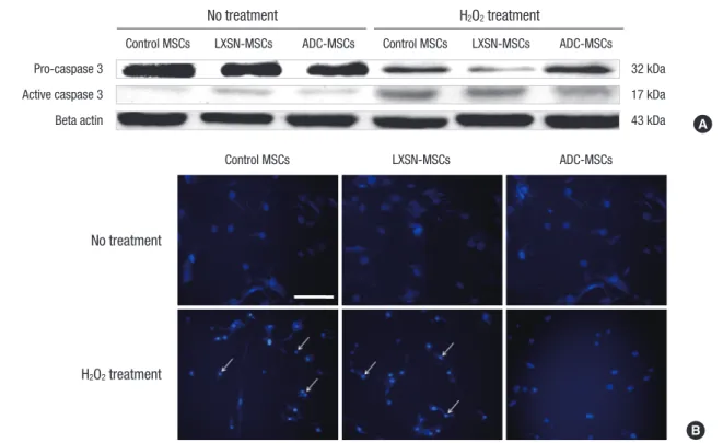
The overexpressed human arginine decarboxylase (ADC) sup- pressed apoptotic events in human mesenchymal stem cells (MSCs) exposed to H2O2. The level of pro-caspase 3 was not
quite different between control MSCs and ADC-MSCs under normal or H2O2 exposure (Fig. 3A). However, the level of cleaved caspase 3 increased in control MSCs treated with H2O2, where- as its level obviously decreased in ADC-MSCs exposed to H2O2
(Fig. 3A). The similar trend also appeared in nuclear integrity. No difference of nuclear morphology was found between control MSCs and ADC-MSCs under normal condition (Fig. 3B). How- ever, a lot of condensed nuclei were observed in control MSCs exposed to H2O2, while their numbers apparently decreased in ADC-MSCs exposed to H2O2 (Fig. 3B). This protective effect of the overexpressed human arginine decarboxylase (ADC) was further substantiated in human mesenchymal stem cells (MSCs) treated with H2O2. That is, the expressions of pro-survival pro- teins P-AKT, P-CREB, and BDNF obviously increased in ADC- MSCs, compared with control MSCs under H2O2 exposure (Fig.
4). However, their expressions were not differed between con- trol MSCs and ADC-MSCs under normal condition (Fig. 4).
DISCUSSION
The current findings demonstrate that the overexpression of human arginine decarboxylase (ADC) stimulates pro-survival factors such as AKT, CREB, and BDNF to protect human mes- enchymal stem cells (MSCs) against H2O2 toxicity. As common- ly known, the low survival of MSCs transplanted is attributed to the unfavorable milieu of host tissues (25, 26). Accordingly, the improvement of MSCs survival following transplantation be- comes the fundamental goal to secure the efficacy of cell thera- Fig. 1. The retroviral overexpression of human ADC in human MSCs and the measurement of agmatine with HPLC. (A) Bands of human ADC protein in control MSCs, LXSN- MSCs, and ADC-MSCs with or without H2O2 treatment. The photograph is a representative from 4 blots with the similar results. (B) Quantification of ADC protein bands demon- strated in Fig. 1A. Asterisks indicate the significant difference at P value less than 0.05 when compared to control and LXSN-MSCs. Data represent the mean ± standard devi- ation (SD) for 4 independent experiments. (C) Quantification of agmatine in control MSCs, LXSN-MSCs, and ADC-MSCs with or without H2O2 treatment. Asterisk indicates the significant difference at P value less than 0.05 when compared to control MSCs exposed to H2O2. Data represent the mean ± standard deviation (SD) for 4 independent exper- iments.
Fold change of ADC protein Fold change of agmatine
Control MSCs LXSN-MSCs ADC-MSCs Control MSCs LXSN-MSCs ADC-MSCs
4
3
2
1
0
3.0 2.5 2.0 1.5 1.0 0.5 0.0
No treatment No treatment
H2O2 treatment H2O2 treatment
B C
* *
*
A ADC
Beta-actin
1 4
No treatment
1, 4 : Control MSCs. 2, 5 : LSXN-MSCs. 3, 6 : ADC-MSCs.
H2O2 treatment
2 3 5 6
43 kDa 43 kDa
py (27, 28). In this perspective, ADC seems to be applicable be- cause it exerts several effects such as resistance against environ- mental insults (5, 6), anti-proliferation of tumors (7), and neu- roprotection (9) in living organisms. In the current study, the potentiality of ADC was explored to enhance the survival of MSCs against hostile environment of host tissues.
The retroviral vector expression system is an efficient tool to deliver genes of interest into target cells (29, 30). In this study, human MSCs (ADC-MSCs) were infected with the retroviral vector (LXSN) containing human ADC gene to overexpress hu- man ADC protein. Western blotting was carried out to confirm the overexpression of human ADC protein (Fig. 1A). The data indicate that human ADC protein level is higher in ADC-MSCs, compared to control MSCs and LXSN-MSCs with or without
H2O2 treatment, respectively (Fig. 1B). It is interesting that ADC protein is up-regulated even in control MSCs exposed to H2O2
although its level is lower than that of ADC-MSCs. Similarly, the increase of ADC protein has been reported in neurons under stress condition (8, 9). Thus, it is presumable that ADC plays the certain roles in the amelioration or exacerbation of stresses. To further confirm the overexpression of ADC, agmatine was ana- lyzed with HPLC. As mentioned earlier, agmatine production is mediated by ADC (1). The data show that the robust elevation of agmatine was found in ADC-MSCs exposed to H2O2 (Fig. 1C).
Overall, the current data demonstrated that human ADC was successfully overexpressed through retroviral vector expression system in human MSCs.
The present findings indicated that the overexpression of hu- Fig. 2. Cell viability in control MSCs, LXSN-MSCs, and ADC-MSCs following H2O2 treat- ment. (A) Formazan formation. (B) Cell membrane integrity. Each photograph is a rep- resentative from 4 fields of microscope. The arrow indicates cells, where plasma mem- brane is damaged. Scale bar represents 200 µm. (a) Control MSCs, (b) LXSN-MSCs, (c) ADC-MSCs. (d) The quantitative graph showing the total number of propidium iodide (PI) positive cells in all experimental groups. Asterisk indicates the significant differ- ence at P value less than 0.05 when compared to LXSN-MSCs exposed to H2O2. (C) Flow cytometric analysis. The data show that the distribution of cells undergoing apop- tosis is reduced in ADC-MSCs compared to control MSCs and LXSN-MSCs under H2O2 exposure. Each panel is a representative from 4 independent experiments with the similar results.
Formazan formation (% control MSCs)
Control MSCs
- + - + - +
H2O2
LXSN-MSCs ADC-MSCs
120
100
80
60
40
20
0
A B
*
a
c
b
d
PI (+) cells (% of control MSC) 120 100 80 60 40 20 0
*
100 101 102 103 104
FL2-H
FL1-H No treatment
104 103 102 101 100
100 101 102 103 104
FL2-H
FL1-H H2O2 treatment
104 103 102 101 100
Control MSCs
100 101 102 103 104
FL2-H
FL1-H 104
103 102 101 100
100 101 102 103 104
FL2-H
FL1-H 104
103 102 101 100
LXSN- MSCs
C
100 101 102 103 104
FL2-H
FL1-H 104
103 102 101 100
100 101 102 103 104
FL2-H
FL1-H 104
103 102 101 100
ADC- MSCs
man ADC supports mitochondrial function (Fig. 2A) and sup- presses apoptotic events (Fig. 3) in MSCs exposed to H2O2. MTT data show that formazan formation was approximately half of untreated group in control MSCs or LXSN-MSCs exposed to H2O2, respectively (Fig. 2A). However, formazan formation was significantly raised in ADC-MSCs, compared with untreated group under H2O2 exposure (Fig. 2A). Therefore, these data im- ply that the overexpression of human ADC preserves mitochon- drial integrity in MSCs against H2O2 toxicity. The fluorescence micrographs show that the numbers of propidium iodide posi- tive cells with condensed nuclei were elevated in control MSCs
and LXSN-MSCs after H2O2 treatment (Fig. 2B, 3B). However, this pathology was significantly attenuated in ADC-MSCs. The similar phenomenon was also found in FACS analysis. The dis- tribution of FITC fluorescent cells was greatly decreased in ADC- MSCs, compared with control and LXSN-MSCs under H2O2 ex- posure (Fig. 2C), suggesting that the overexpressed ADC inhib- ited apoptotic death of MSCs. To understand the implication of apoptotic protein in this pathology, caspase 3 into which apop- totic signals are converged (31), was examined. As expected, cas- pase 3 was activated in control MSCs and LXSN-MSCs exposed to H2O2. However, its activation was obviously inhibited in ADC- Fig. 3. Apoptotic events in control MSCs, LXSN-MSCs, and ADC-MSCs following H2O2 treatment. (A) Caspase 3 activation. Western blotting data show that H2O2 treatment in- creases active form of caspase 3 in control MSCs and LXSN-MSCs. However, such an activation of caspase 3 was alleviated in ADC-MSCs exposed to H2O2. The less or no acti- vation of caspase 3 was observed in control MSCs, LXSN-MSCs, and ADC-MSCs without H2O2 treatment. Each photograph is a representative from 4 blots with the similar re- sults. (B) Nuclear morphology. Each photograph is a representative from 4 fields of microscope. Scale bar represents 200 µm. The data indicate that nuclear condensation (ar- row) is lessened in ADC-MSCs compared to control MSCs and LXSN-MSCs following H2O2 treatment.
Control MSCs
No treatment
H2O2 treatment
LXSN-MSCs ADC-MSCs
B A Control MSCs LXSN-MSCs ADC-MSCs Control MSCs LXSN-MSCs ADC-MSCs
No treatment H2O2 treatment
Pro-caspase 3 32 kDa
Active caspase 3 17 kDa
Beta actin 43 kDa
Fig. 4. AKT, CREB, and BDNF in control MSCs, LXSN-MSCs, and ADC-MSCs following H2O2 treatment. Western blotting was performed to examine AKT p-AKT, CREB, p-CREB, and BDNF in control MSCs, LXSN-MSCs, and ADC-MSCs before and after H2O2 treatment. Results show that the levels of p-AKT, p-CREB and BDNF were greatly increased in ADC-MSCs following H2O2 treatment, compared to those of control MSCs and LXSN-MSCs. Each photograph is a representative from 4 blots with the similar results.
Control MSCs LXSN-MSCs ADC-MSCs Control MSCs LXSN-MSCs ADC-MSCs
No treatment H2O2 treatment
t-AKT 60 kDa
p-AKT 60 kDa
p-CREB 43 kDa
t-CREB 43 kDa
BDNF 15 kDa
Beta-actin 43 kDa
MSCs exposed to H2O2 (Fig. 3A). Therefore, current data suggest that the overexpression of human ADC suppresses the pro-apop- totic executioner in human MSCs treated with H2O2.
The current findings show that the overexpression of human ADC increases the phosphorylation of v-akt murine thymoma viral oncogene homolog (AKT) and cAMP response element binding protein (CREB) and the protein level of brain-derived neurotrophic factor (BDNF) in human MSCs exposed to H2O2
(Fig. 4). AKT (also known as PKB) is a serine/threonine protein kinase that plays the important roles in cell survival (32). Once activated, it phosphorylates CREB (33), which in turn, switches on the expression of BDNF (34). Conversely, BDNF per se phos- phorylates AKT and CREB to promote cell survival against the insults (35). In support, all of AKT, CREB, and BDNF have shown to suppress the activation of caspase 3 and the occurrence of apoptotic death in various pathologic conditions (36-38). Im- portantly, one study demonstrates that BDNF is produced in MSCs (39). Therefore, considering all of these, it is probable that AKT, CREB, and BDNF pathway might contribute to the enhance- ment of survival in ADC-MSCs exposed to H2O2. The support- ing explanation of it may be relevant to agmatine, since AKT is activated by agmatine (40). In this study, agmatine was greatly raised in ADC-MSCs exposed to H2O2 (Fig. 1C) and thus, this elevation of agmatine has supposedly influenced AKT, CREB, and BDNF. However, the details of underlying mechanism re- main to be elucidated. Overall, current data suggest that the overexpression of human ADC activates cell survival-related proteins to enhance the survival of human MSCs against H2O2
toxicity.
Taken together, our data suggest that the overexpression of human ADC stimulates pro-survival factors to protect human MSCs against H2O2 toxicity. In conclusion, the present findings support the idea that human ADC is able to enhance the surviv- al of human MSCs against hostile environment of host tissues.
ACKNOWLEDGMENTS
The authors have no conflicts of interest to disclose.
REFERENCES
1. Morris SM Jr. Enzymes of arginine metabolism. J Nutr 2004; 134: 2743S-7.
2. Cunin R, Glansdorff N, Piérard A, Stalon V. Biosynthesis and metabo- lism of arginine in bacteria. Microbiol Rev 1986; 50: 314-52.
3. Borrell A, Culianez-Macia FA, Altabella T, Besford RT, Flores D, Tibur- cio AF. Arginine decarboxylase is localized in chloroplasts. Plant Physiol 1995; 109: 771-6.
4. Morrissey J, McCracken R, Ishidoya S, Klahr S. Partial cloning and char- acterization of an arginine decarboxylase in the kidney. Kidney Int 1995;
47: 1458-61.
5. Kasinathan V, Wingler A. Effect of reduced arginine decarboxylase activ- ity on salt tolerance and on polyamine formation during salt stress in
Arabidopsis thaliana. Physiol Plant 2004; 121: 101-7.
6. Alvarez-Ordóñez A, Fernández A, Bernardo A, López M. Arginine and lysine decarboxylases and the acid tolerance response of Salmonella Ty- phimurium. Int J Food Microbiol 2010; 136: 278-82.
7. Wolf C, Brüss M, Hänisch B, Göthert M, von Kügelgen I, Molderings GJ.
Molecular basis for the antiproliferative effect of agmatine in tumor cells of colonic, hepatic, and neuronal origin. Mol Pharmacol 2007; 71: 276-83.
8. Zhu MY, Wang WP, Huang J, Regunathan S. Chronic treatment with glucocorticoids alters rat hippocampal and prefrontal cortical morphol- ogy in parallel with endogenous agmatine and arginine decarboxylase levels. J Neurochem 2007; 103: 1811-20.
9. Zhu MY, Wang WP, Huang J, Feng YZ, Regunathan S, Bissette G. Re- peated immobilization stress alters rat hippocampal and prefrontal cor- tical morphology in parallel with endogenous agmatine and arginine decarboxylase levels. Neurochem Int 2008; 53: 346-54.
10. Regunathan S, Reis DJ. Characterization of arginine decarboxylase in rat brain and liver: distinction from ornithine decarboxylase. J Neuro- chem 2000; 74: 2201-8.
11. Parekkadan B, Milwid JM. Mesenchymal stem cells as therapeutics. Annu Rev Biomed Eng 2010; 12: 87-117.
12. Ryan JM, Barry FP, Murphy JM, Mahon BP. Mesenchymal stem cells avoid allogeneic rejection. J Inflamm (Lond) 2005; 2: 8.
13. Strauer BE, Kornowski R. Stem cell therapy in perspective. Circulation 2003; 107: 929-34.
14. Hodgkinson CP, Gomez JA, Mirotsou M, Dzau VJ. Genetic engineering of mesenchymal stem cells and its application in human disease thera- py. Hum Gene Ther 2010; 21: 1513-26.
15. Zhu W, Chen J, Cong X, Hu S, Chen X. Hypoxia and serum deprivation- induced apoptosis in mesenchymal stem cells. Stem Cells 2006; 24: 416-25.
16. Yao EH, Yu Y, Fukuda N. Oxidative stress on progenitor and stem cells in cardiovascular diseases. Curr Pharm Biotechnol 2006; 7: 101-8.
17. Ko E, Lee KY, Hwang DS. Human umbilical cord blood-derived mesen- chymal stem cells undergo cellular senescence in response to oxidative stress. Stem Cells Dev 2012; 21: 1877-86.
18. Chen HY, Zhang X, Chen SF, Zhang YX, Liu YH, Ma LL, Wang LX. The protective effect of 17β-estradiol against hydrogen peroxide-induced apoptosis on mesenchymal stem cell. Biomed Pharmacother 2012; 66:
57-63.
19. Li S, Bian H, Liu Z, Wang Y, Dai J, He W, Liao X, Liu R, Luo J. Chlorogenic acid protects MSCs against oxidative stress by altering FOXO family genes and activating intrinsic pathway. Eur J Pharmacol 2012; 674: 65-72.
20. Ebert R, Ulmer M, Zeck S, Meissner-Weigl J, Schneider D, Stopper H, Schupp N, Kassem M, Jakob F. Selenium supplementation restores the antioxidative capacity and prevents cell damage in bone marrow stro- mal cells in vitro. Stem Cells 2006; 24: 1226-35.
21. Giustarini D, Dalle-Donne I, Tsikas D, Rossi R. Oxidative stress and hu- man diseases: origin, link, measurement, mechanisms, and biomarkers.
Crit Rev Clin Lab Sci 2009; 46: 241-81.
22. Satoh T, Sakai N, Enokido Y, Uchiyama Y, Hatanaka H. Free radical-in- dependent protection by nerve growth factor and Bcl-2 of PC12 cells from hydrogen peroxide-triggered apoptosis. J Biochem 1996; 120: 540-6.
23. Moon SU, Kwon KH, Kim JH, Bokara KK, Park KA, Lee WT, Lee JE. Re- combinant hexahistidine arginine decarboxylase (hisADC) induced en- dogenous agmatine synthesis during stress. Mol Cell Biochem 2010; 345:
53-60.
24. Denizot F, Lang R. Rapid colorimetric assay for cell growth and survival:
modifications to the tetrazolium dye procedure giving improved sensi- tivity and reliability. J Immunol Methods 1986; 89: 271-7.
25. Müller-Ehmsen J, Krausgrill B, Burst V, Schenk K, Neisen UC, Fries JW, Fleischmann BK, Hescheler J, Schwinger RH. Effective engraftment but poor mid-term persistence of mononuclear and mesenchymal bone mar- row cells in acute and chronic rat myocardial infarction. J Mol Cell Car- diol 2006; 41: 876-84.
26. Burst VR, Gillis M, Pütsch F, Herzog R, Fischer JH, Heid P, Müller-Ehm- sen J, Schenk K, Fries JW, Baldamus CA, et al. Poor cell survival limits the beneficial impact of mesenchymal stem cell transplantation on acute kidney injury. Nephron Exp Nephrol 2010; 114: e107-16.
27. Mohammadzadeh M, Halabian R, Gharehbaghian A, Amirizadeh N, Jahanian-Najafabadi A, Roushandeh AM, Roudkenar MH. Nrf-2 over- expression in mesenchymal stem cells reduces oxidative stress-induced apoptosis and cytotoxicity. Cell Stress Chaperones 2012; 17: 553-65.
28. Li Z, Wei H, Liu X, Hu S, Cong X, Chen X. LPA rescues ER stress-associat- ed apoptosis in hypoxia and serum deprivation-stimulated mesenchy- mal stem cells. J Cell Biochem 2010; 111: 811-20.
29. Ausubel FM, Brent R, Kingston RE, Moore DD, Seidman JG, Smith JA, Struhl K. Current protocols in molecular biology. New York: John Wiley
& Sons, 1995.
30. Coffin JM, Hughes SH, Varmus HE. Retroviruses. Plainview: Cold Spring Harbor Laboratory Press, 1997.
31. Porter AG, Jänicke RU. Emerging roles of caspase-3 in apoptosis. Cell Death Differ 1999; 6: 99-104.
32. Manning BD, Cantley LC. AKT/PKB signaling: navigating downstream.
Cell 2007; 129: 1261-74.
33. Du K, Montminy M. CREB is a regulatory target for the protein kinase Akt/PKB. J Biol Chem 1998; 273: 32377-9.
34. Riccio A, Ahn S, Davenport CM, Blendy JA, Ginty DD. Mediation by a CREB family transcription factor of NGF-dependent survival of sympa- thetic neurons. Science 1999; 286: 2358-61.
35. Xia Y, Wang CZ, Liu J, Anastasio NC, Johnson KM. Brain-derived neuro- trophic factor prevents phencyclidine-induced apoptosis in developing brain by parallel activation of both the ERK and PI-3K/Akt pathways.
Neuropharmacology 2010; 58: 330-6.
36. Matsui T, Li L, del Monte F, Fukui Y, Franke TF, Hajjar RJ, Rosenzweig A.
Adenoviral gene transfer of activated phosphatidylinositol 3’-kinase and Akt inhibits apoptosis of hypoxic cardiomyocytes in vitro. Circulation 1999; 100: 2373-9.
37. Nagai-Kusuhara A, Nakamura M, Mukuno H, Kanamori A, Negi A, Sei- gel GM. cAMP-responsive element binding protein mediates a cGMP/
protein kinase G-dependent anti-apoptotic signal induced by nitric ox- ide in retinal neuro-glial progenitor cells. Exp Eye Res 2007; 84: 152-62.
38. Makar TK, Trisler D, Sura KT, Sultana S, Patel N, Bever CT. Brain derived neurotrophic factor treatment reduces inflammation and apoptosis in experimental allergic encephalomyelitis. J Neurol Sci 2008; 270: 70-6.
39. Chen X, Li Y, Wang L, Katakowski M, Zhang L, Chen J, Xu Y, Gautam SC, Chopp M. Ischemic rat brain extracts induce human marrow stro- mal cell growth factor production. Neuropathology 2002; 22: 275-9.
40. Santhanam AV, Viswanathan S, Dikshit M. Activation of protein kinase B/Akt and endothelial nitric oxide synthase mediates agmatine-induced endothelium-dependent relaxation. Eur J Pharmacol 2007; 572: 189-96.