INTRODUCTION
Amyloidosis is a connective tissue disease characterized by the deposition of insoluble, fibrous amyloid proteins, primar- ily in the extracellular space of organ tissues (1). Although median nerve compression, or carpal tunnel syndrome, is a frequent finding of AL amyloidosis, femoral nerve compres- sion caused by hypertrophic amyloid myopathy has not been previously described (2).
We report a case of a patient with AL amyloidosis who developed bilateral lower extremity weakness in association with femoral nerve compression due to iliopsoas pseudo- hypertrophy.
CASE REPORT
A 54-yr-old man visited our clinic because of lower extremi- ty weakness and polyarthritis involving the shoulders, wrists, and hands. He had been in good health until 2 months prior to visiting our clinic, when he began to experience anorexia and fatigue. At that time, he was diagnosed at a local hospital as having end stage renal disease (ESRD) of unknown etiology and was started on regular hemodialysis. About a week after initiating hemodialysis, he had pain and swelling in both shoulders, wrists and metacarpophalangeal (MCP) joints, fol- lowed by progressive bilateral lower extremity weakness,
which caused him to visit our clinic. He also began to have a tingling sensation in both hands.
On admission, his blood pressure was 135/88 mmHg, heart rate 94/min, respiration rate 20/min, and body temperature 36.2℃. A physical examination showed severe swelling in both hands and feet, and tenderness at wrists, MCP joints, and shoulders. Bilateral shoulder pads were also present. Se- vere, isolated bilateral knee extensor weakness (muscle streng- th grade I) was detected, with relatively preserved strength of knee flexors and of hip and ankle flexors and extensors (grade IV to V). A decreased sensation in medial thighs was noted.
Diffuse, firm swelling of bilateral inguinal area was detected.
Deep tendon reflex in both knees was reduced, as was grasp- ing power in both hands. Laboratory data showed anemia (hemoglobin of 8.3 g/dL), azotemia (creatinine of 6.7 mg/dL), and elevated ionized calcium level of 1.55 mEq (normal 1.05- 1.35). The serum total protein and albumin levels were 5.7 g/dL (normal 6.0-8.0) and 3.4 g/dL (normal 3.3-5.2), respec- tively. The serum creatine kinase level was 117 IU/L (normal 20-270), lactate dehydrogenase, 287 IU/L (normal 100-225), and beta 2-microglobulin, 52.9 mg/L (normal 1-3). Erythro- cyte sedimentation rate of 76 mm/hr (normal 0-10) and C- reactive protein level of 2.0 mg/dL (normal 0-0.5) were mea- sured. Urinalysis with microscopy showed three positive amount of albumin excretion and normal sediment findings.
Serum anti-nuclear antibody and rheumatoid factor were negative. An electrocardiogram showed normal sinus rhythm
Eun Ha Kang, Eun Bong Lee, Churl Hyun Im, Jin Hyun Kim, Jeong Jin Park, Jung Chan Lee, Sung Hwan Kim, Yeong Wook Song
Department of Internal Medicine, Seoul National University Hospital, Seoul, Korea
Received : 21 April 2004 Accepted : 4 June 2004
Address for correspondence Eun Bong Lee, M.D.
Department of Internal Medicine, Seoul National University Hospital, 28 Yongon-dong, Jongno-gu, Seoul 110-744, Korea
Tel : +82.2-2072-3944, Fax : +82.2-762-9662 E-mail : [email protected]
*The authors have no vested interest of any kind in the materials or services referred to in this article.
524 J Korean Med Sci 2005; 20: 524-7
ISSN 1011-8934
Copyright � The Korean Academy of Medical Sciences
A Case of Femoral Compressive Neuropathy in AL Amyloidosis
We describe a case of a 54-yr-old AL amyloidosis patient who developed femoral compressive neuropathy due to iliopsoas pseudohypertrophy. The patient, who pre- sented with end stage renal disease, was referred to our clinic because of lower extremity weakness and polyarthritis. Finally, he was diagnosed as having kappa- AL amyloidosis, complicated by femoral compressive neuropathy, hypertrophic amy- loid myopathy, amyloid arthropathy, carpal tunnel syndrome, and end stage renal dis- ease. Femoral compressive neuropathy has never been reported in association with amyloid induced iliopsoas hypertrophic myopathy. This report expands the clinical spectrum of AL amyloidosis.
Key Words :Amyloidosis; Muscle Weakness; Femoral Neuropathy
Femoral Compressive Neuropathy in AL Amyloidosis 525
without any particular abnormalities. Simple radiographic films of the shoulders, hands, feet and the whole spine failed to show any specific bony abnormalities, but did show diffuse systemic osteopenia and soft tissue swelling in both hands and feet. Ultrasonography revealed bilaterally contracted kidneys.
Amyloidosis was confirmed by the shoulder pad biopsy, which showed an apple green birefringence under polarized light when stained with Congo red and the characteristic non- branching fibrillar structure of amyloid by electron micro- scopy. Immunofluorescent staining (IF) for kappa light chain
was strongly positive (Fig. 1). Negative M spike in serum/
urine protein electrophoresis, abnormal kappa chain arc in serum/urine immunoelectrophoresis, and bone marrow plas- macytosis (12.9%) suggested kappa-AL amyloidosis associ- ated with plasma cell dyscrasia. There was no evidence of muscular amyloid deposition in the quadriceps biopsy, ruling out amyloid deposition in quadriceps muscle as the cause of knee extensor weakness. Subsequent bilateral femoral nerve conduction study (NCS) and vastus lateralis/medialis electro- myography showed typical neuropathic findings (decreased compound muscle action potential (CMAP) amplitude, abnor- mal spontaneous activity, discrete interference pattern). Median NCS revealed no formation of CMAPs and sensory nerve action potentials, in contrast with intact ulnar NCS. These findings indicated bilateral femoral and median nerve palsies as the causes of the weakness in knee extensors and of the grasping difficulty in hands. To identify the culprit lesions causing nerve palsies and to evaluate the painful shoulder jo- ints, we obtained magnetic resonance images (MRI) of the wrists, pelvis and shoulders. The MRI findings showed bilat- eral wrist joint fluid accumulation, median nerve compaction in a swollen carpal tunnel, intrinsic hand muscle swelling, marked iliopsoas hypertrophy, and prominent subdeltoid bursa thickening. Iliopsoas muscle showed heterogeneous inten- sity in both T1 and T2 weighted images with irregular gado- linium enhancement in T1 weighted images (Fig. 2). The neurovascular bundle containing the femoral nerve was medi- ally adjacent to the iliopsoas muscle, which seemed to com- press the bundle. We were unable to find subcutaneous fat reticulation at the mid-thigh level, previously reported in the few cases of MRI findings in amyloid myopathy.
A month after admission, we performed bilateral carpal
Fig. 1.Immunofluorescent staining of the shoulder pad biopsy shows diffuse, strong deposits of kappa light chains (×200).
Fig. 2.Pelvis MRI. Enhanced T1-weighted (A) and T2-weighted (B) images, demonstrating pseudohypertrophied iliopsoas muscles (arrow heads) compressing femoral nerves (arrows).
A B
526 E.H. Kang, E.B. Lee, C.H. Im, et al.
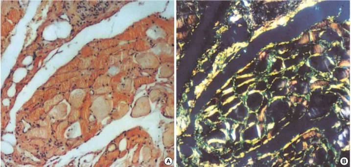
tunnel release and psoas muscle excision, to decompress the nerves. During the operation, tight adhesion was found be- tween the femoral nerves and the psoas muscles. All surgical specimens gained from carpal tunnel release and psoas muscle excision showed an apple green color under polarized light with Congo red staining and showed the same characteristic fibrillar structure of amyloid by electron microscopy, as was found in the shoulder pad biopsy. In muscle specimens, de- posits were primarily distributed in the perimysium, endomy- sium and perivascular space with scanty inflammatory cells (Fig. 3). Variable-sized muscle fibers were frequently separated by a thickened interstitium. About three weeks after the oper- ation, he could grasp with both hands and his knee extensor strength improved gradually to grade III. We planned to try chemotherapy consisting of dexamethasone and cyclophos- phamide after the wound healed.
A month after the operation, he began to suffer profuse diar- rhea due to intestinal amyloidosis. His diarrhea was refractory despite one cycle of chemotherapy. Subsequently, serious mal- nutrition ensued. He died of severe cachexia and combined infection six weeks after the chemotherapy.
DISCUSSION
The involvement of skeletal muscles by AL amyloidosis can be divided into two phenotypes. One is characterized by prox- imal muscle atrophy and presents with inflammatory myosi- tis-like features (3, 4). The other is characterized by muscle pseudohypertrophy, macroglossia, or palpable intramuscular masses without inflammatory infiltration or elevated muscle
enzymes (5, 6). The present case belongs more to the latter form. However, it is distinctive in that muscle weakness was associated with compressive neuropathy rather than amyloid myopathy itself.
Amyloid arthropathy in multiple myeloma is known to be rare and presents in the form of shoulder hypertrophic arthro- pathy or of rheumatoid arthritis-like peripheral arthritis (7).
The polyarthritis in this case, resulting from amyloid depo- sition in the periarticular tissues, showed both forms of arthro- pathy by involving both shoulders, wrists, and MCP joints.
Although the marked periarticular amyloid deposition seen in this case, together with his ESRD status, reminded us of dialysis associated amyloidosis, this seemed to be unlikely because the duration of dialysis (< 2 months) was too short for this type of amyloidosis and because clinically relevant vis- ceral involvement, which manifested as intestinal amyloido- sis in this patient, is known to be rare in dialysis associated amyloidosis, and to be largely confined to long term patients with more than 15 yr of dialysis (8). The positive IF for kappa light chain in the shoulder pad biopsy confirmed this spec- ulation.
Carpal tunnel syndrome seen in this case, is not only a fre- quent finding of AL amyloidosis, but also one of the most common features of hereditary amyloidosis which is charac- terized by a family history (autosomal dominant), and in most cases, sensorimotor polyneuropathy (2). The diagnosis of hereditary amyloidosis is often missed even though different forms of hereditary systemic amyloidoses has been found in as many as 9.7% of patients who were initially classified as having AL amyloidosis and though the prognosis can be im- proved with liver transplantation (9, 10). A Korean case of
Fig. 3.Endomysial and perimysial deposition of amyloid in the iliopsoas muscle. Congo red with light microscopy (A) and with polarizing microscopy (B) (×100).
A B
Femoral Compressive Neuropathy in AL Amyloidosis 527
hereditary amyloidosis, as a form of familial amyloid polyneu- ropathy, has been reported (11). In our case, however, the defi- nite documentation of plasma cell dyscrasia, no clinical pen- etrance of polyneuropathy in a family history as well as in his manifestations, and finally, the positive IF demonstrating kappa light chains as a precursor protein made it possible to exclude hereditary amyloidosis as the underlying process of the disease.
In this case, light chain deposition disease might have been co-present with AL amyloidosis, and played a role in renal failure, because strong and diffuse immunofluorescence, which is unusual in AL amyloiosis, is a finding consistent with light chain deposition disease, as is kappa rather than lamda light chain involvement (12).
In summary, we present a rare case of AL amyloidosis with femoral nerve compressive neuropathy, hypertrophic amyloid myopathy and amyloid arthropathy. It should be emphasized that various musculoskeletal involvements including polyar- thritis and proximal muscle weakness constitute elusive man- ifestations of amyloidosis and that not only amyloid myo- pathy but also compressive neuropathy should be included in the differential diagnosis of muscle weakness in amyloido- sis, especially when muscular hypertrophy is present.
REFERENCES
1. Cohen AS. Amyloidosis. Bull Rheum Dis 1991; 40: 1-12.
2. Falk RH, Comenzo RL, Skinner M. The systemic amyloidoses. N Engl J Med 1997; 337: 898-909.
3. Mandl LA, Folkerth RD, Pick MA, Weinblatt ME, Gravallese EM.
Amyloid myopathy masquerading as polymyositis. J Rheumatol 2000;
27: 949-52.
4. Hull KM, Griffith L, Kuncl RW, Wigley FM. A deceptive case of amyloid myopathy: clinical and magnetic resonance imaging features.
Arthritis Rheum 2001; 44: 1954-8.
5. Metzler JP, Fleckenstein JL, White CL 3rd, Haller RG, Frenkel EP, Greenlee RG Jr. MRI evaluation of amyloid myopathy. Skeletal Radiol 1992; 21: 463-5.
6. Gertz MA, Kyle RA. Myopathy in primary systemic amyloidosis. J Neurol Neurosurg Psychiatry 1996; 60: 655-60.
7. Fautrel B, Fermand JP, Sibilia J, Nochy D, Rousselin B, Ravaud P.
Amyloid arthropathy in the course of multiple myeloma. J Rheuma- tol 2002; 29: 1473-81.
8. Floege J, Ketteler M. Beta2-microglobulin-derived amyloidosis: an update. Kidney Int Suppl 2001; 78: S164-71.
9. Lachmann HJ, Booth DR, Booth SE, Bybee A, Gilbertson JA, Gill- more JD, Pepys MB, Hawkins PN. Misdiagnosis of hereditary amy- loidosis as AL (primary) amyloidosis. N Engl J Med 2002; 346: 1786- 91.
10. Holmgren G, Ericzon BG, Groth CG, Steen L, Suhr O, Andersen O, Wallin BG, Seymour A, Richardson S, Hawkins PN. Clinical impro- vement and amyloid regression after liver transplantation in hered- itary transthyretin amyloidosis. Lancet 1993; 341: 1113-6.
11. Yoon Y, Chang S, Ham D, Lee K, An B, Kwon O, Park E. A case of familial amyloid neuropathy presenting as autonomic failure. J Korean Neurol Assoc 1995; 13: 341-6.
12. Buxbaum JN, Jacobson DR. The amyloidoses. In: Beutler E, Licht- man MA, Coller BS, Kipps TJ, Seligsohn, U, editors, Williams hema- tology, 6th edition. New York: McGraw-Hill, 2001; 1305-16.