© 2012 The Korean Academy of Medical Sciences.
This is an Open Access article distributed under the terms of the Creative Commons Attribution Non-Commercial License (http://creativecommons.org/licenses/by-nc/3.0) which permits unrestricted non-commercial use, distribution, and reproduction in any medium, provided the original work is properly cited.
pISSN 1011-8934 eISSN 1598-6357
Protective Effect of Sauchinone Against Regional Myocardial Ischemia/Reperfusion Injury: Inhibition of p38 MAPK and JNK Death Signaling Pathways
Sauchinone has been known to have anti-inflammatory and antioxidant effects. We determined whether sauchinone is beneficial in regional myocardial ischemia/reperfusion (I/R) injury. Rats were subjected to 20 min occlusion of the left anterior descending coronary artery, followed by 2 hr reperfusion. Sauchinone (10 mg/kg) was administered intraperitoneally 30 min before the onset of ischemia. The infarct size was measured 2 hr after resuming the perfusion. The expression of cell death kinases (p38 and JNK) and reperfusion injury salvage kinases (phosphatidylinositol-3-OH kinases-Akt, extra-cellular signal-regulated kinases [ERK1/2])/ glycogen synthase kinase (GSK)-3β was determined 5 min after resuming the perfusion. Sauchinone significantly reduced the infarct size (29.0% ± 5.3% in the sauchinone group vs 44.4% ± 6.1% in the control, P < 0.05).
Accordingly, the phosphorylation of JNK and p38 was significantly attenuated, while that of ERK1/2, Akt and GSK-3β was not affected. It is suggested that sauchinone protects against regional myocardial I/R injury through inhibition of phosphorylation of p38 and JNK death signaling pathways.
Key Words: Sauchinone; Cardioprotection; Ischemia/reperfusion Injury; Cell Death Signaling Pathway; Reperfusion Injury Salvage Kinase Pathway
Seok Jai Kim1, Cheol Won Jeong1, Hong Beom Bae1, Sang Hyun Kwak1, Jong-Keun Son2, Chang-Seob Seo2,3, Hyun-Jung Lee1, JongUn Lee4, and Kyung Yeon Yoo1
1Department of Anesthesiology and Pain Medicine, Chonnam National University Medical School, Gwangju; 2College of Pharmacy, Yeungnam University, Gyongsan; 3Korea Institute of Oriental Medicine, Daejeon; 4Department of Physiology, Chonnam National University Medical School, Gwangju, Korea
Received: 12 April 2011 Accepted: 1 July 2011 Address for Correspondence:
Kyung Yeon Yoo, MD
Department of Anesthesiology and Pain Medicine, Chonnam National University Medical School, 671 Jebong-ro, Dong-gu, Gwangju 501-757, Korea
Tel: +82.62-220-6893, Fax: +82.62-232-6294 E-mail: [email protected]
This work was supported by a research grant from the Research Institute of Medical Sciences, Chonnam National University (2009-CURIMS-DR002).
http://dx.doi.org/10.3346/jkms.2012.27.5.572 • J Korean Med Sci 2012; 27: 572-575
BRIEF COMMUNICATION
Cardiovascular Disorders
A myocardial ischemia/reperfusion (I/R) injury is a complex process involving the generation and release of reactive oxygen species and inflammatory cytokines (1). An urgent restoration of coronary blood flow to the ischemic area is the most effective strategy to improve outcomes of myocardial I/R injury. Upon resuming the perfusion, reperfusion injury salvage kinases (RISK) survival signaling pathways such as phosphatidylinositol-3-OH kinases (PI3K)-Akt, extra-cellular signal-regulated kinases (ERK1/
2), and their downstream target glycogen synthase kinase (GSK)- 3β may be activated to confer tissue protection (2). However, at the same time, the reperfusion may also activate ‘stress-respon- sive’ mitogen-activated protein kinase (MAPK) subfamilies (so- called cell death signaling pathway), i.e., c-Jun N-terminal kinases (JNK) and p38 MAPKs, resulting in necrosis (3). Therefore, the degree of I/R injury may be determined by a relative extent of death and survival kinases activation (4).
Sauchinone is a biologically active lignan isolated from Saururus chinensis which has been long used in folk medicine for the treat- ment of edema, jaundice, and several inflammatory diseases (5).
It has been known to inhibit apoptotic death of C6 glioma cells induced by staurosporine (6) and to reduce ischemia-induced neuronal death via suppression of intracellular radical produc- tion (7). We have previously shown that sauchinone inhibits lipo- polysaccaride-induced tumor necrosis factor-α (TNF- α) expres- sion in macrophages via suppression of NF-κB activation (8).
The present study aimed at examining whether sauchinone would provide a protection against regional myocardial I/R in- jury. The expression of molecules related with cell survival such as Akt and ERK1/2/GSK-3β as well as that related with cell death such as JNK and p38 MAPKs was determined.
The procedures were carried out after the approval by the in- stitutional animal care and use committee, Research Institute of Medical Science at Chonnam National University Medical School (Gwangju, Korea). Male Sprague-Dawley rats weighing 250-350 g were used. They were housed in a vivarium maintained at 20°C-23°C with 12-hr light/dark cycle and given food and water ad libitum. On the experimental day, the rats were anesthetized with phenobarbital (100 mg/kg, intraperitoneal), and mechani-
Kim SJ, et al. • Sauchinone and Cardioprotection
http://jkms.org 573
http://dx.doi.org/10.3346/jkms.2012.27.5.572
cally ventilated with an air-oxygen mixture. The polyethylene catheter was cannulated into the common carotid artery for the arterial blood pressure monitoring and into the internal jugular vein for drug administration (9). Core body temperature was monitored with a rectal temperature probe and maintained at 37.5°C-38.5°C using heating pad. Left thoracotomy was per- formed at the fifth intercostal space, and 6-0 silk ligature was placed around left anterior descending coronary artery (LAD).
After stabilization period of 20 min, the LAD was occluded for 20 min and then reperfused for 2 hr. LAD occlusion was con- firmed by ST segment changes in electrocardiogram and the presence of regional cyanosis in the myocardial surface. There were three I/R groups as shown in Fig. 1: 1) control group, 2) sauchinone group which received sauchinone (10 mg/kg, dis- solved in dimethyl subfoxide [DMSO]), and 3) DMSO group which received vehicle only. Sauchinone was isolated from the n-hexane fraction of Saururus chinensis by successive silica gel chromatography and reverse-phase high-pressure liquid chro- matography, and was > 97% pure (10). The dosage of sauchi- none adopted was effective to increase survival rates and to decrease the plasma level of TNF-α in mice administered lipo- polysaccharide/D-galactosamine (10). Sauchinone and DMSO were administered intraperitoneally 30 min before the onset of ischemia. At the end of 2 hr reperfusion, the heart was quickly excised and mounted on a Langendorff apparatus. The LAD was reoccluded, and then fluorescent polymer microspheres were infused into the aorta to demarcate the risk zone as the tis- sue without fluorescence. The heart was sliced into 2 mm trans- verse sections, which were incubated at 37°C for 20 min in tri- phenyl tetrazolium chloride in sodium phosphate buffer. The slices were immersed in 10% formalin to enhance the contrast between stained (viable) and unstained (necrotic) tissue and then squeezed between glass plates spaced exactly 2 mm apart.
The myocardium at risk was identified by illuminating the slices with ultraviolet light. The infracted and risk zone regions were traced on a clear acetate sheet and quantified with Image Tool (San Antonio, TX, USA). The areas were converted into volumes by multiplying the areas by slice thickness. Infarct size is expressed as a percentage of the risk zone (11).
For immunoblotting, the heart was excised 5 min after reper- fusion, and the risk area in the left ventricle was separated in
each group. Myocardial samples were homogenized and equiv- alent amounts of proteins were loaded to 10% Tris-HCl SDS poly- acrylamide gel. Proteins were electrotransferred to a polyvinyli- dene difluoride membrane. The membrane was then incubated overnight at 4°C with rat polyclonal specific primary antibodies to phosphorylated p38, JNK, Akt, ERK1/2 and GSK-3β (Ser9) (Cell Signaling Technology; Danvers, MA, USA). It was subsequently incubated with anti-rabbit or anti-rat immunoglobulin HRP- coupled secondary antibodies. Immunoreactive bands were visualized using ECL Western blotting detection reagents. The membranes were then stripped using a stripping buffer, and reprobed with antibodies specific for total p38, JNK, Akt, ERK1/2, GSK-3β and β-actin (Cell Signaling Technology). Data were ex- pressed as means ± SD. Differences in infarct size and immunob- lot data between groups were tested by one-way analysis of vari- ance (ANOVA). A Scheffé test was used for multiple pair-wise comparisons when a significant difference was indicated with ANOVA. P < 0.05 was considered to be statistically significant.
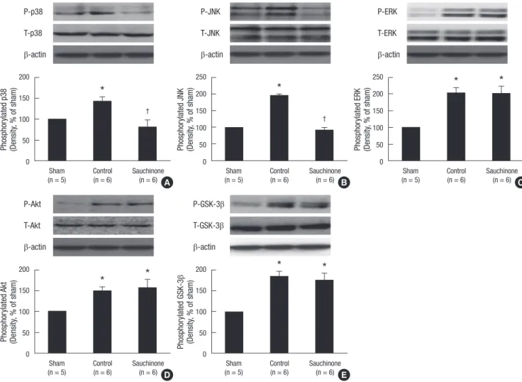
The heart rate and arterial pressure did not significantly differ among the groups at any time point (data not shown). Nor did the ratio of the area at risk to left ventricular mass differ among the groups. Sauchinone significantly reduced the infarct size (Fig. 2). Sauchinone significantly attenuated the degree of phos- phorylation of p38 and JNK (P < 0.05), without affecting that of ERK1/2, Akt and GSK-3β (Fig. 3).
Our study demonstrated that sauchinone has a novel protec- tive effect against regional myocardial I/R injury in an open-chest rodent model. The protective effect was associated with attenu- ated phosphorylation of cell death signaling pathways (p38 and JNK), without effects on that of RISK pathways (ERK1/2, Akt) and GSK-3β. Several recent studies have suggested that activa- tion of p38 MAPK and JNK, known enhancers of the expression of adhesion molecules and the production of cytokines (12), plays a major role in cell death following I/R injury. The concept has
Fig. 1. Experimental protocol. Sauchinone (10 mg/kg) and dimethyl sulfoxide (DMSO) were administered intraperitoneally 30 min before ischemia.
Ischemia Reperfusion
Control (n = 6)
30 min 20 min 120 min
Ischemia Reperfusion
Sauchinone (n = 7)
Ischemia Reperfusion
DMSO (n = 6)
Fig. 2. Effects of sauchinone on the infarct size. All rats were subjected to 20 min regional ischemia followed by 2 hr of reperfusion. The infarct size is expressed as a percentage of area at risk. Sauchinone reduced infarct size, while DMSO alone was without effects. Values are mean ± SD. *P < 0.05 vs control.
Infarct size (% of area at risk)
*
Control Sauchinone DMSO
(n = 6) (n = 7) (n = 6)
60
40
20
0
Kim SJ, et al. • Sauchinone and Cardioprotection
574 http://jkms.org http://dx.doi.org/10.3346/jkms.2012.27.5.572
been supported by the observations in which an inhibition of p38 activation reduced the caspase-3 activity and protected car- diac myocytes following I/R insult in vitro (13), and improved postischemic cardiac function in vivo (14). It was also shown that an inhibition of JNK decreased ischemia-induced necrosis and apoptosis in myocardial I/R injury (15, 16). However, an acti- vation of p38 MAPK or JNK has been also shown to play a pro- tective role in I/R injury (17, 18). The distinction of beneficial versus detrimental role of p38 and/or JNK signaling remains to be further addressed.
On the other hand, the extent of phosphorylation of Akt, ERK and GSK-3β induced by I/R was not affected by sauchinone in the present study, suggesting that RISK/GSK-3β pathways were not involved. The disturbance of ion homeostasis resulting from ATP depletion following the opening of the mitochondrial per- meability transition pore (mPTP) can lead to necrotic and apop- totic cell death (19). Both ischemic/pharmacological pre- and post-conditioning are generally perceived to protect the heart against I/R injury via activation of the RISK pathway involving
PI3K-Akt and ERK1/2 at the time of reperfusion (2). Its activa- tion may prevent cellular damages by converging on the mPTP through GSK-3β, which in turn inhibits its opening (19). Sauchi- none (EC50= 1 μM) has been shown to prevent mitochondrial dysfunction and apoptosis induced by iron plus arachidonic acid in the hepatocyte, possibly through inhibition of reactive oxygen species production (20). Further studies defining the role of mPTP in sauchinone-induced myocardial protection against I/R injury may be needed.
In summary, the present study demonstrates a cardioprotec- tive effect of sauchinone against regional I/R injury in the rat, which may be related to an attenuation of cell death signaling pathways involving p38 and JNK but not to activation of RISK/
GSK-3β pathways.
REFERENCES
1. Vinten-Johansen J. Involvement of neutrophils in the pathogenesis of lethal myocardial reperfusion injury. Cardiovasc Res 2004; 61: 481-97.
Fig. 3. Effects of sauchinone on the expression of total and phosphorylated p38 (A), JNK (B), ERK1/2 (C), Akt (D), and GSK-3β (E). Sauchinone (10 mg/kg) attenuated the phos- phorylation of p38 and JNK, while it did not affect that of ERK1/2, Akt and GSK-3β. The expression of total p38, JNK, ERK1/2, Akt, or GSK-3β was not altered by sauchinone.
Upper panels show representative immunoblots, and lower panels show densitometric data. Sham group was without I/R. Values are mean ± SD. *P < 0.05 vs sham; †P <
0.05 vs control.
P-p38 T-p38 β-actin
Phosphorylated p38 (Density, % of sham)
Sham Control Sauchinone (n = 5) (n = 6) (n = 6) 200
150 100 50 0
*
†
A
P-ERK T-ERK β-actin
Phosphorylated ERK (Density, % of sham)
Sham Control Sauchinone (n = 5) (n = 6) (n = 6) 250
200 150 100 50 0
* *
C P-JNK
T-JNK β-actin
Phosphorylated JNK (Density, % of sham)
Sham Control Sauchinone (n = 5) (n = 6) (n = 6) 250
200 150 100 50 0
*
†
B
P-Akt T-Akt β-actin
Phosphorylated Akt (Density, % of sham)
Sham Control Sauchinone (n = 5) (n = 6) (n = 6) 200
150 100 50 0
* *
D
P-GSK-3β T-GSK-3β β-actin
Phosphorylated GSK-3β (Density, % of sham)
Sham Control Sauchinone (n = 5) (n = 6) (n = 6) 200
150 100 50 0
* *
E
Kim SJ, et al. • Sauchinone and Cardioprotection
http://jkms.org 575
http://dx.doi.org/10.3346/jkms.2012.27.5.572
2. Hausenloy DJ, Yellon DM. New directions for protecting the heart against ischaemia-reperfusion injury: targeting the Reperfusion Injury Salvage Kinase (RISK)-pathway. Cardiovasc Res 2004; 61: 448-60.
3. Yin T, Sandhu G, Wolfgang CD, Burrier A, Webb RL, Rigel DF, Hai T, Whelan J. Tissue-specific pattern of stress kinase activation in ischemic/
reperfused heart and kidney. J Biol Chem 1997; 272: 19943-50.
4. Yue TL, Wang C, Gu JL, Ma XL, Kumar S, Lee JC, Feuerstein GZ, Thom- as H, Maleeff B, Ohlstein EH. Inhibition of extracellular signal-regulated kinase enhances ischemia/Reoxygenation-induced apoptosis in cultured cardiac myocytes and exaggerates reperfusion injury in isolated perfused heart. Circ Res 2000; 86: 692-9.
5. Sung SH, Kim YC. Hepatoprotective diastereomeric lignans from Sauru- rus chinensis herbs. J Nat Prod 2000; 63: 1019-21.
6. Song H, Kim YC, Moon A. Sauchinone, a lignan from Saururus chinen- sis, inhibits staurosporine-induced apoptosis in C6 rat glioma cells. Biol Pharm Bull 2003; 26: 1428-30.
7. Choi IY, Yan H, Park YK, Kim WK. Sauchinone reduces oxygen-glucose deprivation-evoked neuronal cell death via suppression of intracellular radical production. Arch Pharm Res 2009; 32: 1599-606.
8. Bae HB, Li M, Son JK, Seo CS, Chung SH, Kim SJ, Jeong CW, Lee HG, Kim W, Park HC, et al. Sauchinone, a lignan from Saururus chinensis, reduces tumor necrosis factor-alpha production through the inhibition of c-raf/MEK1/2/ERK 1/2 pathway activation. Int Immunopharmacol 2010; 10: 1022-8.
9. Kim SJ, Yoo KY, Jeong CW, Kim WM, Lee HK, Bae HB, Kwak SH, Li M, Lee J. Urinary trypsin inhibitors afford cardioprotective effects through activation of PI3K-Akt and ERK signal transduction and inhibition of p38 MAPK and JNK. Cardiology 2009; 114: 264-70.
10. Seo CS, Lee YK, Kim YJ, Jung JS, Jahng Y, Chang HW, Song DK, Son JK.
Protective effect of lignans against sepsis from the roots of Saururus chi- nensis. Biol Pharm Bull 2008; 31: 523-6.
11. Song DK, Jang Y, Kim JH, Chun KJ, Lee D, Xu Z. Polyphenol (-)-epigallo- catechin gallate during ischemia limits infarct size via mitochondrial K(ATP) channel activation in isolated rat hearts. J Korean Med Sci 2010;
25: 380-6.
12. Srivastava S, Weitzmann MN, Cenci S, Ross FP, Adler S, Pacifici R. Estro- gen decreases TNF gene expression by blocking JNK activity and the re- sulting production of c-Jun and JunD. J Clin Invest 1999; 104: 503-13.
13. Mackay K, Mochly-Rosen D. An inhibitor of p38 mitogen-activated pro- tein kinase protects neonatal cardiac myocytes from ischemia. J Biol Chem 1999; 274: 6272-9.
14. Li Z, Ma JY, Kerr I, Chakravarty S, Dugar S, Schreiner G, Protter AA. Selec- tive inhibition of p38α MAPK improves cardiac function and reduces myocardial apoptosis in rat model of myocardial injury. Am J Physiol Heart Circ Physiol 2006; 291: H1972-7.
15. Ferrandi C, Ballerio R, Gaillard P, Giachetti C, Carboni S, Vitte PA, Got- teland JP, Cirillo R. Inhibition of c-Jun N-terminal kinase decreases car- diomyocyte apoptosis and infarct size after myocardial ischemia and reperfusion in anaesthetized rats. Br J Pharmacol 2004; 142: 953-60.
16. Milano G, Morel S, Bonny C, Samaja M, von Segesser LK, Nicod P, Vas- salli G. A peptide inhibitor of c-Jun NH2-terminal kinase reduces myo- cardial ischemia-reperfusion injury and infarct size in vivo. Am J Physiol Heart Circ Physiol 2007; 292: H1828-35.
17. Weinbrenner C, Liu GS, Cohen MV, Downey JM. Phosphorylation of tyrosine 182 of p38 mitogen-activated protein kinase correlates with the protection of preconditioning in the rabbit heart. J Mol Cell Cardiol 1997;
29: 2383-91.
18. Dougherty CJ, Kubasiak LA, Prentice H, Andreka P, Bishopric NH, Web- ster KA. Activation of c-Jun N-terminal kinase promotes survival of car- diac myocytes after oxidative stress. Biochem J 2002; 362: 561-71.
19. Nishihara M, Miura T, Miki T, Tanno M, Yano T, Naitoh K, Ohori K, Hotta H, Terashima Y, Shimamoto K. Modulation of the mitochondrial perme- ability transition pore complex in GSK-3beta-mediated myocardial pro- tection. J Mol Cell Cardiol 2007; 43: 564-70.
20. Kim YW, Lee SM, Shin SM, Hwang SJ, Brooks JS, Kang HE, Lee MG, Kim SC, Kim SG. Efficacy of sauchinone as a novel AMPK-activating lig- nan for preventing iron-induced oxidative stress and liver injury. Free Radic Biol Med 2009; 47: 1082-92.