INTRODUCTION
Intracranial schwannomas are relatively common tumors comprising 8% of all primary intracranial neoplasms (1), and most frequently located in the cerebello-pontine angle originating from the vestibular component of the eighth cranial nerve. Intracranial schwannomas not related to cra- nial nerves are very rare and especially intrasellar location of schwannomas has been reported in only four publications (2-5). The authors present a primary intrasellar schwannoma that mimicked a pituitary tumor both clinically, and radio- graphically.
CASE REPORT
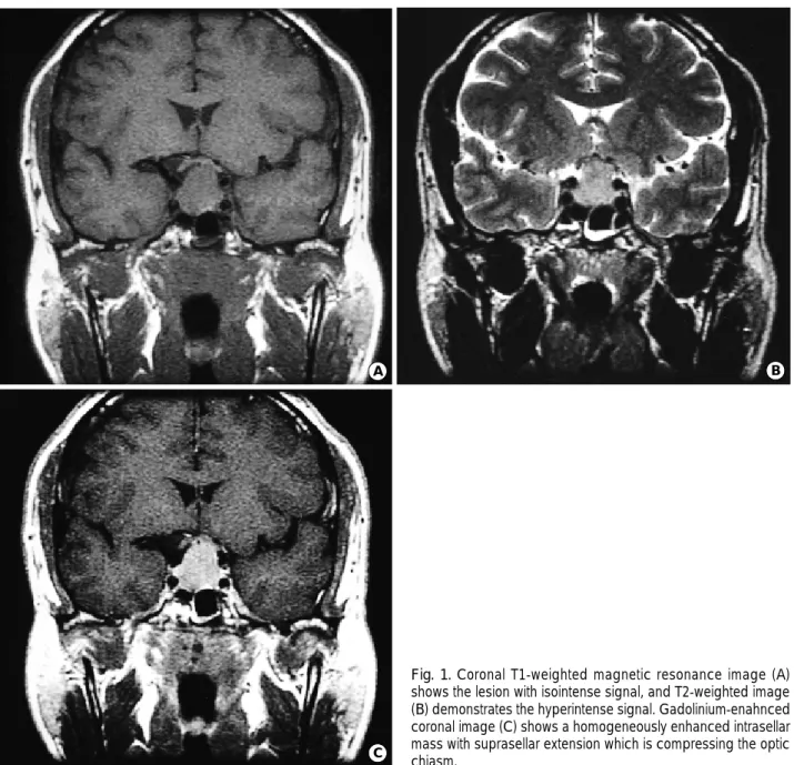
A 39-yr-old man visited the outpatient clinic due to de- creased visual acuity of both eyes for 10 months. In addition, he complained of the loss of libido and impotence. Visual testing showed 20/60 and 20/160 acuity in each side. Visual field examination showed a bitemporal hemianopsia. Other- wise, no remarkable findings were found on the complete physical and neurological examinations. Magnetic resonance imaging (MRI) revealed enlarged sellar turcica and sellar mass with suprasellar extension (Fig. 1). The signal of the lesion was isointense to cerebral cortex in T1-weighted image, and slightly hyperintense in T2-weighted image. The mass was homogeneously enhanced after administration of gadolin-
ium. Detailed preoperative endocrine testing revealed hypo- pituitarism. The patient showed elevated prolactin level 41.7 ng/mL (normal, less than 16.1 ng/mL) and depressed GH 0.27 ng/mL (normal, 1-5 ng/mL), LH 1.0 mIU/mL (normal, 1.4-11.1 mIU/mL), FSH 1.0 mIU/mL (normal, 1.6-17.8 mIU/mL), testosterone 0.03 ng/mL (normal, 2.79- 8.76 ng/mL), and cortisol 0.6 g/mL (normal, 5.9-26.1 g/
mL). A TRH stimulation test was performed giving a base- line TSH of 1.94 IU/mL to a peak TSH of 6.61 IU/mL at 60 min, which was consistent with a normal response.
Insulin tolerance test demonstrated an impaired response of plasma cortisol to lowered blood sugar with a peak of 1.6 g/mL. The responses of LH and FSH values after LHRH stimulation were significantly depressed. The pre-operative diagnosis of pituitary adenoma was made. A trans-sphenoidal approach was selected for the removal of the sellar mass. We noticed a hypervascularized fibrous tumor with a harder con- sistency than that of usual pituitary adenomas. The tumor was removed incompletely because of its tight attachment to the optic nerves and chiasm. The final pathological diag- nosis was schwannoma. Microscopic examination showed a spindle cell tumor consistent with schwannoma. Immuno- histochemistry for S-100 protein was strongly positive, whereas that for glial fibrillary acidic protein (GFAP) was negative (Fig. 2).
Postoperative endocrinological test revealed panhypopi- tuitarism basically similar to the preoperative state with a little aggravation. In addition, the patient experienced a tran-
Sung Mock Whee, Jung-Il Lee, Jong Hyun Kim
Department of Neurosurgery, Samsung Medical Center, Sungkyunkwan University School of Medicine, Seoul, Korea
Received : 9 February 2001 Accepted : 20 April 2001
Address for correspondence Jong Hyun Kim, M.D.
Department of Neurosurgery, Samsung Medical Center, Sungkyunkwan University School of Medicine, 50 Ilwon-dong, Kangnam-gu, Seoul 135-710, Korea
Tel : +82.2-3410-3499, Fax : +82.2-3410-0048 E-mail : [email protected]
147 J Korean Med Sci 2002; 17: 147-50
ISSN 1011-8934
Copyright � The Korean Academy of Medical Sciences
Intrasellar Schwannoma Mimicking Pituitary Adenoma
: A Case Report
Intrasellar location of schwannoma is extremely rare, although intracranial schwan- nomas account for up to 8% of all primary brain tumors. An unusual case of an intrasellar schwannoma radiographically and clinically simulating a pituitary ade- noma is reported. A 39-yr-old man presented a 10-month history of visual distur- bance and decreased libido. Neurological examination showed poor visual acu- ity of both eyes with bitemporal hemianopsia. Computed tomography and mag- netic resonance imaging showed a sellar tumor with suprasellar extension. Pitu- itary adenoma was considered as a preoperative diagnosis. The tumor was re- moved through a trans-sphenoidal approach. Microscopic examination of the tumor revealed schwannoma.
Key Words : Schwannoma, Pituitary Neoplasms
148 S.M. Whee, J.-I. Lee, J.H. Kim
sient diabetes insipidus. Postoperative visual testing showed a remarkable improvement in the visual field with an acuity of 20/25 in the right eye and 20/60 in the left eye. The patient underwent a replacement therapy of cortisol and thyroxine for 14 months following the surgery. A follow-up MRI scan 14 months after the operation showed no evidence of tumor regrowth.
DISCUSSION
The differential diagnoses of sellar and suprasellar lesions include pituitary adenomas, craniopharyngiomas, menin- giomas, and many others. However, schwannoma is not usu-
ally included, because the occurrence of schwannoma in the sellar or suprasellar region is exceedingly rare. Only four cases of intrasellar schwannomas have been reported previously, all of which presented a suprasellar extension similar to that of our case (2-5). Enlargement of the sella turcica and an enhancing sellar mass with suprasellar extension are most often associated with pituitary adenomas (6) and radiologi- cal findings in these cases were not significantly different from the usual pituitary macroadenomas. In T1-weighted MRI scan usually the schwannoma has an isointense or hypo- intense signal compared to brain cortex as like pituitary adenoma. The schwannoma in T2-weighted image shows more frequently hyperintense signal than pituitary adeno- ma. However, it is possible for pituitary adenoma to have
C
A B
Fig. 1.Coronal T1-weighted magnetic resonance image (A) shows the lesion with isointense signal, and T2-weighted image (B) demonstrates the hyperintense signal. Gadolinium-enahnced coronal image (C) shows a homogeneously enhanced intrasellar mass with suprasellar extension which is compressing the optic chiasm.
Intrasellar Schwannoma 149
variable findings including hyperintense signal which is not different from usual schwannomas. So MRI findings could not provide information favoring schwannoma even if the images were reviewed retrospectively. Clinically three out of four cases developed with visual change and two out of four showed pituitary insufficiency. Varying degrees of hypopi- tuitarism have been found with the empty sella syndrome, intracranial aneurysms, pituitary adenomas, craniophary- giomas, chordomas, and meningiomas. So, the presence of pituitary insufficiency is not helpful for the differential diag- noses either, and the diagnosis is established by light micro- scopic examination of surgical specimens. Immunohistochem- istry is of value to rule out other tumors such as fibroblastic meningioma, pilocytic astrocytoma, glioma, and metastatic tumors which may have palisading structures mimicking a schwannoma (7).
In our case the lesion was removed through the trans-sphe- noidal approach as in the previous four reports (2-5). Surgi- cal removal of these lesions by the trans-sphenoidal approach seems tougher than operation of pituitary adenoma due to the harder consistency and greater vascularity of schwanno- ma.
Total removal of the tumor was possible in only one of the previous cases. In the others including our case, the tumor could not be removed completely. Though none of the report- ed cases required a second operation for remnant tumors, additional treatment such as radiosurgery needs to be con- sidered because the follow-up period in previous cases was too short to determine the prognosis.
The origin of intrasellar schwannoma is still being debat- ed. Schwann cells are not normally found in the central ner-
vous system. Three histopathological hypotheses have been previously discussed. Russell and Rubinstein suggested that ectopic Schwann cells could have been the origin of the tumors (1). Also, they suggested that schwannomas with a tight attachment to the dura may arise from Schwann cells ensheathing the small nerve twigs that innervate the dura.
Other authors suggested that perivascular Schwann cells could have been the origin of the tumors (8, 9). In our case, the tumor was located anteriorly to the normal pituitary gland and stalk. Based on the operative findings, it seemed that the tumor developed in the area around dorsum sellae.
Although the exact origin of the tumor in our case is still unclear, it is suggested that intrasellar schwannoma should be considered in the differential diagnosis of sellar lesions mimicking a pituitary adenoma.
REFERENCES
1. Russell DS, Rubinstein LJ. Pathology of the tumours of the nervous system, 5th ed. London: Edward Arnold 1989; 533-89.
2. Perone TP, Robinson B, Holmes SM. Intrasellar schwannoma:
case report. Neurosurgery 1984; 14: 71-3.
3. Wilberger JE Jr. Primary intrasellar schwannoma: case report.
Surg Neurol 1989; 32: 156-8.
4. Guenot M, Bataille B, Wager M. Intrasellar neurinoma. Apropos of a case and review of the literature. Neurochirurgie 1994; 40: 263-6.
5. Civit T, Pinelli C, Klein M, Auque J, Baylac F, Hepner H. Intrasel- lar schwannoma. Acta Neurochir 1997; 139: 160-1.
6. Johnsen DE, Woodruff WW, Allen IS, Cera PJ, Funkhouser GR, Coleman LL. MR imaging of the sellar and juxtasellar regions.
A B
Fig. 2.(A) Photomicrogragh of pathologic specimen shows a typical pattern of schwannoma consisting of spindle cells (H&E, ×100).
(B) Immunohistochemical staining for S-100 protein is shown as positive (×100).
150 S.M. Whee, J.-I. Lee, J.H. Kim
Radiographics 1991; 11: 727-58.
7. Sobel RA, Michaud J. Microcytic meningioma of the falx cerebri with numerous palisading structures: an unusual histological pat- tern mimicking schwannoma. Acta Neuropathol 1985; 68: 256-8.
8. Gibson AA, Hendrick EB, Conen PE. Intracerebral schwannoma.
Report of a case. J Neurosurg 1966; 24: 552-7.
9. New PF. Intracerebral schwannoma. Case report. J Neurosurg 1972; 36: 795-7.