Archives of Craniofacial Surgery
www.kcpca.or.kr ISSN 2287-1152
50
Copyright © 2012 The Korean Cleft Palate-Craniofacial AssociationThis is an Open Access article distributed under the terms of the Creative Commons Attribution Non-Commercial License (http://creativecommons.org/
licenses/by-nc/3.0/) which permits unrestricted non-commercial use, distribution, and reproduction in any medium, provided the original work is properly cited.
Dyke-Davidoff-Masson 증후군 환자의 두개골 변형:
증례보고
이승현·이혜경·정희선
관동대학교 의과대학 성형외과학교실
Purpose: The Dyke-Davidoff-Masson syndrome is a rare disease entity that was first reported in 1993, and it is characterized by not only the cerebral hemiatrophy that is accompanied by the ipsilateral ventriculomegaly and ipsilateral compensatory osseous hypertrophy, but also the overgrowth of the paranasal sinuses. No studies have attempted to examine it from perspectives of the skull deformity and plastic surgery. Here, we report our case with a review of the literatures.
Methods: A 45-year-old man with Dyke-Davidoff-Masson visited our medical institution with nasal bone fracture. Based on the previously taken brain MRI scans, we measured the degree of craniofacial deformity, and the horizontal distance, which is based on the margin of the skull, as well as the falx cerebri.
Results: We made a comparison of the degree of craniofacial deformity. This showed that the mean horizontal distance on the axial view was shorter by approximately 28.46%, as compared with that of the left unaffected side.
Conclusion: The Dyke-Davidoff-Masson is characterized by a concurrent presence of the atrophy of the cerebral hemisphere, with the cranial deformity. For the reconstruction of the bone and soft-tissue deformity with Dyke-Davidoff-Masson syndrome, it is needed to perform objective assessments.
Keywords: Dyke-Davidoff-Masson syndrome, Craniofacial deformity
Craniofacial Deformity in a Patient with Dyke-Davidoff-Masson Syndrome:
A Case Report
Seung-Hyun Lee, Hye-Kyung Lee, Hii-Sun Jeong
Department of Plastic and Reconstructive Surgery, Myongji Hospital, Kwandong University College of Medicine, Goyang, Korea
서 론
Dyke–Davidoff–Masson 증후군은 1933년 Dyke 등1에 의 해 처음 보고된 질환으로 두개골의 편위축을 보이며 편마 비 증세가 있는 9명의 환자를 보고하면서 처음 알려졌다.
이후 이 증후군에 대한 연구가 이루어지면서 대뇌 반구의
편위축과 동측의 뇌전위 및 뇌실 확장 소견과 함께 동측의 대상성 골비후, 부비동의 발육과다 등을 특징으로 하는 것 이 보고되었다.2 Dyke–Davidoff–Masson 증후군은 현재까 지 국내문헌에서 7례 보고되었으나 대부분이 소아, 청소년 에 국한되었다. 기존의 증례들은 마비 등의 신경학적 증상 에 대해서만 주로 언급하였고, 두개골 변형에 대한 언급은 없었기 때문에 이에 대해 소개하고자 한다.
증 례
좌측 편마비를 동반한 45세 남자환자로 비골골절을 주
Arch Craniofac Surg Vol.13 No.1, 50-53 http://dx.doi.org/10.7181/acfs.2012.13.1.50
Correspondence: Hii-Sun Jeong
Department of Plastic and Reconstructive Surgery, Myongji Hospital, Kwandong University College of Medicine, 697-24 Hwajung-dong, Deokyang-gu, Goyang-si, Gyeonggi-do 412-270, Korea
Tel: +82-31-810-6830 / Fax: +82-31-810-6837 / E-mail: [email protected] Received February 11, 2012 / Revised March 8, 2012 / Accepted March 16, 2012
Case Report
51
www.kcpca.or.kr
소로 내원하였다. 가족력상 특이사항은 없었다. 좌반신 편 마비는 5세경 부터 시작되었고 연령이 증가하면서 편마비 의 정도가 심해져 중등도의 좌반신 편마비 소견이 관찰되 었다. 경도의 지능발달 지연과 구음장애 및 간헐적인 두통 소견을 보였고 반복적인 재발성 경련의 과거력이 있었다. 신 체적 발육 상태는 정상적이었다. 그 외 Dyke–Davidoff–
Masson 증후군 환자에게서 나타날 수 있는 내상사위, 과활 동, 정류고환, 수지상 형성 부전, 무정위 운동증 등의 소견 은 관찰되지 않았다. 고혈압, 위–식도 역류질환, 불면증 등 을 주소로 경구약을 복용 중이었으며 외견상 좌측 전두골
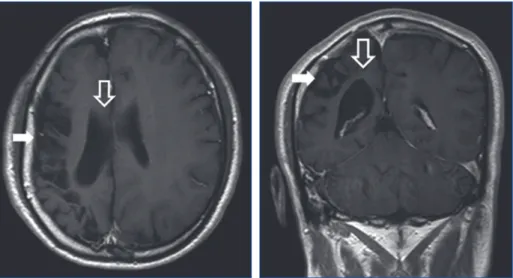
에 비해 우측 전두골이 상대적으로 함몰되어 있었으나 안 면 비대칭 소견은 보이지 않았다(Fig. 1). 뇌자기공명영상촬 영상 우측 대뇌 위축, 동측 뇌실의 확장 및 대상성 골비후 가 보였고(Fig. 2), 컴퓨터단층촬영에서는 유양동 함기세포 의 발육과다 소견이 나타났다(Fig. 3). 또한 뇌혈관 촬영상 우측 중간대뇌동맥과 후대뇌동맥이 좁아진 모습이 관찰되 었다(Fig. 4).
저자들은 두부의 뇌자기공명영상촬영 사진을 이용해 양 측 두개골을 측정하여 정상적인 좌측 두개골과 변형된 우 측 두개골의 수평 거리를 비교해 보았다. 사진의 axial view에
Fig. 2. Magnetic resonance image shows right cerebral hemiatrophy (arrow), secondary dilatation of the ipsilateral lateral ventricle (empty arrow).
Seung-Hyun Lee, et al. Dyke-Davidoff-Masson syndrome
Fig. 1. A 45-year-old man showing depression of right forehead and scalp.
Archives of Craniofacial Surgery Vol. 13, No. 1, 2012
www.kcpca.or.kr
52
서 앞, 뒤 내겸을 이은 중심선상에 동일한 간격으로 4개의 지점을 잡고, 각 지점으로부터 양측 두개골까지의 거리를 측정하여 정상적인 좌측에 비해 변형된 우측 두개골의 거 리를 측정하였다. 세 번 이상 측정하여 두 번 같은 값이 나 오면 이것을 측정치로 선택하였으며 통계학적 유의성을 알 아보기 위하여 SPSS ver. 12.0 (SPSS Inc., Chicago, IL, USA) 를 이용한 Mann–Whitney Test로 측정값을 검증하였고, p- value 0.05 미만에서 통계학적으로 유의한 것으로 분석하였 다. 그 결과, 변형된 우측의 경우 각각 40.23 mm/56.85 mm/5 8.13 mm/55.27 mm, 정상적인 좌측의 경우 69.65 mm/74.66 mm/72.86 mm/68.36 mm로 측정되었다(Fig. 5).
고 찰
Dyke–Davidoff–Masson 증후군 환자는 보통 생후 일찍 대뇌의 편위축으로 인해 반대측 체간의 부전마비가 나타 나면서 동시에 경련, 지능발육 부전, 동측성 반맹, 외안근 마비, 언어장애, 환미, 반신 감각소실, 두통 등이 동반되며 신체 발육상태는 일반적으로 정상이다.3 대뇌의 편위축은 우측보다 좌측에서 흔한 것으로 알려져 있으며4 본 증례의 경우에는 우측 대뇌위축 소견을 보였다. 현재 국내에서 보 고된 Dyke–Davidoff–Masson 증후군 환자의 대부분은 소 아의 증례이며 20세 이상 성인의 경우는 단 2례에 불과하 다. 대뇌반구 편위축 및 동측 뇌실 확장 등의 영상학적인 소 견 및 편마비는 공통적으로 나타나는 소견이었으나 그 외 의 소견은 증례마다 차이가 있었으며 특히 본 증례에서처 럼 현저한 두개골 변형에 대한 보고는 없었다.
발생 원인은 본 증례의 경우처럼 대개 선천적인 경우가 많은데 이 경우 혈관 자체의 원인 또는 자궁내 환경에 의해 뇌의 손상이 발생하는 것으로 알려져 있으며 후천적인 경 우는 분만 시 생기는 분만손상, 감염, 주산기 두개강내 출혈 Fig. 5. The cranium was divided to 2 part by falx cerebri, comparing the horizontal distance between the normal and deformity part. The horizontal distance was statistically significant reduced in right deformity part.
Fig. 3. Computed tomography shows a hyperpneumatization of the mastoid cells (arrow).
Fig. 4. MR angiography shows stenosis of right middle (arrow) and posterior cerebral artery (empty arrow).
53
www.kcpca.or.kr
Seung-Hyun Lee, et al. Dyke-Davidoff-Masson syndrome
에 기인한 것으로 생각되나 정확한 원인은 아직까지 밝혀 지지 않은 상태이다. 두부 X–선 촬영상 동측의 대상성 골비 후, 부비동 특히 전두동의 발달과다, 접형골익 및 추체부 융 선의 거상 등의 소견이 나타나는데 본 증례에서는 전두동 의 발달 과다를 제외한 다른 소견은 뚜렷하게 보이지 않았 다.5,6 Dyke–Davidoff–Masson 증후군의 가장 대표적인 소견 이라 할 수 있는 대뇌 반구의 편위축 소견을 관찰하기 위해 서는 뇌전산화단층촬영 또는 뇌자기공명영상촬영이 유용 한데, 본 증례의 경우 우측 반구의 편위축 소견 뿐만아니라 동측 뇌실의 확장, 대상성 골비후 및 유양동 함기세포의 발 육과다 소견도 볼 수 있었다. 또한 드물게 뇌혈관의 협착 등 의 이상 소견이 나타날 수 있는데 증례의 경우 우측 중간대 뇌동맥과 후대뇌동맥이 협착되어 있었으며,7 이는 우측 대 뇌 반구의 편위축에 의한 관류 감소로 생기는 이차적 변화 로 생각되어 진다. 이외에도 뇌수종에 의한 뇌하수체 기능 저하증과 그에 따른 여러 내분비적인 합병증이 발생 할 수 있다.8
저자들은 두부의 뇌자기공명영상촬영 사진을 이용해 정상적인 좌측 두개골과 함몰된 우측 두개골의 길이를 측 정하였는데 X–선 사진은 머리자세에 따라 측정치가 달라 질 수 있고, 또한 정확한 측정값을 얻기 위해 내겸을 기준으 로 좌, 우를 구분하였기 때문에 뇌자기공명영상촬영 사진 이 적합하였다. 그 결과, 수평적으로는 정상 좌측 두개골에 비해 평균 약 28.46% 짧은 것으로 측정되었고 이는 통계학 적으로 유의하였다(p=0.048). 저자들이 사용한 네 지점을 통한 비모수적 통계방법으로도 유의한 차이가 있었기 때 문에 그 함몰 정도가 상당하다는 것을 알 수 있었다.
Dyke–Davidoff–Masson 증후군은 그 치료 지침이 아직 명확하게 제시되고 있지 않을 만큼 드문 질환이다. 뇌수종 발병 시 이에 대한 일반적 치료 및 반신마비 등의 신체 장애 에 대한 재활훈련, 유전 상담 등을 포함한 고식적 치료가
시행 되고 있을 뿐이다. 특히 증례에서처럼 두개골 비대칭 등과 같은 변형이 동반될 수 있으므로 이에 대한 교정을 위 해 성형외과적 접근이 필요할 수 있다. 부위와 정도에 따라 지방 이식술, 보형물을 이용한 재건술, 피판술 등을 이용할 수 있을 것이다.9 따라서 두개골 또는 안면 비대칭 및 변형 을 주소로 내원한 환자들 중에 외상 등에 의한 후천적인 원 인이 아닌 경우 Dyke–Davidoff–Masson 증후군에 대한 가 능성을 생각해야 하겠고 영유아의 경우 후일 변형이 더욱 진행될 수 있으므로 장기적인 경과관찰이 필요하겠다.
REFERENCES
1. Dyke CG, Davidoff LM, Masson CB: Cerebral hemiatrophy with ho- molateral hypertrophy of the skull and sinuses. Surg Gynecol Obstet 57: 588, 1933
2. Parker CE, Harris N, Mavalwala J: Dyke–Davidoff–Masson syndrome.
Five case studies and deductions from dermatoglyphics. Clin Pediatr (Phila) 11: 288, 1972
3. Sener RN, Jinkins JR: MR of craniocerebral hemiatrophy. Clin Imaging 16: 93, 1992
4. Unal O, Tombul T, Cirak B, Anlar O, Incesu L, Kayan M: Left hemi- sphere and male sex dominance of cerebral hemiatrophy (Dyke–Davi- doff–Masson Syndrome). Clin Imaging 28: 163, 2004
5. Zilkha A: CT of cerebral hemiatrophy. AJR Am J Roentgenol 135: 259, 1980
6. Ono K, Komai K, Ikeda T: Dyke–Davidoff–Masson syndrome mani- fested by seizure in late childhood: a case report. J Clin Neurosci 10:
367, 2003
7. Solomon GE, Hilal SK, Gold AP, Carter S: Natural history of acute hemiplegia of childhood. Brain 93: 107, 1970
8. Park SY, Lee MY, Kim JH, Kim SY, Shin JY, Shin YG, Chung CH: A case of dyke–davidoff–masson syndrome associated with hypopituitarism and diabetes mellitus. Korean J Med 79: 316, 2010
9. Mehrara BJ, McCarthy JG: Repair and grafting of bone. In Mathes SJ, Weingerger SE, Hentz VR (eds): Plastic Surgery. 2nd ed, Philadelphia, Saunders, 2006, p 639