K yu -S u k L e e
1 ,5, W o n -S u k L e e
1, S eong-Il S uh
2, S ang-P yo K im
3,
S ung-R yong Lee
4, Y oung-W ook R yoo
1, and B yung-C hun K im
11Institute of Medical Science and Department of Dermatology
2Department of Microbiology
3Department of Pathology
4Department of Pharmacology
School of Medicine, Keimyung University, Daegu 700-120, Korea
5Corresponding author: Tel, 82-53-250-7624;
Fax, 82-53-250-7626; E-mail, [email protected] Accepted 2 June 2003
Abbreviations: GAPDH, glyceraldehyde-3-phosphate dehydroge- nase; ODC, ornithine decarboxylase
Abstract
UV radiation is known to cause photoaging of the skin and is considered one of the leading cause of developing skin carcinogenesis. Melatonin which has a highly lipophilic m olecular structure facilita- ting penetration of cell m em branes and serving as an extra- and intracellular free radical scavenger has been dem onstrated to protect photodam age of skin affected by UV exposure. In this study, w e have exam ined the role of m elatonin in response to UVB induced photodam aging process, using hum an skin fibroblasts in vitro. Cell survival cur- ves after UVB irradiation showed dose-dependent decrease. Only 60% of fibroblasts w ere survived at 140 m J/cm
2UVB irradiation. By pre-cultivation of cells w ith m elatonin (100 nM ), a significant num ber of cells rem ained unaffected. After UVB irradiation with 70 m J/cm
2, the level of putrescine w as 1.7±0.3 fold increased com pared to m ela- tonin pre-treated group. In Northern analyses, the transcriptional level of ornithine decarboxylase (ODC) gene expression w as increased by UVB ir- radiation and prohibited by m elatonin. These re- sults indicated that m elatonin was effectively able to neutralize m em brane peroxidation when present in relevant concentration during UVB irradiation and dim inishes the UVB-induced increase of poly-
am ine synthesis and O DC gene expression. Col- lectively, O DC response to UVB induced changes are possibly involves a m elatonin or antioxidant sensitive regulatory pathw ay in norm al hum an skin fibroblast.
Keywords: antioxidants; cell death; melatonin; ornithine decarboxylase; polyamine; ultraviolet rays
Introduction
Exposure to UV results in photoaging of the skin and is considered an initiating event of skin carcino- genesis (Chainiaux et al., 2002). UVB radiation not only causes inducing DNA damage and cell muta- genecity, but can also modulate the expression of several genes at dose levels to natural solar exposure (Shindo and Hashimoto, 1998). Some of these genes are regulated by an oxidative mechanism (Lee et al., 1998; Takeda et al., 2002). It has been reported that the effect of UVB radiation on the in vitro regulation of important enzymes that participate in inflammation and cancer progression (Kulms et al., 2002). The polyamines, (putrescine, spermidine, and spermine) have been shown to be essential for mammalian cell growth and function (Ahmad et al., 2001). Intracellular polyamine concentrations are highly regulated by the enzyme, ornithine decarboxylase (ODC), which ca- talyzes the conversion of ornithine to putrescine, the initial and often rate limiting step in polyamine bio- synthesis (van Weelden et al., 1990; O'Brien et al., 1997). UVB light induces overexpression of ODC an enzyme that plays a critical role in photocarcino- genesis (Jansen et al., 2001). Therefore, the ODC activity served as a marker of the mutagenic and carcinogenic effects of UV light, because the activity of ODC in quiescent cells is extremely low and readily induced by UV light (Manzow et al., 2000). Fur- thermore, ODC is also activated by many other stimuli including tumor promoters and growth factors (Oguro and Yoshida, 2001), as well. Melatonin (N-acetyl- 5-methoxytrptamine) is a hormone with multiple func- tions in human, produced by the pineal gland and stimulated by beta-adrenergic receptors (Ryoo et al., 2001). Areas of greater interest and potential impor- tance include the antimitotic effects of melatonin on some types of tumor cells in culture and potent ra- dical scavenging effects, but there has been little progress toward identifying the specific mechanisms
Melatonin reduces ultraviolet-B induced cell damages and
polyamine levels in human skin fibroblasts in culture
of its action. In the present study, we investigated whether the photoprotective actions of melatonin can attenuate the changes of polyamine levels following UVB induced cell damages in cultured skin fibro- blasts.
M aterials and M ethods
Fibroblast culture
Primary cultures of dermal fibroblasts were establi- shed from children (n = 3, mean age = 5 yr) skin left over from cosmetic surgery and cultured on plastic culture dishes in DMEM supplemented with 10% FBS, penicillin (100 U/ml), streptomycin (100 µg/ml), and amphotericin B (1 µg/ml). The cells were maintained in a humidified 5% CO
2, 95% air incubator at 37
oC.
Analyses of confluent fibroblast cultures were carried out at 3-6 passages of subcultivation, 1×10
5/ml. Cell viability was determined by trypan blue exclusion.
UVB irradiation
Immediately before irradiation, the medium was re- placed by PBS. UVB was supplied by a closely spaced array of seven Westinghouse FS-40 sun- lamps, which delivered uniform irradiation at a dis- tance of 30 cm. The energy output of UVB (290-320 nm) was measured with a UVB photometer (IL 1350 photometer, International Light, Mass). The output of the FS-40 sunlamps was 0.1 mW/cm
2at 38 cm. UVB irradiation doses were 0, 50, 70, 140, and 200 mJ/
cm
2.
M elatonin treatm ent
Melatonin was purchased from Sigma (St Louis, MO) and dissolved in 95% ethanol. We treated cultured fibroblasts with 10-1,000 nM melatonin 3 min before UV irradiation and after UV irradiation for 24 h.
Propidium iodide staining
After 2 or 5 days of culture, the cells were fixed with 4% paraformaldehyde in 0.1% PBS (pH 7.4) for 10 min at room temperature, followed by 70% ethanol containing 1% HCl for 10 min at -20
oC, and then stained with 1 g/ml propidium iodide containing 100
µg/ml DNAase free RNase A for 30 min at 37oC to visualize the nuclei. The cells were examined with a confocal laser scanning microscopy. More than 100 cells in several microscopic fields were counted under the microscope to determine the percentage of apop- totic cells.
Polyam ine extraction and HPLC analysis
The extraction procedure was carried out in ice-chilled conditions. Derivation and HPLC analysis of polya- mines were based upon the methods of Spragg and Hutchings (1983) with some modification. Each cul- tured cells were lysed in 10 volumes of ice-chilled 0.4 M perchloric acid containing 2 mM EDTA and 40
µM1,8-diaminooctane as an internal standard. And cen- trifuged at 15,000 g for 10 min, at 4
oC. 200 µl of the supernatant was evaporated by a vacuum drier.
The dried samples were dissolved in 100 µl of 1 M sodium bicarbonate then derived with 300
µl of 4-fluoro-3-nitrobenzo-trifluoride (FNBT) reagent (a mix- ture of 10 µl of FNBT and 1 ml of DMSO at 60
oC for 20 min. At the end of derivation, 40 µl of 1 M histidine in 1 M sodium bicarbonate was added to the reaction mixture then the derivation continued for another 5 min to scavenge excess FNBT. After cool- ing the mixture in an ice basket, the N-2-nitro-4- trifluoromethylphenyl derivatives of polyamines were extracted twice with 2 ml of 2-methylbutane. After centrifugation at 3,000 g for 10 min, the organic phase was evaporated under nitrogen gas flow and the residue was reconstituted with 1 ml of HPLC grade methanol. The 20 µl of the methanol solution was applied to the isocratic reversed phase HPLC system (Gilson Medical Electronics, Villiers-le-Bel, France), then the separation of NTP-polyamines was accomplished by elution of acetonitrile-water (85:15, v/v) mobile phase at the flow rate of 1.0 ml/min within 30 min. The eluent was monitored by UV/VIS detector set at 242 nm and a MicrosorbTM C18 column (5 M, 4.6 mm, 25 cm, Rainin instrument Co., Woburn, Mass) was used.
cDNA probe preparation
The following human-sequence-specific cDNAs were utilized in this study: a 3.2 kb cDNA for ODC; a 1.2 kb cDNA for GAPDH. The cDNAs were labeled with [
32P]-dCTP (NEG 036H, New England Nuclear, USA) by nick translation (Rigby et al., 1977) to a specific activity of approximately 1×10
8cpm/µg.
Q uantitation of m RNA levels by Northern blot analysis
Total RNA was isolated by the methods of Chom- zynski and Sacchi (1987) from cultured normal skin fibroblasts. Total RNA (20 µg) was fractionated by 1%
agarose gel electrophoresis (85 V, 5 h) after dena-
turating the samples with formaldehyde and forma-
mide (Wahl et al., 1979). RNA transcripts obtained
were transferred to the charged nylon filter (Zeta-
probe, BioRad, CA) in 20×SSC overnight at 4
oC
(Theiss et al., 2002). The filters was prehybridized for
12-18 h at 42
oC with prehybridization mixture (50%
formamide, 0.1% SDS, 3×SSC, 1×Denhart's solu- tion, 50 g/ml ss-DNA) and hybridized with [
32P]- labeled cDNA by nick translation at 42
oC for 24 to 36 h. Following hybridization, the filters were washed and autoradiography was performed.
R esults
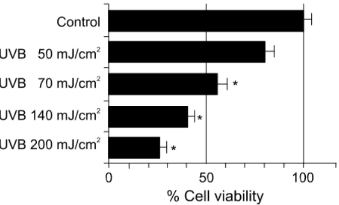
UVB induced cytotoxicity and m elatonin defense To examine the effect of UVB irradiation, cultured dermal fibroblasts were exposed to varying doses of UVB 50, 70, 140, and 200 mJ/cm
2and checked their cell viability after 24 h later. Such treatment result in 80 ± 9.2%, 56 ± 6.9%, 43 ± 5.2%, 26 ± 5.8% of sur- viving cells, respectively (Figure 1). To clarify the effect of melatonin on dermal fibroblasts under UVB irradiation (140 mJ/cm
2), we treated with 10-1000 nM of melatonin. The protective effect was obtained from 1
µM and 100 nM of melatonin, the cell viability inthe pre-treated groups with 1
µM and 100 nM ofmelatonin were 58% and 64.5%, respectively. The pre-treated groups with 100 nM of melatonin are significantly different from only UVB 140 mJ/cm
2irradiated groups at P<0.01 (Figure 2).
Apoptotic changes by UVB irradiation and m elatonin defense
When cells were stained with propidium iodide and observed under confocal laser scanning microscopy, normal cells appear as homogenous fluorescent with oval nuclei. Using this staining method, we found that the UVB irradiated cells were irregular shaped with intensely fluorescent and contained fragmented round bodies characteristics of apoptotic cells after 24 h
after treatment (Figure 3). The degree of apoptosis induced by UVB irradiation and the protective effects of melatonin were measured by counting the number of apoptotic cells among 10 high power field (×400).
More apoptotic cells were observed in UVB irradiated group than melatonin treated group. The percentage of apoptotic cells in UVB treated group was 35.1 ±
8.21%, whereas those in melatonin protected group was 10.4 ± 5.95%. In control group, less than 2% of attached cells showed apoptotic features.
Effects of UVB and m elatonin on polyam ine level The changes in polyamine level were examined 24 h after UVB and melatonin treatment by HPLC analysis. The level of putrescine of UVB irradiated
Figure 1. Viability of the dermal fibroblast following UVB treatment.
The cells were treated with UVB (50, 70, 140 and 200 mJ/cm2) and the number of viable cells was countered by trypan blue exclusion assay. Data shown are mean values ±standard errors determined from three independent experiments. *P<0.01.
Figure 2. The effects of melatonin on cultured skin fibroblast viability under UVB irradiation (140 mJ/cm2). Cell viability is represent as percent viable cells, where the untreated cells are regarded as 100%.
*Significantly different from control and **significantly different from only UVB 140 mJ/cm2 irradiated groups at P<0.01.
Figure 3. Detection of apoptotic cells by propidium iodide among adherent dermal fibroblasts treated with UVB and/or melatonin. A:
untreated control, B: UVB (140 mJ/cm2) irradiation, C: cells pretreated with melatonin before UVB irradiation and incubated for 24 h in DMEM containing melatonin (10-7 M). Apoptotic cells appear in B as con- densed, brightly fluorescent, oval particles (arrow) that are easily distin- guished from the controls (A) and these changes were moderately protected by melatonin (C).
group was 9.0±0.79 nmol per ml which was increa- sed compared to the untreated control (5.2±0.44 nmol). When pre-treated with melatonin before UVB irradiation, the level of putrescine was 6.9±0.55 nmol (Figure 4).
Expression of O DC gene
In northern blot analysis of cultured normal skin fibroblasts, [
32P]-labeled ODC and GAPDH cDNA probes specifically hybridized with each mRNA. ODC revealed 9.0 kb sized mRNA transcript, while GAPDH revealed 1.3 kb. There were no changes in size, indicating no alteration in quality. Northern analysis with cDNA probe for ODC after UVB (70, 140 mJ/
cm
2) irradiation resulted in a signal markedly in- creased (by about 1.7±0.07, 2.2±0.79 folds respec- tively) compared to control, after correction of the signal by GAPDH mRNA levels. And these over-ex- pressions were decreased up to 1.4±0.06, and 1.45
±0.02 folds under melatonin pretreatment at concen- tration of 100 nM compared to control. This means melatonin pretreatment decreased UVB induced ODC mRNA levels up to 35% (Figure 5). Relative quan- titation of ODC mRNA levels were measured, where the untreated cells are regarded as 100 densitometric absorbance units (Table 1).
D iscussion
Polyamines are present in living cells and play a pivo- tal role in cellular growth and developmental process (Theiss et al., 2002). The naturally occurring polya-
mines in mammamlian cells are putrescine, spermi- dine, and spermine (Megosh et al., 2002). ODC plays a critical role in the biosynthesis of those polyamines (Rebel et al., 2002). Actually, the level of ODC activity in quiescent cells is extremely low, but readily in- duced by a wide variety of growth-promoting agents or UV light (Nilsson et al., 2000; Tanaka et al., 2001).
Also rapidly proliferating cells and neoplastic cells express elevated levels of ODC, therefore the level of ODC activity is served as a marker of the initial mutagenic and carcinogenic process. Ultraviolet radia- tion activates the expression of a wide variety of genes by pathways which differ between the short non-solar UVC wavelengths, which are strongly ab- sorbed by nucleic acids, and the long solar UVA (320-380 nm) wavelengths, which generate active oxygen intermediates. Intermediate solar UV wave- lengths in the UVB (290-320 nm) range also contain
Figure 4. Quantitation of putrescine levels under UVB and melatoninby HPLC. A, control; B, UVB 140 mJ/cm2; C, UVB 140 mJ/cm2+ melatonin 10-7 M. Data are expressed as mean ± SD of three independent experiments. *Significantly different from control and
**significantly different from only UVB 140 mJ/cm2 irradiated groups at P<0.01.
Figure 5. Quantitation of ODC gene expression by northern blot analyses, where the untreated cells are regarded as 100 densitometric absorbance unit (DAU). A, Control; B, C, UVB 70/140 mJ/cm2; D, E, UVB 70/140 mJ/cm2+ melatonin 10-7 M. Data are expressed as mean
± SD of three independent experiments. *Significantly different from control and **significantly different from only UVB 140 mJ/cm2 ir- radiated groups at P<0.01.
Table 1. Steady-state levels of ODC mRNA.
ꠚꠚꠚꠚꠚꠚꠚꠚꠚꠚꠚꠚꠚꠚꠚꠚꠚꠚꠚꠚꠚꠚꠚꠚꠚꠚꠚꠚꠚꠚꠚꠚꠚꠚꠚꠚꠚꠚꠚꠚꠚꠚꠚꠚꠚꠚꠚꠚꠚꠚꠚꠚꠚ
Control 49 ± 9 (1)
UVB 70 mJ/cm2 83 ± 8 (1.7)
UVB 140 mJ/cm2 110 ± 11 (2.25)
UVB 70 mJ + Melatonin 10-7 M 68 ± 9 (1.4) UVB 140 mJ + Melatonin 10-7 M 71 ± 6 (1.45) ꠏꠏꠏꠏꠏꠏꠏꠏꠏꠏꠏꠏꠏꠏꠏꠏꠏꠏꠏꠏꠏꠏꠏꠏꠏꠏꠏꠏꠏꠏꠏꠏꠏꠏꠏꠏꠏꠏꠏꠏꠏꠏꠏꠏꠏꠏꠏꠏꠏꠏꠏꠏꠏ The values are mean ± SD and expressed as densitometric absor- bance unit which are the percentage of the value of GAPDH. Fold difference is shown in parethesis, *Significantly different from control and **significantly different from only UVB 140 mJ/cm2 irradiated groups at P<0.01
an oxidative component, but also closely resemble UVC in their gene activating properties. Short wave- length UV, in common with other extracellular stimuli including growth factors, activates signal transduction events that involve both stress- and mitogen-activated protein kinase cascades (Reeve et al., 2000; Soriani
et al., 2000; Tyrrell, 2000; Kim et al., 2001). Thisstudy describes the protective effects of melatonin in skin fibroblast against the UVB induced apoptotic changes. UVB induced cytotoxicity was examined 24 h after treatment by trypan blue exclusion test. A distinct dose response relationship was observed between the dose of UVB and the degree of survival rate. About 26% of cultured dermal fibroblasts were only survived at 200 mJ/cm
2. The UVB induced cyto- toxicity is a rapid event, and melatonin prevents such damage when present at concentration of 1 nM (Fischer et al., 1999). In our study, the protective effect of melatonin was observed at concentration of 1
µM and 100 nM. This discrepancy may result fromdifferent proctocol (in vivo vs in vitro). Moreover, melatonin treated cells showed marked suppression of UVB induced apoptotic changes. Like the other methods, this one also has some limitations in that it does not include all the floating cells. Therefore, these values are an underestimation of the actual degree of apoptosis. It was our interest to know whether the photoprotective actions of melatonin can attenuate the changes of polyamine levels and ODC activities following UVB irradiation in cultured skin fibroblasts. Putrescine is the first product from orni- thine by the action of ODC and known as a marker of cell injury. The level of putrescine of UVB irradiated group was 1.7 times increased compared to the un- treated control. When pre-treated with melatonin before UVB irradiation, the level of putrescine was de- creased up to 75%. The expression of ODC gene after UVB irradiation resulted in markedly increased with dose dependent fashion. This result is supported by other previous reports (Bornman et al., 1999;
Soriani et al., 1999). And these overexpressions were decreased up to 65% under melatonin pretreatment at concentration of 100 nM. The suppressive effect of melatonin to increased ODC mRNA can be re- sulted from increasing turn-over time of mRNA or decreasing transcription. Additionally, that effect of melatonin may be related with role of melatonin as antioxidant or not. Therefore, to elucidate the mecha- nisms of action of melatonin to polyamine and ODC gene, further experiments are needed. In conclusion, our results demonstrate that melatonin not only direct- ly acts as a protector from cell damage induced by UVB irradiation only when present in relevant con- centration at the site of action beginning but dimini- shes the UVB induced increase of polyamine syn- thesis and ODC gene expression. Collectively, ODC
response to UVB induced changes are possibly in- volves melatonin sensitive regulatory pathway in nor- mal human skin fibroblast.
Acknow ledgem ent
The present research has been supported by the Attached Research Institute Research Grant of Kei- myung University in 2001.
R eferences
Ahmad N, Gilliam AC, Katiyar SK, O'Brien TG, Mukhtar H.
A definitive role of ornithine decarboxylase in photocar- cinogenesis. Am J Pathol 2001;159:885-92
Bornman L, Baladi S, Richard MJ, Tyrrell RM, Polla BS.
Differential regulation and expression of stress proteins and ferritin in human monocytes. J Cell Physiol 1999;178:1-8 Chainiaux F, Magalhaes J, Eliaers F, Remacle J, Toussaint O. UVB-induced premature senescence of human diploid skin fibroblasts Int J Biochem Cell Biol 2002;34:1331-4 Chomczynski P, Sacchi N. Single-step method of RNA iso- lation by acid guanidium thiocyanate-phenol-chloroform ex- tract. Anal Biochem 1987;162:156-9
Fischer T, Bangha E, Elsner P, Kistler GS. Suppression of UV-induced erythema by topical treatment with melatonin.
Influence of the application time point. Biol Signals Recept 1999;8:132-5
Jansen AP, Dreckschmidt NE, Verwiebe EG, Wheeler DL, Oberley TD, Verma AK. Relation of the induction of epi- dermal ornithine decarboxylase and hyperplasia to the different skin tumor-promotion susceptibilities of protein ki- nase C alpha, -delta and -epsilon transgenic mice. Int J Can- cer 2001;93:635-43
Kim BC, Shon BS, Ryoo YW, Kim SP, Lee KS. Melatonin reduces X-ray irradiation-induced oxidative damages in cul- tured human skin fibroblasts. J Dermatol Sci 2001;26:194- 200
Kulms D, Zeise E, Poppelmann B, Schwarz T. DNA damage, death receptor activation and reactive oxygen species con- tribute to ultraviolet radiation-induced apoptosis in an essen- tial and independent way. Oncogene 2002;29:5844-51 Lee KS, Kim SJ, Ryoo YW, Kim BC. All-trans-retinoic acid down-regulates elastin promoter activity elevated by ultra- violet B irradiation in cultured skin fibroblasts. J Dermatol Sci 1998;17:182-9
Manzow S, Richter KH, Stempka L, Furstenberger G, Marks F. Evidence against a role of general protein kinase C down- regulation in skin tumor promotion. Int J Cancer 2000;85:503-7 Megosh LC, Hu J, George K, O'Brien TG. Genetic control of polyamine-dependent susceptibility to skin tumorigenesis.
Genomics 2002;79:505-12
Nilsson J, Gritli-Linde A, Heby O. Skin fibroblasts from sper- mine synthase-deficient hemizygous gyro male (Gy/Y) mice overproduce spermidine and exhibit increased resistance to
oxidative stress but decreased resitance to UV irradiation.
Biochem J 2000;352:381-7
O'Brien TG, Megosh LC, Gilliard G, Soler AP. Ornithine decarboxylase overexpression is a sufficient condition for tu- mor promotion in mouse skin. Cancer Res 1997;57:2630-7 Oguro T, Yoshida T. Effect of ultraviolet A on ornithine decarboxylase and metallothionein gene expression in mouse skin. Photodermatol Photoimmunol Photomed 2001;17:71-8 Rebel H, van Steeg H, Beems RB, Schouten R, de Gruijl FR, Terleth C. Suppression of UV carcinogenesis by difluo- romethylornithine in nucleotide excision repair-deficient Xpa knockout mice. Cancer Res 2002;62:1338-42
Reeve VE, Tyrrell RM, Shen J. Interactions between the UVA and UVB wavebands relevant for immune function and carcinogenesis. Redox Rep 2000;5:50-1
Rigby PW, Dieckmann M, Rhodes C, Berg P. Labeling de- oxyribonucleic acid to high specific in vitro by nick-translation with DNA polymerase I. J Mol Biol 1977;113:237-51 Ryoo YW, Suh SI, Mun KC, Kim BC, Lee KS. The effects of the melatonin on ultraviolet-B irradiated cultured dermal fibroblasts. J Dermatol Sci 2001;27:162-9
Shindo Y, Hashimoto T. Ultraviolet B-induced cell death in four cutaneous cell lines exhibiting different enzymatic anti- oxidant defences: involvement of apoptosis. J Dermatol Sci 1998;17:140-50
Soriani M, Luscher P, Tyrrell RM. Direct and indirect modul- ation of ornithine decarboxylase and cyclooxygenase by UVB radiation in human skin cells. Carcinogenesis 1999;20:727-32 Soriani M, Hejmadi V, Tyrrell RM. Modulation of c-jun and c-fos transcription by UVB and UVA radiations in human dermal fibroblasts and KB cells. Photochem Photobiol 2000;
71:551-8
Spragg BP, Hutchings AD. High-performance liquid chro- matographic determination of putrescine, spermidine, and spermine after deprivation with 4-fluoro-3-nitroben-zotrifluoride.
J Chromatogr 1983;258:289-92
Takeda J, Ito Y, Maeda K, Ozeki Y. Assignment of UVB- responsive cis-element and protoplastization-(dilution-) and elicitor-responsive ones in the promoter region of a carrot phenylalanine ammonia-lyase gene (gDcPAL1). Photochem Photobiol 2002;76:232-8
Tanaka K, Kamiuchi S, Ren Y, Yonemasu R, Ichikawa M, Murai H, Yoshino M, Takeuchi S, Saijo M, Nakatsu Y, Miyauchi-Hashimoto H, Horio T. UV-induced skin carcinoge- nesis in xeroderma pigmentosum group A (XPA) gene- knockout mice with nucleotide excision repair-deficiency.
Mutat Res 2001;477:31-40
Theiss C, Bohley P, Voigt J. Regulation by polyamines of ornithine decarboxylase activity and cell division in the uni- cellular green alga Chlamydomonas reinhardtii. Plant Physiol 2002;128:1470-9
Tyrrell RM. Role for singlet oxygen in biological effects of ultraviolet A radiation. Methods Enzymol 2000;319:290-6 van Weelden H, van der Putte SC, Toonstra J, van der Leun JC. Ultraviolet B-induced tumors in pigmented hairless mice, with an unsuccessful attempt to induce cutaneous melanoma.
Photodermatol Photoimmunol Photomed 1990;7:68-72 Wahl GM, Stern M, Starck GR: Efficient transfer of large DNA fragments from agarose gel to diabenzyloxymethyl-paper and rapid hybridization by using dextran sulfate. Proc Natl Sci USA 1979;76:3683-7