Clin Endosc 2013;46:662-665
662 Copyright © 2013 Korean Society of Gastrointestinal Endoscopy CASE REPORT
Gastrostomy in a Patient with Situs Inversus Totalis
Hyung Ki Lee, Kwang Bum Cho, Eun Soo Kim and Kyung Sik Park
Department of Internal Medicine, Keimyung University School of Medicine, Daegu, Korea
Situs inversus totalis (SIT) is a rare condition in which there is complete right to left reversal of the abdominal and thoracic organs. SIT generally does not bear any pathophysiological significance, and the survival rate of patients with SIT does not differ from that of healthy individuals. However, patients with SIT require a thorough radiological examination to identify the presence of associated anatomic variations before undergoing invasive procedures such as surgery or hemostasis of gastrointestinal hemorrhage because they may have accompanying abnormalities in anatomical structures along with reversed organs. Percutaneous endoscopic gastrostomy (PEG) is a rela- tively safe procedure that is most commonly performed for the enteral feeding of patients with dysphagia and a normal gastrointestinal function. However, the procedure requires extracaution because minor complications may lead to life-threatening situations due to the underlying illnesses. Here, we report the case of a patient with SIT who underwent a PEG procedure without complications, and review the existing literature on this subject.
Key Words: Situs inversus totalis; Percutaneous endoscopic gastrostomy; Anatomic variation Open Access
Received: December 14, 2012 Revised: February 13, 2013 Accepted: April 18, 2013
Correspondence: Kwang Bum Cho
Department of Internal Medicine, Keimyung University School of Medicine, 56 Dalseong-ro, Jung-gu, Daegu 700-712, Korea
Tel: +82-53-250-7088, Fax: +82-53-250-7088, E-mail: [email protected]
cc This is an Open Access article distributed under the terms of the Creative Commons Attribution Non-Commercial License (http://creativecommons.org/
licenses/by-nc/3.0) which permits unrestricted non-commercial use, distribution, and reproduction in any medium, provided the original work is properly cited.
Print ISSN 2234-2400 / On-line ISSN 2234-2443 http://dx.doi.org/10.5946/ce.2013.46.6.662
INTRODUCTION
Situs inversus is a congenital condition in which the major organs within the abdomen or the thoracic cavities are re- versed from their normal positions. Situs inversus totalis (SIT) is a rare condition in which the abdominal and thoracic or- gans are completely reversed. Its incidence is approximately one in every 10,000 to 50,000 live births. Although the exact cause is unknown, SIT generally does not cause inconve- nience in daily life. SIT has no pathophysiological signifi- cance, and the survival rate of patients with SIT does not dif- fer from that of healthy persons.1,2 However, patients with SIT require a thorough radiological examination to identify the presence of associated variations before undergoing invasive procedures such as surgery or hemostasis of gastrointestinal hemorrhage because they may have an abnormal spleen or other abnormalities in anatomic structures3,4 along with re- versed organs.5
Percutaneous endoscopic gastrostomy (PEG) is one of the most frequently performed enteral feeding methods for pa- tients with dysphagia and a normal gastrointestinal function, and is generally highly successful, with a low death rate. This procedure can be easily implemented in patients with serious underlying diseases.6 The reported complications are surgical site infection, bleeding, gastrocolic fistula, and displacement of the feeding tube. In severe cases, more critical and life- threatening complications are seen, including aspiration pneumonia, intestinal perforation, peritonitis, and necrotiz- ing fasciitis, requiring extracaution during the procedure.7 In particular, caution should be exercised concerning the differ- ences in anatomical location when performing PEG on a pa- tient with SIT.
Here, we report the case of a patient with SIT who under- went a PEG procedure because of dysphagia because of intra- cerebral hemorrhage (ICH), and include a review of the liter- ature.
CASE REPORT
A 65-year-old woman was transferred to our department for PEG insertion during nasogastric tube feeding because of dysphagia caused by ICH. She was taking medication for hy- pertension and osteoporosis for the last 7 years. She was diag- nosed with SIT during a colonoscopy performed 2 years pri-
Lee HK et al.
663 or; she has no family history of this condition. At the time of
transfer, she was maintaining a blood pressure of 130/80 mm Hg, heart rate of 65 to 80 beats per minute, body temperature of 36.5°C, and respiratory rate of 18 breaths per minute with alert consciousness. Her heart rate was normal without heart murmur. No difference was found from the previous exami- nation of the chest, and no abnormal findings were detected in the abdomen, limbs, and skin. Analysis of peripheral blood showed a white blood cell count, platelet count, and hemoglo- bin of 5,170/mm3, 9.7 g/dL, and 290,000/mm3, respectively.
The liver function test results were as follows: aspartate aminotransferase/alanine aminotransferase, 13/9 IU/L; pro- thrombin time-international normalized ratio, 1.08; and par- tial thromboplastin time, 32.4 seconds (reference range, 20 to 38). The renal function tests were within the reference ranges.
Dextrocardia was observed on a chest radiograph (Fig. 1), and complete right to left reversal of internal organs was detected on abdominal computed tomography (CT) scan, indicating a finding of SIT (Fig. 2). No abnormal findings were found on the CT scans of the spleen and major blood vessels. Upper gastrointestinal endoscopy was conducted identical to a gen- eral endoscopic procedure, with the patient in a left lateral de- cubitus position. Along with preoperative monitoring of pulse rate and oxygen saturation, midazolam (3 mg) and propofol (50 mg) were administered via intravenous injection as a pre- treatment. The endoscope was turned counterclockwise when it was passed from the bulb to the descending part of the duo- denum. The gastric angle, when observed with a J shaped ret- roflection at the antrum, was mirrored from its normal posi- tion. No specific findings were detected during the endoscopy
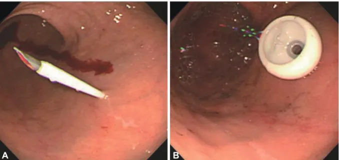
except for a mild chronic superficial gastritis. A puncture site was chosen with the help of an endoscopic light after manual- ly pressing the stomach wall to find the thinnest site. Subse- quently, a gastrostomy feeding tube (PEG-24-Pull; Wilson- Cook Medical GI Endoscopy, Winston-Salem, NC, USA) was inserted into the anterior wall of the lower gastric body, which looks like the posterior wall of the lower gastric body in a normal person (Fig. 3). The nutrition was delivered directly into the stomach the next morning after PEG insertion, and no specific complications were observed.
DISCUSSION
SIT is a rare congenital condition in which the abdominal and thoracic cavity structures are completely opposite of their normal positions. The exact cause of this condition has not been clarified, except for a genetic predisposing factor. Ac- cording to Layton,8 the allele regulating the asymmetric ex- pression of normal organ placement is a complete dominant one, and the reversal of organ position results from the ab- sence of that allele. A patient with SIT requires a thorough ra- diological examination to identify the presence of associated anatomic variations before undergoing invasive procedures such as surgery or hemostasis of gastrointestinal hemorrhage because the condition may be associated with an abnormal spleen and other abnormalities in anatomical structures. Lee et al.9 performed endoscopic hemostasis on a SIT patient with Dieulafoy gastric bleeding. However, several attempts with endovascular embolization and hemoclips failed, so they per- formed a suture and ligation of the Dieulafoy lesion as well as total resection of the accessory spleen with devascularization of prominently developed vessels around the upper stomach.
Kang et al.10 reported a case study of an early gastric cancer
Fig. 1. Chest radiograph (posteroanterior view) showing a left-
right shifted heart. Fig. 2. Abdominal computed tomography showing left-right shift-
ed internal organs.
664 Clin Endosc 2013;46:662-665
Gastrostomy in a Patient with Situs Inversus Totalis
patient with SIT in whom surgery was completed safely by identifying the anatomic variations of the right gastric artery directly arising from the aorta and the splenic artery directly arising from the celiac artery on a preoperative abdominal CT scan.
PEG insertion is an effective and safe procedure for patients having difficulty in swallowing, with the placement of a feed- ing tube into the stomach. However, complications continue to be reported despite advancements in the procedure.11 The most common complication of PEG is surgical site infection, followed by gastric bleeding, pneumoperitoneum, displace- ment of the feeding tube, aspiration pneumonia, intestinal perforation, peritonitis, necrotizing fasciitis, buried bumper syndrome, and others, accounting for 5% to 40% depending on the reports.12 Although PEG insertion is relatively safe and effective, the procedure requires extra caution because minor complications may lead to life-threatening situations due to underlying illnesses in the presence of malnutrition and old age. In particular, when performing PEG in patients with SIT, the procedure requires added caution because of the unfamil- iar structures associated with anatomical placement and vari- ations, as well as because of postoperative complications.
Endoscopic retrograde cholangiopancreatography (ERCP) and endoscopy are generally performed in the right lateral decubitus position in patients with SIT, taking into consider- ation the reversed abdominal organs.13 However, ERCP could also be performed without difficulty in the left lateral decubi- tus position, according to Chowdhury et al.14 In a previous re- port, when endoscopy and colonoscopy were carried out in a patient with SIT, with careful preparation and in keeping with the general principles, in the left lateral decubitus position as with ordinary patients, the procedures were successfully per- formed without pain or complications except for a prolonged
examination time.15
In conclusion, PEG insertion was performed in a patient with SIT with the help of an endoscope, by puncturing the anterior wall of the lower gastric body, which looks like the posterior wall of the lower gastric body in a normal person, after identifying the presence of associated anatomic varia- tions. No complications occurred. Although PEG insertion in a patient with SIT may be rare and accompanied by different complications, the procedure could be implemented without difficulty if the anatomic variations as well as the reversed or- gans in these patients are carefully considered.
Conflicts of Interest
The authors have no financial conflicts of interest.
REFERENCES
1. Douard R, Feldman A, Bargy F, Loric S, Delmas V. Anomalies of later- alization in man: a case of total situs inversus. Surg Radiol Anat 2000;
22:293-297.
2. Fujiwara Y, Fukunaga Y, Higashino M, et al. Laparoscopic hemicolecto- my in a patient with situs inversus totalis. World J Gastroenterol 2007;
13:5035-5037.
3. Yi SQ, Tanaka S, Tanaka A, Shimokawa T, Ru F, Nakatani T. An ex- tremely rare inversion of the preduodenal portal vein and common bile duct associated with multiple malformations. Report of an adult cadav- er case with a brief review of the literature. Anat Embryol (Berl) 2004;
208:87-96.
4. Marta MJ, Falcão LM, Saavedra JA, Ravara L. A case of complete situs inversus. Rev Port Cardiol 2003;22:91-104.
5. Lee SE, Kim HY, Jung SE, Lee SC, Park KW, Kim WK. Situs anomalies and gastrointestinal abnormalities. J Pediatr Surg 2006;41:1237-1242.
6. Hull MA, Rawlings J, Murray FE, et al. Audit of outcome of long-term enteral nutrition by percutaneous endoscopic gastrostomy. Lancet 1993;
341:869-872.
7. Calton WC, Martindale RG, Gooden SM. Complications of percutane- ous endoscopic gastrostomy. Mil Med 1992;157:358-360.
8. Layton WM Jr. Random determination of a developmental process: re- versal of normal visceral asymmetry in the mouse. J Hered 1976;67:
336-338.
Fig. 3. Endoscopic findings. (A) The needle is passed through the nearest site of the abdominal wall. (B) The internal bolster of the percuta- neous endoscopic gastrostomy tube is fixed in the stomach.
A B
Lee HK et al.
665
9. Lee JR, Kim MS, Kim DJ, Choi SJ. Gastric bleeding arisen in a patient with situs inversus totalis and large accessory spleen. J Korean Surg Soc 2010;78:258-261.
10. Kang BH, Lee SL, Hur H, Kim JY, Cho YK, Han SU. Laparoscopy as- sisted subtotal gastrectomy in gastric cancer patient with situs inversus in Korea. J Korean Surg Soc 2010;79:513-517.
11. Choi KW, Rhee JC, Jang JK, et al. Complications of percutaneous endo- scopic gastrostomy and predictors of wound infection. Korean J Gas- troenterol 2001;38:23-28.
12. Kirby DF, Delegge MH, Fleming CR. American Gastroenterological
Association technical review on tube feeding for enteral nutrition. Gas- troenterology 1995;108:1282-1301.
13. Kim SH, Kong ON, Ha JK, et al. A case of endoscopic removal of cho- ledocholithiasis in a patient with situs inversus totalis. Korean J Gastro- intest Endosc 2002;24:59-61.
14. Chowdhury A, Chatterjee BK, Das U, Dutta P, Dhali GK, Banerjee PK.
ERCP in situs inversus: do we need to turn the other way? Indian J Gastroenterol 1997;16:155-156.
15. Cho SH. Endoscopy and colonoscopy in a situs inversus totalis patient:
a case report. Korean J Gastrointest Endosc 2009;38:98-102.