380
www.jkns.or.kr
Pseudoaneurysm of Ulnar Artery
after Endoscopic Carpal Tunnel Release
Sung-Joo Ryu, M.D., In-Soo Kim, M.D., Ph.D.
Department of Neurosurgery, Dongsan Medical Center, Keimyung University School of Medicine, Daegu, Korea J Korean Neurosurg Soc 48 : 380-382, 2010
The authors present an extremely rare case of a pseudoaneurysm of the ulnar artery as a complication of a two-portal endoscopic carpal tunnel release (ECTR). A 70-year-old man with chronic renal failure and on maintenance hemodialysis with a left arteriovenous fistula presented with paresthesia of his right hand. A clinical diagnosis of right carpal tunnel syndrome was confirmed by ultrasonography and an electrophysiologic study. He underwent two-portal ECTR, and the paresthesia was much improved. However, he presented to us one month after operation with severe pain, a tender mass distal to the right wrist crease and more aggravation of the paresthesia in the ulnar nerve distribution. Doppler ultrasound was performed and revealed a hypoechoic lesion 20 mm in diameter in the right palm, with arterial Doppler flow inside connected to the palmar segment of the ulnar artery. An ulnar artery pseudoaneurysm was diagnosed and treated by ultrasound-guided percutaneous thrombin injection. Transverse color Doppler ultrasound image showed complete thrombosis of the pseudoaneurysm and flow cessation after a total injection of 500 units of thrombin. The symptoms were also improved.
KEY WORDS : Endoscopic carpal tunnel release
˙
Complication˙
Pseudoaneurysm.10.3340/jkns.2010.48.4.380
Case Report
Copyright ©2010 The Korean Neurosurgical Society Print ISSN 2005-3711 On-line ISSN 1598-7876
INTRODUCTION
Endoscopic carpal tunnel release (ECTR) has been used to decrease postoperative complication rates, to reduce posto- perative pain, to speed up recovery, and to improve the pre- servation of grip strength compared with open carpal tunnel release. However, various complications may occur such as incomplete release of the retinaculum, and injury to the me- dian nerve and vessels
14). A literature review was conducted of the PubMed database. “Neurovascular complication”, “en- doscopic carpal tunnel release”, “complications related carpal tunnel release” and “pseudoaneurysm” were used as medical subject headlines in combination with “carpal tunnel synd- rome” as a key word in the abstracts or the title in the Pub- Med database. In our search, there was only one report of an ulnar artery pseudoaneurysm after ECTR. Here, we report on a case with a pseudoaneurysm of the ulnar artery as a
complication of two-portal ECTR.
CASE REPORT
A 70-year-old, right hand dominant man with chronic renal failure and on maintenance hemodialysis with a left arteriovenous fistula for 10 years presented with a 10 year history of paresthesia involving the palms of his right hand. A clinical diagnosis of right carpal tunnel syndrome was con- firmed by ultrasonography and an electrophysiologic study.
Ultrasonography showed mild thickening of the flexor reti- naculum, mild flattening of the median nerve, and no defi- nite anatomic malformations. In the electrophysiologic study, absence of sensory nerve action potential and compound motor action potential at the abductor pollicis brevis with median nerve stimulation were compatible with severe-grade median nerve neuropathy at the wrist. The patient under- went two-portal ECTR under lidocaine IV anesthesia, and the paresthesia was much improved. However, he presented to us one month after the operation with severe pain, a tender mass distal to the right wrist crease and more aggravation of the paresthesia in the ulnar nerve distribution. Doppler ultrasound was performed and revealed a hypoechoic lesion 20 mm in diameter in the right palm, with arterial Doppler
•Received : February 19, 2010 •Revised : October 5, 2010
•Accepted : October 6, 2010
•Address for reprints : In-Soo Kim, M.D., Ph.D.
Department of Neurosurgery, Dongsan Medical Center, Keimyung University School of Medicine, 216 Dalseong-ro, Jung-gu, Daegu 700-712, Korea
Tel : +82-53-250-7730, Fax : +82-53-250-7820 E-mail : [email protected]
flow inside connected to the palmar segment of the ulnar artery and compressing the ulnar nerve distally. The ulnar artery pseudoaneurysm was diagnosed and treated by ultra- sound-guided percutaneous thrombin injection. An echoge- nic needle was percutaneously injected with a side delivery port to the middle of the pseudoaneurysm, away from the neck. Arteriography after direct puncture of the pseudoaneu- rysm showed the unilobulated pseudoaneurysm originating from the palmar segment of the ulnar artery through a nar- row neck. (Fig. 1). Transverse color Doppler ultrasound ima- ge showed complete thrombosis of the pseudoaneurysm and flow cessation after a total injection of 500 units of thrombin.
(Fig. 2). The symptoms were improved.
DISCUSSION
Carpal tunnel syndrome is the most common nerve entrap- ment disorder of the upper extremity with an estimated life time risk of 10% and an overall prevalence of 2.1% in the general population
11). The syndrome is usually related to compression of the median nerve within the carpal tunnel because of a variety of etiologic factors
6). A number of medi- cal conditions also increase the risk of carpal tunnel synd- rome. The incidence and recurrence of carpal tunnel synd- rome in long-term hemodialysis patients are higher than in the general population
12). In our case, the patient had chronic renal failure and was receiving hemodialysis with the arterio- venous fistula on the unaffected side.
With typical clinical presentation and characteristic electro- physiologic findings, the diagnosis is made in most cases
16). Initially, conservative treatments such as medication and steroid injection are considered. In patients receiving long term hemodialysis, open carpal tunnel release has been considered as the operative procedure of choice because of the anatomic deterioration of the carpal tunnel tissue caused by beta-2 microglobulin amyloid deposition and the need for neuroly- sis of the median nerve. However, Gelberman et al.
5)re- ported that neurolysis is no longer indicated for routine or severe carpal tunnel syndrome. In the current reports, long term hemodialysis patients who suffered from carpal tunnel syndrome have almost the same or better recovery rate than idiopathic patients using ECTR
7,10,17).
The most common complication is neuroma of the palmar cutaneous branch of the median nerve
1). Other complica- tions are hypertrophic scar, pillar pain, scar tenderness, failure to relieve symptoms and neuromas of the dorsal sensory branch of the radial nerve
3,8,15). Various complications after carpal tunnel decompression have been reported. However, in our knowledge, there is only one report of an ulnar artery pseudoaneurysm after ECTR. ECTR usually demonstrates a
Pseudoaneurysm of Ulnar Artery after Endoscopic Carpal Tunnel Release |SJ Ryu and IS Kim
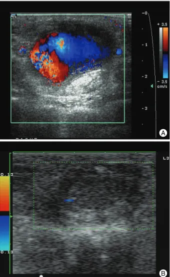
381 Fig. 1. Transverse color Doppler ultrasound image showing the distal ulnar artery showing a pseudoaneurysm with turbulent flow in its lumen (A) and complete thrombosis of the pseudoaneurysm and flow cessation after a total injection of 500 units of thrombin (B).
B A
Fig. 2. Arteriography after direct puncture of the pseudoaneurysm showing the unilobulated pseudoaneurysm originating from the palmar segment of the ulnar artery through a narrow neck.
J Korean Neurosurg Soc 48|October 2010
382
reduction of postoperative pain and a faster recovery of strength compared with open carpal tunnel release
9). Leon et al. reported that structural complications to nerves, arteries, or tendons are 0.19% for the endoscopic technique compared with 0.49% for the open approach
2). Despite the success of this procedure, complications may occur, such as incomplete release of the retinaculum, injury to the palmar branch of the median nerve and injuries to adjacent vessels
13).
Ultrasonography is widely used in the preoperative exami- nation. Although an electrophysiologic study is the gold standard in the diagnosis, localization and typing of a nerve lesion, it may not yield enough information
4). Ultrasonogra- phy may confirm the diagnosis and offer some reliable infor- mation about the morphological status of the affected nerve and surrounding structures prior to surgery. Although we preo- peratively confirmed that there were no anatomical variations or other local pathologies using ultrasonography, and posto- peratively excluded complete injury of the palmar branch of the median nerve and adjacent vessels by careful observation after tourniquet release, an ulnar artery pseudoaneurysm de- veloped. We believe this injury occurred because the ulnar artery is a mobile structure and is positioned around the me- dian nerve. The ulnar artery runs along the ulnar border to the wrist and crosses the transverse carpal ligament on the radial side of the pisiform bone adjacent to the median nerve. We thought the injury to the ulnar artery could also been due to the endoscope, which was advanced medial to the ulnar neu- rovascular bundle. We thought that minimal incomplete arter- ial injury developed after ECTR, and hemodynamic factors might play a role in pseudoaneurysm formation in addition to amyloidosis. If there is a pulsatile mass around the opera- tion site after ECTR, a pseudoaneurysm should be considered.
CONCLUSION
Two-portal ECTR is a safe and effective method for carpal tunnel release. However, it limits the visual field and leads to operation-related complications such as pseudoaneurysm as in this case. Also, patients with chronic renal failure are sus- ceptible to vascular and nerve damage because of underlying pathophysiology. So, more careful procedures are needed in high risk patients.
References
1. Akhtar S, Arenas Prat J, Sinha S : Neuropraxia of the palmar cutaneous branch of the ulnar nerve during carpal tunnel decompression. Ann R Coll Surg Engl 87 : W1-W2, 2005
2. Benson LS, Bare AA, Nagle DJ, Harder VS, Williams CS, Visotsky JL : Complications of endoscopic and open carpal tunnel release. Arthro- scopy 22 : 919-924, 2006
3. Boya H, Ozcan O, Oztekin HH : Long-term complications of open carpal tunnel release. Muscle Nerve 38 : 1443-1446, 2008
4. Cokluk C, Aydin K : Ultrasound examination in the surgical treatment for upper extremity peripheral nerve injuries : part I. Turk Neurosurg 17 : 277-282, 2007
5. Gilbert MS, Robinson A, Baez A, Gupta S, Glabman S, Haimov M : Carpal tunnel syndrome in patients who are receiving long-term renal hemodialysis. J Bone Joint Surg Am 70 : 1145-1153, 1988
6. Katz JN, Simmons BP : Clinical practice. Carpal tunnel syndrome. N Engl J Med 346 : 1807-1812, 2002
7. Kim SJ, Shin SJ, Kang ES : Endoscopic carpal tunnel release in patients receiving long-term hemodialysis. Clin Orthop Relat Res : 141-148, 2000
8. Louis DS, Greene TL, Noellert RC : Complications of carpal tunnel surgery. J Neurosurg 62 : 352-356, 1985
9. Mackenzie DJ, Hainer R, Wheatley MJ : Early recovery after endosco- pic vs. short-incision open carpal tunnel release. Ann Plast Surg 44 : 601-604, 2000
10. Okutsu I, Hamanaka I, Ninomiya S, Takatori Y, Shimizu K, Ugawa Y : Results of endoscopic management of carpal-tunnel syndrome in long- term haemodialysis versus idiopathic patients. Nephrol Dial Transpl- ant 8 : 1110-1114, 1993
11. Pingree MJ, Bosch EP, Liu P, Smith BE : Delayed ulnar neuropathy at the wrist following open carpal tunnel release. Muscle Nerve 31 : 394- 397, 2005
12. Staub F, Dombert T, Assmus H : [Carpal tunnel syndrome in haemo- dialysis patients : analysis of clinical and electrophysiological findings in 268 patients (395 hands).] Handchir Mikrochir Plast Chir 37 : 150- 157, 2005
13. Subasi M, Ay S, Tuzuner T : Transection of the ulnar nerve as a com- plication of two-portal endoscopic carpal tunnel release. Isr Med Assoc J 6 : 443-444, 2004
14. Thoma A, Veltri K, Haines T, Duku E : A systematic review of reviews comparing the effectiveness of endoscopic and open carpal tunnel decompression. Plast Reconstr Surg 113 : 1184-1191, 2004 15. Uygur F, Sever C, Yüksel F : Comparing the results of limited incision
technique and standard longitudinal incision technique for carpal tunnel decompression by numerical grading system. Turk Neurosurg 19 : 51-57, 2009
16. Vogt T, Mika A, Thömke F, Hopf HC : Evaluation of carpal tunnel syndrome in patients with polyneuropathy. Muscle Nerve 20 : 153- 157, 1997
17. Yoshida A, Okutsu I, Hamanaka I, Motomura T : Results of endosco- pic management of primary versus recurrent carpal tunnel syndrome in long-term haemodialysis patients. Hand Surg 9 : 165-170, 2004