Anatomical resection of hepatocellular carcinoma: A critical review of the procedure and its benefits on survival
Koo Jeong Kang, Keun Soo Ahn
Koo Jeong Kang, Keun Soo Ahn, Division of Hepatobiliary and Pancreatic Surgery, Department of Surgery, School of Medicine, Keimyung University Dong-san Medical Center, Daegu 41931, South Korea
Author contributions: Kang KJ and Ahn KS designed the review and collected and analyzed data; Kang KJ wrote the manuscript; Ahn KS revised the manuscript.
Conflict-of-interest statement: No potential conflicts of interest.
Open-Access: This article is an open-access article which was selected by an in-house editor and fully peer-reviewed by external reviewers. It is distributed in accordance with the Creative Commons Attribution Non Commercial (CC BY-NC 4.0) license, which permits others to distribute, remix, adapt, build upon this work non-commercially, and license their derivative works on different terms, provided the original work is properly cited and the use is non-commercial. See: http://creativecommons.org/
licenses/by-nc/4.0/
Manuscript source: Invited manuscript
Correspondence to: Koo Jeong Kang, MD, PhD, Division of Hepatobiliary and Pancreatic Surgery, Department of Surgery, School of Medicine, Keimyung University Dong-san Medical Center, 56 Dalseong-Ro, Jung-Gu, Daegu 41931,
South Korea. [email protected] Telephone: +82-53-2507655 Fax: +82-53-2507322 Received: August 27, 2016
Peer-review started: August 31, 2016 First decision: September 20, 2016 Revised: October 18, 2016 Accepted: November 14, 2016 Article in press: November 16, 2016 Published online: February 21, 2017
Abstract
Hepatocellular carcinoma (HCC) is the sixth most common type of cancer and the third most frequent cause of cancer-related death. Advances in preoperative
assessment of HCC (e.g., imaging studies and liver function tests), surgical techniques, and postoperative care have improved the surgical outcomes and survival of patients who undergo hepatic resection for HCC.
However, in the last 20 years, the long-term survival after hepatectomy has remained unsatisfactory owing to the high rates of local recurrence and multicentric occurrence. Anatomical liver resection (AR) was introduced in the 1980s. Although several studies have revealed tangible benefits of AR for HCC, these benefits are still debated. Because most HCCs occur in patients with liver cirrhosis and poor hepatic function, there are many factors that affect survival, including the surgical method. Nevertheless, many studies have documented the perioperative and long-term benefits of AR in various conditions. In this article, we review the results of several recently published, well-designed comparative studies of AR, to investigate whether AR provides real benefits on survival outcomes. We also discuss the potential pitfalls associated with this approach.
Key words: Hepatocellular carcinoma; Cirrhosis; Curative;
Anatomical resection; Prognosis
© The Author(s) 2017. Published by Baishideng Publishing Group Inc. All rights reserved.
Core tip: Anatomical liver resection (AR) has been widely used for two decades in hepatocellular car- cinoma and although many studies have shown the perioperative benefits, long term survival benefit of AR is still debated. For evaluation of benefits of AR, many factors should be considered, such as degree of cirrhosis, anatomical variation and surgical techniques.
Moreover, critical review of previous studies considering bias is necessary. In this article, we review the results of several recently published, well-designed comparative studies of AR to investigate whether AR provides real benefits on survival outcomes. We also discuss the potential pitfalls associated with this approach.
MINIREVIEWS
DOI: 10.3748/wjg.v23.i7.1139 ISSN 1007-9327 (print) ISSN 2219-2840 (online)
Kang KJ, Ahn KS. Anatomical resection of hepatocellular carcinoma: A critical review of the procedure and its benefits on survival. World J Gastroenterol 2017; 23(7): 1139-1146 Available from: URL: http://www.wjgnet.com/1007-9327/full/
v23/i7/1139.htm DOI: http://dx.doi.org/10.3748/wjg.v23.i7.1139
INTRODUCTION
Primary liver cancer is the second leading cause of cancer death worldwide[1]. The most common histologic types of liver cancer are hepatocellular carcinoma (HCC) and intrahepatic cholangiocarcinoma (ICC). HCC is the sixth most common type of cancer and the third most frequent cause of cancer-related death[2]. Advances in preoperative assessment of HCC (e.g., imaging studies and liver function tests), surgical techniques, and postoperative care have markedly improved the surgical outcomes and survival of patients who undergo hepatic resection for HCC.
However, in the last 20 years, the long-term survival after hepatectomy has remained unsatisfactory owing to the high rates of local and multicentric recurrence.
The oncologic principles of hepatic resection, especially in cirrhotic liver, are to resect all of the malignant tissue (tumor, satellite nodules, and portal vein territory) completely with effective clearance while preserving enough nontumorous liver parenchyma to prevent postoperative liver failure[3]. Regarding hepatectomy, modern surgical techniques were developed and important insight was gained in the late 1980s and through the 1990s. In the mid-1980s, hepatic surgeons gained more knowledge about liver anatomy, including its segments, leading to the introduction of systematic segmentectomy. Several strategies for reducing blood loss during hepatic transection were also introduced. They included keeping central venous pressure (CVP) as low as possible, the Pringle maneuver, and establishment of ischemia and reperfusion injury[4-6]. As a consequence of these developments, the surgical outcomes improved markedly in this era and the mortality rate decreased from 5%-10% to < 1%[7].
Although most hepatic surgeons estimate the optimal tumor-free margin during hepatic resection of the primary hepatic tumor, the concept of anatomical resection (AR) was introduced, in which the tumor-free margin is independent of margin length. This approach may improve the survival rate by reducing local recurrence[8]. AR was first proposed by the distinguished Japanese hepatic surgeon, Dr. Makuuchi[9,10]. The concept involves resection of the entire hepatic parenchymal tissue supplied by the portal venous system draining the tumor tissue. To achieve this, the liver surface is tattooed by injecting indigo carmine dye into the portal vein under intraoperative ultrasound guidance. A similar approach, Glissonean pedicle transection, was introduced by Dr. Takasaki[11]. Both groups suggested that AR confers a survival benefit.
However, the outcomes of AR reported for other case- series differed between institutions when compared with non-anatomical resection (NAR). Accordingly, the benefits of AR are still debated. Different results may be due to patient selection bias and the use of different surgical techniques at each institution. To date, no prospective randomized studies have compared the outcomes of AR and NAR, for example. Several case-controlled studies have been published in which cases and controls were matched by propensity score matching. Although this statistical method is retrospective, it may provide valuable data in lieu of randomized studies.
Here, we perform a review of well-designed comparative studies, including case-series, meta- analyses, and case-control studies with propensity score matching, to investigate whether AR confers real clinical benefits relative to other resection methods. We also discuss the possible pitfalls of AR in this setting.
DEFINITION AND THEORETICAL BACKGROUND
AR is defined as resection of the tumor together with the portal veins draining the tumor and the corresponding hepatic territory, as determined by dye injection into the feeding portal vein, or Glissonean pedicle transection[8,11]. AR involves either segmentectomy or sectionectomy, which includes bisectionectomy, hemihepatectomy, and trisectionectomy. NAR is defined as resection of a lesion regardless of the anatomical segment or section of the lobar anatomy, and includes limited resection or enucleation[8,11-15]. In the case of subsegmentectomy, which involves resection of the hepatic parenchyma fed by the fourth-order portal venous branch or one of several third-order branches, it is debated whether resection of parenchyma fed by one or several fourth-order branches should be classified as either AR or NAR.
Liver tumors are thought to invade the portal venous branches, allowing tumor cells to be carried to other regions of the liver in the portal venous flow.
These disseminated tumor cells grow into microscopic tumor thrombi and then into daughter nodules[8]. Accordingly, it is theorized that AR confers a survival benefit by removing possible microscopic tumor thrombi or hitherto undetected daughter nodules in other parts of the liver. Several rigorous comparative analyses of the survival benefits of patients who underwent hepatic resection by AR or NAR have been conducted in many different centers. Most of these studies were performed in Eastern countries, and the results were inconsistent. This theory to explain the survival benefit of AR was subsequently reinforced by a well-designed imaging study, in which the authors used CT angiography to monitor the intratumoral hemodynamic changes associated with hepatocarcinogenesis[16]. The study showed that early
deterioration of arterial blood flow and an increase in neovascularized arterial blood flow was followed by a decrease in portal flow. Therefore, these intratumoral vascular and hemodynamic changes allow intratumoral blood containing free cancer cells to flow into the portal vein.
SURGICAL TECHNIQUES OF ANATOMICAL RESECTION
There are two main types of AR technique, the Glis- sonean pedicle transection method and transection guided by dye injected into the portal venous branches.
Systematic segmentectomy with ultrasound-guided dye injection
In segmentectomy or subsegmentectomy, it is important that the ligations are made at a point inside the liver parenchyma. In this method, the tumor- bearing portal pedicle is punctured and dye is injected under ultrasound guidance[8]. There are some tips to consider when performing ultrasound-guided puncture.
The tip of the 21-gauge needle used in percutaneous ethanol injection therapy is easily visible on ultrasound.
The needle tip has three holes, and its visibility can be improved by moving the needle forward and backward by millimeters, which sends vibrations towards the target portal vein. If the tip does not reach the target portal vein, the angle of the needle should be changed.
Once the needle has punctured the vein, a blue dye (indigocarmine) should be injected very slowly without regurgitation. To stain the liver surface, the blood flow of the hepatic artery should be temporarily clamped with a Bulldog-type clamp[10]. The stained area must be carefully marked with electrocautery, and transection should gradually proceed from the liver surface towards the portal pedicle stained by the dye.
Finally, the target segment should be removed after cutting the pedicle[8,9].
Glissonean pedical transection
This procedure is based on the three ramifications of the Glissonean pedicle, namely the left, middle, and right, as initially proposed by Takasaki[11]. The hepatic parenchymal territory of each ramification includes the relevant sector or segment, and is now referred to by section according to the Brisbane terminology[17]. Resection of the hepatic area corresponding to one ramus was originally referred to as “systematized hepatectomy”[11,18]. In this procedure, right posterior sectionectomy and anterior sectionectomy correspond to the right ramus and middle ramus, respectively.
Meanwhile, left hemihepatectomy of the left ramus can be divided into left medial and lateral sectionectomy.
The ramifications of the Glissonean pedicles are located outside the liver. The sheath can be easily detached from the liver tissue without injuring the hepatic parenchyma or the portal vein or duct inside
the pedicle, especially when the surgeon uses a Yankauer suction device plus a periosteal elevator. The trunks of the secondary or tertiary branches run inside of the liver. The area fed by each tertiary branch is cone-shaped, and is termed a “cone unit”.
Anatomical variation of the portal pedicles
The pattern and number of third-order portal branches in each ramification differ between patients. In a study of 16 cadavers, it was found that the segmental branches of the anterior pedicle arose from the main trunk of the anterior pedicle, while branches of the posterior pedicle arose from the main trunk of the posterior pedicle in 55.9% of the cadavers[19]. Meanwhile, anterior pedicle branches arose from the posterior dominant branches in 26.5% of cadavers and posterior pedicle branches arose from the ante- rior dominant branch in 17.6% of cadavers. These findings were confirmed in multidetector computed tomographic (MD-CT) study of 20 liver donors[19]. In most cases, the tertiary branches of the portal pedicle are supplied by one pedicle in a cone unit. However, in 33%-70% of cases, a single Couinaud segment is supplied by ≥ 2 tertiary branches arising from the same or different secondary branches, especially in segments 7 and 8[20,21]. In these variations, the sliding branch is not included in one Couinaud segment, instead crossing into a neighboring segment. In anatomical segmentectomy with ultrasound-guided dye injection, 54.8% percent of lesions were fed by a single main portal vein and 23.3% by adjacent double portal vein branches. In addition, 15.1% of lesions were partially stained and the opposite side could not be distinguished after dye injection because the resection line was demarcated through counterstaining of the adjacent segment in some cases. Furthermore, 7.1% of lesions were supplied by several small distributed portal veins or the lesions were difficult to see, in which case it is impossible to stain the tributaries. In these patients, AR is unfeasible[22]. Selection of appropriate surgical method for AR
Glissonean pedicle transection is a feasible and safe method for AR, especially in cases requiring hemi- hepatectomy and sectionectomy. However, in cases requiring segmentectomy, the Glissonean approach is an invasive and technically demanding modality for patients with complicated portal anatomical variations or tumors located in the posterosuperior segments (segments Ⅶ and Ⅷ). In these cases, a larger volume of parenchyma in the noninvolved segment needs to be dissected to determine the involved Glissonean pedicle.
Ultrasound-guided dye injection might be superior to Glissonean pedicle transection in cases requiring systematic segmentectomy, especially for tumors located farther from the main portal branches, tumors located in superioposterior segments (Ⅶ or Ⅷ), and if the tumor is fed by several branches. However, the ultrasound-guided dye injection method is a technically
Glissonean pedicle cross over a segment included in AR? If the tumor is fed by two or more different segmental branches, is resection of all of the feeding Glissonean pedicles, including the parenchymal territory for AR? We suggest that AR procedures also include resection of one cone unit, whether or not the target tissue is smaller than a segment (i.e., subsegmentectomy), is larger than the anatomical segment, or is fed by several branches when resection includes the hepatic parenchyma with the tumor- feeding Glissonean pedicle.
BENEFICIAL OUTCOMES OF AR
Perioperative results
HCC is usually associated with liver cirrhosis and many patients with HCC have poor liver function.
Therefore, for curative resection with preservation of liver function, an adequate amount of tumor- bearing hepatic parenchyma should be resected while preserving a sufficient remnant liver volume.
Considering these features of hepatic resection in cirrhotic liver, AR typically involves resection of the tumor-bearing portal pedicles, correspond to the liver parenchyma, without disrupting the blood flow through and the biliary drainage from the remnant segments. This approach may be ideal for limited liver resection. Although the benefits of this procedure on patient survival are still controversial, AR is often considered the gold standard procedure based not only on the pattern of intrahepatic spread of HCC but also because it preserves as much of the remnant liver tissue as possible, thereby reducing the risk of postoperative hepatic failure. It has been reported to offer several benefits in terms of achieving adequate surgical margins, reducing intraoperative blood loss, and reducing perioperative biliary complications by preserving the vascular and biliary structures in the remnant liver[15,22-29]. The advantages of AR are further magnified in patients with deep-seated tumors.
Therefore, in most reports, AR was usually preferred in patients with HCC, whenever possible.
Long-term results
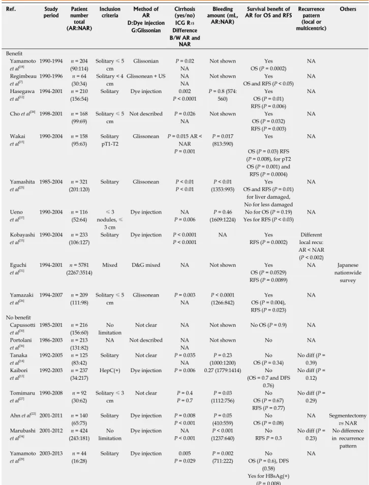
Dozens of well-designed studies have compared the benefits of AR versus NAR using the systematic segmentectomy technique or the Glissonean pedicle transection method. Most of the prior studies were published between 1990 and 2005. All of the studies were conducted retrospectively, and most were case- series, including one large nationwide survey in Japan and three case-controlled studies with propensity score matching. The results of these studies are summarized in Table 1 for case-series and Table 2 for propensity score matching studies[7,12-15,18,22,25-38].
Of 18 studies evaluated, 8 reported that AR was beneficial while 8 revealed no benefits of AR.
Meanwhile, one report described benefits of AR in demanding procedure that may be invasive, cause
bleeding and prick the tumor. In addition, it is possible to inject the dye into the wrong portal vein branch.
Aside from AR of the portal vein, some tumors span two or more segments and sections. If major hepatectomy is not indicated, a combination of the Glissonean pedicle approach and ultrasound-guided dye injection may allow the surgeon to remove the tumor with a cone- shaped resection. Therefore, the surgical method(s) can be selected based on tumor location and the surgeon’s preference. Moreover, a combination of both techniques might be helpful in complex cases.
For safe AR, it is essential that the surgeon has good understanding of the anatomical variability of the Glisson pedicle, particularly the right hepatic vein, which serves as the reference structure for dividing the right anterior and right posterior sections. Careful imaging studies are very important; in addition to intraoperative ultrasound, careful review of the relation between portal venous branches and tumor looking at MD-CT that taken before hepatectomy.
Other issues regarding AR and what is real AR?
There is another issue of importance to segmen- tectomy. For segmentectomy of Couinaud’s segments 7 or 8, it is essential to expose the adjacent hepatic vein(s): the right hepatic vein for segment 7, and the right and mid-hepatic veins for segment 8[9,12]. This is reasonable when we consider the concept of Couinaud’s classification of liver segments in patients with normal anatomy. However, in cases with sliding pedicles, for example the anterior pedicular branch has slid from the posterior ramus and posterior pedicle branch from the anterior ramus, the slid branch may bypass the hepatic venous limitation of the corresponding segment[19]. In patients with these variations, hepatic segmentectomy should include partial resection of the neighboring segment, including the relevant peripheral branch of the hepatic vein.
Some questions arise regarding how to perform AR and what technique should be used if the bran- ching pattern of the portal vein does not match the Couinaud’s segment and the tumor location is not confined to the Couinaud’s anatomical segment.
Does cone-shaped parenchymal resection of tissue fed by one or several fourth-order branches represent AR or NAR? If the sliding branches cross over another neighboring segment[19], resection of the entire cone unit of the Glissonean pedicular branch, including partial resection of the neighboring segment, can be included in AR. If one cone unit partially fills a segment, resection of the cone unit without exposing the neighboring hepatic vein (for right upper segments) corresponds to resection of less than one segment. Therefore, does this constitute NAR? Should we always expose the segment bordering the hepatic veins in anatomical segmentectomy of segments 7 or 8? Does resection of the cone unit fed by the
Table 1 Summary of studies comparing the outcomes of anatomical and non-anatomical resection in patients with hepatocellular carcinoma
Ref. Study
period Patient number total (AR:NAR)
Inclusion
criteria Method of
AR Cirrhosis
(yes/no) Bleeding amount (mL,
AR:NAR)
Survival benefit of
AR for OS and RFS Recurrence pattern (local or multicentric)
Others D:Dye injection ICG R15
G:Glissonian Difference B/W AR and
NAR Benefit
Yamamoto et al[18]
1990-1994 n = 204 (90:114)
Solitary ≤ 5 cm
Glissonian P = 0.02 Not shown Yes NA
NA OS (P = 0.0002)
Regimbeau et al[7]
1990-1996 n = 64 (30:34)
Solitary < 4 cm
Glissonean + US NA Not shown Yes NA
NA OS and RFS (P < 0.05)
Hasegawa et al[12]
1994-2001 n = 210 (156:54)
Solitary Dye injection 0.002 P = 0.8 (574:
560)
Yes NA
P < 0.0001 OS (P = 0.01)
RFS (P = 0.006) Cho et al[38] 1998-2001 n = 168
(99:69)
Solitary ≤ 5 cm
Not described P = 0.026 Not shown Yes NA
NA OS (P = 0.032)
RFS (P = 0.003) Wakai
et al[15]
1990-2004 n = 158 (95:63)
Solitary pT1-T2
Glissonean P = 0.015 AR <
NAR
P = 0.017 (813:590)
Yes NA
P = 0.001 OS (P = 0.03) RFS
(P = 0.008), for pT2 OS (P = 0.001) and RFS (P = 0.0004) Yamashita
et al[25]
1985-2004 n = 321 (201:120)
Solitary Glissonean P < 0.01 P < 0.01 (1353:993)
Yes NA
P < 0.01 OS and RFS (P = 0.01)
for liver damaged, No for less damaged Ueno
et al[37]
1990-2004 n = 116 (52:64)
≤ 3 nodules, ≤
3 cm
Dye injection NA P = 0.46
(1609:1224)
No for OS (P = 0.19) NA
P = 0.006 Yes for RFS (P < 0.03)
Kobayashi et al[33]
1990-2004 n = 233 (106:127)
Solitary Dye injection P < 0.0001 NA Yes Different
P < 0.0001 RFS (P = 0.0002) local recu:
AR < NAR (P < 0.002) Eguchi
et al[31]
1994-2001 n = 5781 (2267:3514)
Mixed D&G mixed NA Not shown Yes NA Japanese
nationwide survey OS (P = 0.0529)
RFS (P = 0.0089) Yamazaki
et al[26]
1994-2007 n = 209 (111:98)
Solitary ≤ 5 cm
Glissonean P = 0.003 P < 0.0001 (1266:842)
Yes NA
NA OS (P = 0.004),
RFS (P = 0.023) No benefit
Capussotti et al[30]
1985-2001 n = 216 (156:60)
No limitation
Not clear NA Not shown No OS (P = 0.9) NA
Portolani et al[36]
1986-2003 n = 213 (131:82)
NA Not described NA Not shown No NA
NA Tanaka
et al[14]
1992-2005 n = 125 (83:42)
Solitary Not clear P = 0.035 P = 0.23 (1000:1200)
No No diff (P =
0.39)
NA OS (P = 0.34)
Kaibori et al[13]
1992-2003 n = 237 (34:217)
HepC(+) Dye injection P = 0.006 0.27 (1779:1414) No No diff (P = 0.12) (OS = 0.7 and DFS
0.76) Tomimaru
et al[27]
1990-2008 n = 92 (30:62)
Solitary ≤ 3 cm
Not clear P = 0.4 P = 0.03 (1112:756)
No No diff (P =
0.29)
P = 0.7 OS (P = 0.67)
RFS (P = 0.77) Ahn et al[22] 2001-2011 n = 140
(65:75)
Solitary Dye injection P = 0.008 P = 0.05 (410:559)
No NA Segmentectomy
vs NAR
P < 0.001 OS (P = 0.08)
Marubashi et al[34]
2001-2012 n = 424 (243:181)
No limitation
Dye injection NA P < 0.001 (1237:640)
No No diff (P =
0.23)
No difference in recurrence
pattern
P < 0.001 RFS P = 0.3
Yamamoto et al[29]
2003-2013 n = 44 (16:28)
Solitary Dye injection 0.005 P = 0.002 No NA
P = 0.029 (711:222) OS (P = 0.6), DFS (0.58) Yes for HBsAg(+)
(P = 0.008)
AR: Anatomical resection; NAR: Non-anatomical resection; D: Dye injection method; G: Glissonean pedicle method; ICG R15: Indocyanine green retention rate at 15 min; B/W: Between; OS: Overall survival; RFS: Recurrence-free survival; NA: Not applicable; US: Ultrasound; DFS: Disease-free survival; diff:
Difference; HBsAg: Hepatitis B surface antigen.
patients with non-cirrhotic HCC but not in patients with cirrhotic HCC[25]. The nationwide survey in Japan revealed that AR was only beneficial in HCC patients whose tumor size within the range of 2 to 5 cm. All three case-controlled studies using the propensity score matching method revealed no benefit of AR.
Critical review of prior studies
The prognosis of HCC is affected by the degree of cirrhosis and tumor stage. It is difficult to assess the degree of cirrhosis and tumor stage, and these classifications have changed over time. Therefore, survival analysis is difficult when considering the degree of cirrhosis and tumor stage. The type of surgical resection (i.e., AR or NAR) may not affect the survival outcomes in cirrhotic patients. Furthermore, it is very difficult to determine whether disease recurrences are due to local recurrence, intrahepatic metastasis, or de novo multicentric recurrence. To determine the impact of AR, it is important to assess the pattern of recurrence, and local recurrence may be influenced by the surgical method. Therefore, in order to determine whether a specific surgical resection method has advantages on patient outcomes over another method, the endpoint should be recurrence- free survival rather than overall survival. However, many of the studies analyzed overall survival. It is also important to consider that there is a lot of bias, especially selection bias, in studies examining the benefits of AR.
First, the background liver functions of patients in both groups were significantly different in most of the studies shown in Table 1. Patients who underwent AR had better liver function in terms of cirrhotic status and/or the indocyanine green retention rate at 15 min, which closely reflects the degree of cirrhosis.
Five studies assessed whether the recurrence pattern was local recurrence, intrahepatic metastasis, or multicentric recurrence. Only one study showed a significantly higher local recurrence rate in NAR than in AR; the other four studies did not find any differences in the recurrence pattern between the two groups[13,14,27,33,34]. If the background liver function
was better in the AR group and the local recurrence rate was similar in the AR and NAR groups, how do we know that the survival benefit of AR was due to superiority of this resection rather than an effect of cirrhosis?
Second, all of the studies were conducted retro- spectively, and the tumor size and T stage were not standardized between AR and NAR in most of the studies, although many studies were limited to patients with a solitary mass of less than 3-5 cm.
In the nationwide survey in Japan, which comprised 5781 patients with a single HCC lesion, the overall and disease-free survival rates were significantly better for AR than for NAR. When the patients were stratified according to tumor size (< 2 cm, 2-5 cm, or > 5 cm), the disease-free survival rate was better in patients who underwent AR, but only in those with a tumor size of 2-5 cm. There was no benefit of AR in patients with tumors of < 2 cm or > 5 cm[31]. It seems reasonable that, in patients with small tumors (i.e., < 2 cm), any type of surgery, even ablative therapy, is associated with favorable survival. By contrast, for larger tumors (i.e., > 5 cm), survival is more likely to be affected by advanced tumor stage rather than the resection method. Therefore, the importance of AR should be emphasized in patients with a tumor of 2-5 cm in size.
Most of the studies did not consider other factors likely to influence the short-term and long-term outcomes.
Finally, many other factors, including an anterior approach and perioperative transfusion, can influence the long-term survival after liver resection. During mobilization of the liver, the surgeon’s or assistant’
s left hand may compress the liver, including the tumor. The degree of compression may affect intrahepatic metastasis differently, and its impact may also differ according to tumor location. This problem can be overcome by using an anterior approach or the hanging maneuver for standard right hepatectomy[39-41]. Unfortunately, this factor was not mentioned in most studies. Regarding transfusion, although the effects of perioperative transfusion on survival after hepatic resection for HCC are controversial, a meta-analysis of 22 studies revealed Table 2 Summary of case-control studies using the propensity score matching method to compare the outcomes of anatomical and non-anatomical resection for hepatocellular carcinoma
Ref. Study
period Patient total number and after propensity score
matched (AR:NAR)
Inclusion
criteria Method of AR ICG R15 Bleeding amount Survival benefit of AR D:Dye injection Difference (mL, AR:NAR)
G:Glissonian B/W AR and NAR Okamura et al[35],
2014
2002-2013 n = 236 (139:97 and 64:64)
Solitary Dye injection P = 0.07 P = 0.008 (551:465)
No RFS (P = 0.52) Ishii et al[32], 2014 2002-2010 n = 268 (110:158 and
44:44)
Solitary ≤ 5 cm
Not Clear P = 0.053 0.9 (400:355) No
OS (P = 0.29) RFS (P = 0.28) Marubashi et al[28],
2015
1981-2012 n = 1102 (577:525 and 329:329)
No limitation
Not clear NA Not shown No
OS (P = 0.7) and RFS (P = 0.4) AR: Anatomical resection; NAR: Non-anatomical resection; D: Dye injection method; G: Glissonean pedicle method; ICG R15: Indocyanine green retention rate at 15 min; B/W: Between; OS: Overall survival; RFS: Recurrence-free survival; NA: Not applicable.
that perioperative blood transfusion was associated with adverse clinical outcomes, including increased mortality, recurrence, and complication rates, but opposite findings were reported in other articles[42,43]. Extensive blood loss in cirrhotic patients, who were more likely to undergo NAR, might be associated with poor prognosis and this factor could represent a bias towards poor prognosis of NAR. A technique comprising low central venous pressure (LCVP) management combined with extrahepatic control of venous outflow enables the surgeon to easily control the hepatic veins before and during parenchymal transection. This LCVP technique combined with the Pringle maneuver reduced bleeding and blood transfusion, and improved the surgical outcomes. The hanging maneuver and LCVP approach was introduced in the late 1990s and its use widened in the early 2000s, a similar period of time over which most of the AR studies were conducted. As indicated in Table 1, although the amount of bleeding was not recorded in all of the studies, the available data are fairly high in the amount of bleeding. This may be possible because the study was performed under development of new surgical and anesthetic methods. Therefore, the changes over time in the surgical techniques, such as use of the hanging maneuver, and management by an anesthesiologist using a low CVP technique, may affect the recurrence rate after hepatectomy.
CONCLUSION
AR in patients with HCC has a theoretical benefit in terms of improving recurrence-free survival, and this is partly observed in clinical practice. However, three recently published, well-designed, case-controlled studies using the propensity score matching method did not show an improvement in recurrence-free survival following AR. Studies examining the benefits of AR displayed considerable bias, including liver function, surgical techniques, anatomical variability, tumor size, tumor location, pathologic heterogeneity and chronology. Because prospective randomized studies are not possible for ethical reasons, it is difficult to reach a conclusion on the benefit of AR in HCC. However, the results of previous studies suggest that AR is associated with favorable perioperative and long-term outcomes in some conditions, including in patients with a tumor of 2-5 cm in size that is located in a deep region of the parenchyma.
REFERENCES
1 Petrick JL, Braunlin M, Laversanne M, Valery PC, Bray F, McGlynn KA. International trends in liver cancer incidence, overall and by histologic subtype, 1978-2007. Int J Cancer 2016;
139: 1534-1545 [PMID: 27244487 DOI: 10.1002/ijc.30211]
2 Forner A, Llovet JM, Bruix J. Hepatocellular carcinoma.
Lancet 2012; 379: 1245-1255 [PMID: 22353262 DOI: 10.1016/
s0140-6736(11)61347-0]
3 Belghiti J, Kianmanesh R. Surgical treatment of hepatocellular
carcinoma. HPB 2005; 7: 42-49
4 Clavien PA, Selzner M, Rüdiger HA, Graf R, Kadry Z, Rousson V, Jochum W. A prospective randomized study in 100 consecutive patients undergoing major liver resection with versus without ischemic preconditioning. Ann Surg 2003; 238:
843-850; discussion 851-852 [PMID: 14631221 DOI: 10.1097/01.
sla.0000098620.27623.7d]
5 Rüdiger HA, Kang KJ, Sindram D, Riehle HM, Clavien PA.
Comparison of ischemic preconditioning and intermittent and continuous inflow occlusion in the murine liver. Ann Surg 2002;
235: 400-407 [PMID: 11882762]
6 Yadav SS, Sindram D, Perry DK, Clavien PA. Ischemic pre- conditioning protects the mouse liver by inhibition of apoptosis through a caspase-dependent pathway. Hepatology 1999; 30:
1223-1231 [PMID: 10534344 DOI: 10.1002/hep.510300513]
7 Regimbeau JM, Kianmanesh R, Farges O, Dondero F, Sauvanet A, Belghiti J. Extent of liver resection influences the outcome in patients with cirrhosis and small hepatocellular carcinoma. Surgery 2002; 131: 311-317 [PMID: 11894036]
8 Makuuchi M, Hasegawa H, Yamazaki S. Ultrasonically guided subsegmentectomy. Surg Gynecol Obstet 1985; 161: 346-350 [PMID: 2996162]
9 Makuuchi M. Surgical treatment for HCC--special reference to anatomical resection. Int J Surg 2013; 11 Suppl 1: S47-S49 [PMID:
24380552 DOI: 10.1016/s1743-919160015-1]
10 Makuuchi M, Mori T, Gunvén P, Yamazaki S, Hasegawa H.
Safety of hemihepatic vascular occlusion during resection of the liver. Surg Gynecol Obstet 1987; 164: 155-158 [PMID: 3810429]
11 Takasaki K. Glissonean pedicle transection method for hepatic resection: a new concept of liver segmentation. J Hepatobiliary Pancreat Surg 1998; 5: 286-291 [PMID: 9880776]
12 Hasegawa K, Kokudo N, Imamura H, Matsuyama Y, Aoki T, Minagawa M, Sano K, Sugawara Y, Takayama T, Makuuchi M. Prognostic impact of anatomic resection for hepatocellular carcinoma. Ann Surg 2005; 242: 252-259 [PMID: 16041216]
13 Kaibori M, Matsui Y, Hijikawa T, Uchida Y, Kwon AH, Kamiyama Y. Comparison of limited and anatomic hepatic resection for hepatocellular carcinoma with hepatitis C. Surgery 2006; 139:
385-394 [PMID: 16546504 DOI: 10.1016/j.surg.2005.08.035]
14 Tanaka K, Shimada H, Matsumoto C, Matsuo K, Nagano Y, Endo I, Togo S. Anatomic versus limited nonanatomic resection for solitary hepatocellular carcinoma. Surgery 2008; 143: 607-615 [PMID:
18436008 DOI: 10.1016/j.surg.2008.01.006]
15 Wakai T, Shirai Y, Sakata J, Kaneko K, Cruz PV, Akazawa K, Hatakeyama K. Anatomic resection independently improves long- term survival in patients with T1-T2 hepatocellular carcinoma. Ann Surg Oncol 2007; 14: 1356-1365 [PMID: 17252289 DOI: 10.1245/
s10434-006-9318-z]
16 Tajima T, Honda H, Taguchi K, Asayama Y, Kuroiwa T, Yoshimitsu K, Irie H, Aibe H, Shimada M, Masuda K. Sequential hemodynamic change in hepatocellular carcinoma and dysplastic nodules: CT angiography and pathologic correlation. AJR Am J Roentgenol 2002; 178: 885-897 [PMID: 11906868 DOI: 10.2214/
ajr.178.4.1780885]
17 IHPBA TCot. Terminology of liver anatomy and resections. HPB Surg 2000; 2: 333-339
18 Yamamoto M, Takasaki K, Ohtsubo T, Katsuragawa H, Fukuda C, Katagiri S. Effectiveness of systematized hepatectomy with Glisson’s pedicle transection at the hepatic hilus for small nodular hepatocellular carcinoma: retrospective analysis. Surgery 2001;
130: 443-448 [PMID: 11562668 DOI: 10.1067/msy.2001.116406]
19 Xu W, Wang HJ, Kim BW, Park YK, Li G. Anatomical Variation of the Glissonean Pedicle of the Right Liver. Korean J Hepatobiliary Pancreat Surg 2011; 15: 101-106 [PMID: 26421024 DOI: 10.14701/kjhbps.2011.15.2.101]
20 Cho A, Okazumi S, Takayama W, Takeda A, Iwasaki K, Sasagawa S, Natsume T, Kono T, Kondo S, Ochiai T, Ryu M. Anatomy of the right anterosuperior area (segment 8) of the liver: evaluation with helical CT during arterial portography. Radiology 2000; 214:
491-495 [PMID: 10671598 DOI: 10.1148/radiology.214.2.r00fe38
21 Kogure K, Kuwano H, Fujimaki N, Ishikawa H, Takada K. 491]
Reproposal for Hjortsjo’s segmental anatomy on the anterior segment in human liver. Arch Surg 2002; 137: 1118-1124 [PMID:
12361415]
22 Ahn KS, Kang KJ, Park TJ, Kim YH, Lim TJ, Kwon JH. Benefit of systematic segmentectomy of the hepatocellular carcinoma:
revisiting the dye injection method for various portal vein branches. Ann Surg 2013; 258: 1014-1021 [PMID: 23478518 DOI:
10.1097/SLA.0b013e318281eda3]
23 Makuuchi M, Imamura H, Sugawara Y, Takayama T. Progress in surgical treatment of hepatocellular carcinoma. Oncology 2002; 62 Suppl 1: 74-81 [PMID: 11868790]
24 Liau KH, Blumgart LH, DeMatteo RP. Segment-oriented approach to liver resection. Surg Clin North Am 2004; 84: 543-561 [PMID:
15062661 DOI: 10.1016/j.suc.2003.12.003]
25 Yamashita Y, Taketomi A, Itoh S, Kitagawa D, Kayashima H, Harimoto N, Tsujita E, Kuroda Y, Maehara Y. Longterm favorable results of limited hepatic resections for patients with hepatocellular carcinoma: 20 years of experience. J Am Coll Surg 2007; 205:
19-26 [PMID: 17617328 DOI: 10.1016/j.jamcollsurg.2007.01.069]
26 Yamazaki O, Matsuyama M, Horii K, Kanazawa A, Shimizu S, Uenishi T, Ogawa M, Tamamori Y, Kawai S, Nakazawa K, Otani H, Murase J, Mikami S, Higaki I, Arimoto Y, Hanba H. Comparison of the outcomes between anatomical resection and limited resection for single hepatocellular carcinomas no larger than 5 cm in diameter: a single-center study. J Hepatobiliary Pancreat Sci 2010;
17: 349-358 [PMID: 20464566 DOI: 10.1007/s00534-009-0253-9]
27 Tomimaru Y, Eguchi H, Marubashi S, Wada H, Kobayashi S, Tanemura M, Umeshita K, Doki Y, Mori M, Nagano H. Equivalent outcomes after anatomical and non-anatomical resection of small hepatocellular carcinoma in patients with preserved liver function.
Dig Dis Sci 2012; 57: 1942-1948 [PMID: 22407377 DOI: 10.1007/
s10620-012-2114-7]
28 Marubashi S, Gotoh K, Akita H, Takahashi H, Ito Y, Yano M, Ishikawa O, Sakon M. Anatomical versus non-anatomical resection for hepatocellular carcinoma. Br J Surg 2015; 102: 776-784 [PMID:
25847111 DOI: 10.1002/bjs.9815]
29 Yamamoto T, Yagi S, Kita R, Masui H, Kinoshita H, Sakamoto Y, Okada K, Miki A, Kondo M, Hashida H, Kobayashi H, Uryuhara K, Kaihara S, Hosotani R. Comparison between anatomical subsegmentectomy and nonanatomical partial resection for hepatocellular carcinoma located within a single subsegment: a single-center retrospective analysis. Hepatogastroenterology 2015;
62: 363-367 [PMID: 25916064]
30 Capussotti L, Muratore A, Amisano M, Polastri R, Bouzari H, Massucco P. Liver resection for hepatocellular carcinoma on cirrhosis: analysis of mortality, morbidity and survival--a European single center experience. Eur J Surg Oncol 2005; 31: 986-993 [PMID: 15936169 DOI: 10.1016/j.ejso.2005.04.002]
31 Eguchi S, Kanematsu T, Arii S, Okazaki M, Okita K, Omata M, Ikai I, Kudo M, Kojiro M, Makuuchi M, Monden M, Matsuyama Y, Nakanuma Y, Takayasu K. Comparison of the outcomes between an anatomical subsegmentectomy and a non-anatomical minor hepatectomy for single hepatocellular carcinomas based on a Japanese nationwide survey. Surgery 2008; 143: 469-475 [PMID:
18374043 DOI: 10.1016/j.surg.2007.12.003]
32 Ishii M, Mizuguchi T, Kawamoto M, Meguro M, Ota S, Nishidate T, Okita K, Kimura Y, Hui TT, Hirata K. Propensity score analysis demonstrated the prognostic advantage of anatomical liver resection in hepatocellular carcinoma. World J Gastroenterol 2014;
20: 3335-3342 [PMID: 24696614 DOI: 10.3748/wjg.v20.i12.3335]
33 Kobayashi A, Miyagawa S, Miwa S, Nakata T. Prognostic impact of anatomical resection on early and late intrahepatic recurrence in patients with hepatocellular carcinoma. J Hepatobiliary Pancreat Surg 2008; 15: 515-521 [PMID: 18836806 DOI: 10.1007/
s00534-007-1293-7]
34 Marubashi S, Gotoh K, Akita H, Takahashi H, Sugimura K, Miyoshi N, Motoori M, Kishi K, Noura S, Fujiwara Y, Ohue M, Nakazawa T, Nakanishi K, Ito Y, Yano M, Ishikawa O, Sakon M. Analysis of Recurrence Patterns After Anatomical or Non- anatomical Resection for Hepatocellular Carcinoma. Ann Surg Oncol 2015; 22: 2243-2252 [PMID: 25373536 DOI: 10.1245/
s10434-014-4214-4]
35 Okamura Y, Ito T, Sugiura T, Mori K, Uesaka K. Anatomic versus nonanatomic hepatectomy for a solitary hepatocellular carcinoma: a case-controlled study with propensity score matching.
J Gastrointest Surg 2014; 18: 1994-2002 [PMID: 25213582 DOI:
10.1007/s11605-014-2646-6]
36 Portolani N, Coniglio A, Ghidoni S, Giovanelli M, Benetti A, Tiberio GA, Giulini SM. Early and late recurrence after liver resection for hepatocellular carcinoma: prognostic and therapeutic implications. Ann Surg 2006; 243: 229-235 [PMID: 16432356 DOI: 10.1097/01.sla.0000197706.21803.a1]
37 Ueno S, Kubo F, Sakoda M, Hiwatashi K, Tateno T, Mataki Y, Maemura K, Shinchi H, Natsugoe S, Aikou T. Efficacy of anatomic resection vs nonanatomic resection for small nodular hepatocellular carcinoma based on gross classification. J Hepatobiliary Pancreat Surg 2008; 15: 493-500 [PMID: 18836803 DOI: 10.1007/
s00534-007-1312-8]
38 Cho YB, Lee KU, Lee HW, Cho EH, Yang SH, Cho JY, Yi NJ, Suh KS. Anatomic versus non-anatomic resection for small single hepatocellular carcinomas. Hepatogastroenterology 2007; 54:
1766-1769 [PMID: 18019714]
39 Lai EC, Fan ST, Lo CM, Chu KM, Liu CL. Anterior approach for difficult major right hepatectomy. World J Surg 1996; 20: 314-317;
discussion 318 [PMID: 8661837]
40 Liu CL, Fan ST, Lo CM, Tung-Ping Poon R, Wong J. Anterior approach for major right hepatic resection for large hepatocellular carcinoma. Ann Surg 2000; 232: 25-31 [PMID: 10862191]
41 Belghiti J, Guevara OA, Noun R, Saldinger PF, Kianmanesh R.
Liver hanging maneuver: a safe approach to right hepatectomy without liver mobilization. J Am Coll Surg 2001; 193: 109-111 [PMID: 11442247]
42 Yang T, Lu JH, Lau WY, Zhang TY, Zhang H, Shen YN, Alshebeeb K, Wu MC, Schwartz M, Shen F. Perioperative blood transfusion does not influence recurrence-free and overall survivals after curative resection for hepatocellular carcinoma: A Propensity Score Matching Analysis. J Hepatol 2016; 64: 583-593 [PMID:
26596543 DOI: 10.1016/j.jhep.2015.10.012]
43 Wei AC, Tung-Ping Poon R, Fan ST, Wong J. Risk factors for perioperative morbidity and mortality after extended hepatectomy for hepatocellular carcinoma. Br J Surg 2003; 90: 33-41 [PMID:
12520572 DOI: 10.1002/bjs.4018]
P- Reviewer: Lam V, Perini MV, Santambrogio R S- Editor: Gong ZM L- Editor: O’Neill M E- Editor: Wang CH
8226 Regency Drive, Pleasanton, CA 94588, USA Telephone: +1-925-223-8242
Fax: +1-925-223-8243 E-mail: [email protected]
Help Desk: http://www.wjgnet.com/esps/helpdesk.aspx http://www.wjgnet.com
I S S N 1 0 0 7 - 9 3 2 7
9 7 7 1 0 07 9 3 2 0 45 0 7