고추 부위별추출물에 의한 종양세포의 세포사유도 - Hepatoma 세포와 MCF-7 세포 -
정 용 자 경성대학교 약학과
(Received March 19, 2003; Revised April 14,2003)
Induction of Cancer Cell Apoptosis by the Extract of Capsicum annuum L. var.
angulosum Mill Sorted According to the Parts in Hepatoma Cells and MCF-7 Cells
Yong-Za Chung
College of Pharmacy, Kyungsung University, 110-1 Daeyeon-dong, Nam-gu, Pusan 608-736,Korea
Abstract — Under the active search for biologically active novel agents for cancer prevention and treatment, some agents have been found from plants which are easily available. Our previous research on them revealed that C. annuum L. var.
angulosum M ill have high antiproliferating effect on cancer cells. However, it has not been known whether the anticancer efficacy is different according to each part of C. annuum L. var. angulosum M ill or whether it can be changed by timing of harvest or solvent for extraction. Thus we compared the efficacy of each part of C. annuum L. var. angulosum M ill and assessed how much difference in the efficacy can be made according to the time of harvest or solvents for extraction. We observed the morphologic change and apoptosis 48 hr after treatment with the extract of each part of C. annuum L. var.
angulosum M ill in MCF-7 mammary gland adenocarcinoma cells and human hepatoma cells. We also counted cancer cells by trypan blue method and MTT method to check the cytotoxicity. The leaf extract showed the highest anticancer effect among all the parts of C. annuum L. var. angulosum M ill; 50% and 70% reduction in the number of cancer cells was observed at 25 [ig/ml and 50 [Lg/ml, respectively. It was more than 2 times as potent as 5-fluorouracil (5-FU). We found chro
mosomal fragmentation, clumping, and destuction by PI staining, and DNA fragmentation by electrophoresis. In conclusion, this study suggests that leaf extraction using water as solvent has the highest antiproliferative and apoptotic activity in can
cer cells compared with other parts of extraction.
Keywords □ Capsicum annuum, morphology, apoptosis
천연물에는항종양효과등여러 생리활성물질이 포함되어있 다. ^ 이에우리들식품중식탁에흔히오르는수종의식물을 대상으로항종양효과를관찰해본결과거특히고추의 항종양효 과가뚜렷하였 0-口■로고추의부위별로항종양효과를관찰해보 았다. 고추를추출용매,계절과, 부위 별; 잎, 청고추(열매),홍고 추(열매), 청고추씨홍고추씨로나누고, 그부위, 계절과용매에 따른간암세포와유방범■세포에서의함종양효과를관찰하기위하 여그증식억제효과를중심으로항암효과를관찰하였다.
세포는세포독성유발물질이나여러인자들의외부자극에의하
#본논문에 관한문의는저자에게로
(전화) 051-620-4887 (팩스) 051-628-6540 (E-mail) [email protected]
여세포사멸이유도되며, 세포사과정에서 세포의 형태변화및축 소또는파괴가유도되고, 핵의농축과변화, 그리고파괴등이 관찰된다. 이에실험방법으로세포성장을관찰하는데는 역상현 미경을이용한세포착상및증식형태관찰, MTT법과 trypan blue 염색에 의한세포수즉정,colcemid를이용한염색체의 banded 모형와전기영동법에의한 DNA 분절관찰,그리고형광현미경하 에서 핵변화를 propodium iodide(PI) 염색등으로관잘하였다. 고추의부위별추출물이 간암세포와유방암세포의 중식에 미치 는영향을관찰한결과모든부위에서증식억제효과가분명히나 타났다. 그중간암세포에서는청고추추출물에서 증식억제작용 이낮게나타났고, 유방암세포에서는홍고추추출물이 비교적 억 제작용이 낮게나타났다. 그들시료중고추잎의 수추출물이 가 장큰증식억제작용을보여주었다.
57
Capsicum annuum L. var. angulosum Mill Leaf Lactuca dentata Makino. var. flaviflora Makino Aerial Part
Capsicum annuum L.var. angulosum Mill Fruit, unripen
Capsicum annuum L.var. angulosum Mill Seed, unripen
Capsicum annuum L.var. angulosum Mill Fruit, ripen
Capsicum annuum L.var. angulosum Mill Seed, ripen
세포배양
Human hepatoma 세포와유방암세포인 MCF-7 세포를실험 대상세포로사용하였으며, 배양조건은 RPM I 1640 배지에 10%
FBS(Gibco사)를 첨가하고 1001.U.penicilin G - 100|ag/m/
strep- tomycin과 0.075% NaHC03를첨가하였다.5)
생존세포수측정
배양한 Hepatoma세포와MCF-7 세포를 0.4% trypan blue를 섞은 다음2분 투 hematocytometer로 광학현미경하에서 세포
수를 측정하였다. 다음의 세포성장및 억제를 관찰하기 위해사
용된 세포수는이방법으로 셈하였다. 이 trypan blue에염색되 지 않은세포수는살아있는 세포 수이다.6)
MTT법에의한세포성장억제도측정
각시료을 용량별로 MCF-7 세포와 hepatoma 세포(4X10³개) 에 첨가하여 37°C 5% C 0 2 배양기 내에서 48시간 배양한 뒤,
MTT 정량법으로 생존세포에 따른흡광도를측정했다. 홉광도의
감소는 살아있는 세포수의 감소,즉 암세포의 성장억제 효과를
나타낸다.7) 96 well plate를사용하여, well당배지총량을 200 |a/
로 하며 배양했다.
측정값은 표준편차 범위 밖의 최소 최대 값은 제외하고,
6회이상측정값을평균하여 얻었으며, 대조군과비교하며 표시
하였다.
실험방법
시료추출및선택
시료는 계절별로 봄과 가을에 채취한 것을 선택하고, 에칠
알콜과 물로 추출하여 용매에 따른 결과를 얻었으며, 고추의
가식부위를 부위별로 분류하여 실험에 용했다. 가식부위는
잎, 청고추,흥고추, 청고추씨 홍고추씨, 별로 나누어 사용하 였고, 5FU와 씀바귀를 비교하여 실험하였다. 각 시료는 먼저 1차 증류수로 수회세척하고, clean bench내에서 멸균한 3차
증류수로 2회 세척하여, 추출용매로는 알콜과 증류수를 각각
사용하여 주 출 했 다. 주출액은 0.45 |_im cellulose acetate membrane filter로 여과시켜 시료로 사용하였다. 시료 1 g당
수 추출물은 15 mg이었다. 실험에 사용된 부위의 명칭은 다
음과 같다.
No. Used sample (Scientific name) Part of use
형태학적관찰
MCF-7 세포와 hepatoma 세포를 각well 당4X10³개씩 접종
하여, 24시간배양으로 세포를 안정화시킨 후각 시료추출물을
농도별로 첨가하여 대조군과실험군의 형태적 변화를 100배율의
역상현미경에서 관찰하였다.
DNA 분절확인 실험
MCF-7 세포를2X10⁵개/m/씩되게 배양병(culture bottle)에서
배양24시간후에 배지를조심스럽게 제거하고, 준비된 각각의
시료를 flask에50ng/m/씩 넣었다.8> 9일후까지 여기에 신선한
배지를 48시간마다 교환해주면서, 그 상등액(세포 파쇄분)은
260nm에서흡광도를 측정하여 비교하였다.9)
또한 DNA 분절분석을 위해, 상기 암세포를 배양병에 이식하
여24시간배양하고안정화한후신선배지와 각추출시료 50 W m/씩첨가하여 24시간, 36시간,또는 48시간배양후 배지를 제 거하고, 그각각을 0.05% trypsin-EDTA로처리한후 부유상태 의세포를취하여 lysis buffer를첨가하고 1시간배양기에 방치 한다. 이것을 DNA 분리침전법10〉에 따라분리하여 침전물을약 10& 정도감압건조히여진여에타놀을제거하고, 1 ng/m/ RNase A로처리하여 1시간 배양한다. 이렇게 만든시료에 bromphenol blue tracking dye를 첨가하고, 이것을 TBE buffer속 1.2%
agarose gel에서 전기영동한다. agarose gel에는 ethidium bromide를첨가하여 apoptosis에의한 DNA fragmentation를비 교관찰하였다.u)
염색체의 banded 모형변화
Hepatoma 세포와 MCF-7 세포를 24시간 배양하여 배지를조
심스럽게 제거하고,준비된각 시료를 씩넣었다.12) 여
기에 신선한배지를 첨가하고,24시간, 40시간,또는 48시간뒤 에colcemid를추가한후 5시간배잉하고배지를제거하였다. 여 기에 0.25% trypsin을 첨가하고 10분간 배양 후 원심분리하여 상등액을 제거하고, 저장액(0.04 M KC1, 0.025 M sodium citrate)에세포를 재현탁시켜, 36.50C에서 15분간방치시켰다. 그 뒤신선한ice-cold 초산■메탄올로 세포를고정시키고, ice에 10 분간 방치한뒤 원심분리하여 초산 ■메탄올을제거하고, 신선한
초산 • 메탄올에 세포를재현탁하여,세포 현탁액을한방울 덜
어내어 cold slide에놓고 고정시켰다.6) 그리고 Giemsa로염색
하여 1000X에서 염색체변화양상을 관찰하였다.
PI 염색후의관찰
상기와동일방법으로 배양된세포를 trypsin 처리하여 세포를
취하고 70% ethanol을첨가하며, 40C에서 60분고정한다. PBS 로 재현탁하여, RNase(250 ng/m/)를 첨가하고, PI, 25|ag/m/로
염색하여 250C에서 15분간 반응시켜 형광현미경하에서 관찰하
①②
③
④⑤
⑥
/. Pharm. Soc. Korea
0 . 4 5
0.4
0 . 3 5
0 . 3
0 . 2 5
0.2
0 . 1 5
0 .7
0.6 0 .5
^ 0 .4
< 0 .3 0.2
0.1
0.1
0 . 0 5
1 0O ug 2 5 ᄋ u g 5 0 0 u g
s a m p le
7 5 ᄋ니 g 1 m g / m l
Fig. 2 - Antiproliferative effects of ripen fruit extract of C. annuum using water as solvent on hepatoma cells. The samples were harvested on spring and autumn, respectively. The cells were cultured at several different concentrations of the extracts. The absorbance was measured by MTT assay.
10Ouo 2 5 0ug 5 0 0ug 7 5 0 u g 1 mg/ml Amount of sample
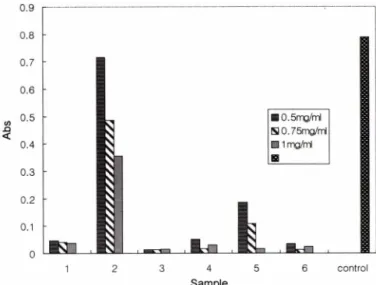
Fig. 3b - Antiproliferative effects of C. annuum extract of each part using water as solvent on MCF-7 cells. The cells were cultured at the various concentrations of the extracts for 48 hr. The absorbance was measured by MTT assay.
1. C. annuum L. var. angulosum M ill, Leaf; 2. Lactuca dentata Makino. var. flaviflora Makino; 3. C. annuum L.
var. angulosum M ill, fruit unripen; 4. C. annuum L. var.
angulosum M ill, fruit ripen.
■ control 國 0.5 m g/m l
□ 0.75m g/m l
■ 1m g/m l
0.6
0.5
0.4
0.3
0.2
sample
Fig. 1 - Antiproliferative effects of C. annuum extract of each part using alcohol as solvent on hepatoma cells. The cells were cultured at several different concentrations of the extracts for 48 hr. The absorbance was measured by MTT assay.
1. Extract on 99% alcohol of C. annuum L. var. angulosum M ill, Leaf; 2. Extract on 75% alcohol of C. annuum L. var.
angulosum M ill, Leaf; 3. Extract on 99% alcohol of C.
annuum L. var. angulosum Mill,fruit unripen; 4. Extract on 75% alcohol of C. annuum L. var. angulosum M ill, fruit unripen; 5. Extract on 99% alcohol of C. annuum L. var.
angulosum M ill, fruit ripen; 6. Extract on 75% alcohol of C.
annuum L. var. angulosum M ill,fruit ripen.
o
1 2 3 4 5 6 control
Sample
Fig. 3a ᅳ Antiproliferative effects of C. annuum extract of each part using water as solvent on MCF-7 cells. The cells were cultured at several different concentrations of the extracts for 48 hr. The absorbance was measured by MTT assay.
1. C. annuum L. var. angulosum M ill, Leaf; 2. Lactuca dentata Makino. var. flaviflora Makino; 3. C. annuum L.
var. angulosum M ill, fruit unripen; 4. C. annuum L. var.
angulosum M ill,seed unripen; 5. C. annuum L. var.
angulosum M ill, fruit ripen; 6. C. annuum L. var.
angulosum M ill, seed ripen.
였다.13)
실험결과 및 고찰
시료의에타놀추출물과시료채취계절에 따른항암효고®찰
MCF-7세포와 hepatoma 세포(4X10³개)에각시료를용량별 로첨가하여 37°C 5% C 02 배양기 내에서 48시간 배양한 뒤 MTT 정량법으로그흡광도를측정했다. MTT 정량법은 MTT 가살아있는세포의 mitochondrial dehydrogenase에의하여 환 원되어청색침전물을형성하는원리를이용한분석방법으로생 존세포수에비례하여 540 nm에서의흡광도가상승된다.14) 각시
료에따른암세포성장억제정도를비교하여 상대적 항암효과를 관찰하였다. 시료에대한추출용매인에타놀농도와수확기에따 른계절별항암효과는 Fig. 1과 2와같다. 봄과가을에취한시 료에서의 실험결과, 가을에채취한시료에서증식억제작^ ^ 약 간높게 관찰되었으나, 채취시기에따른작용정도차이로결론지 을수는없었다. 왜냐하면동시기의다른시료에서 얻은결과차 이가더크게 관찰되었다. 따라서 계절에 따른 항암효과차이
0.9
0.8
0.7
[j"|,
c f , [111 era. [우오[ h i.o.
o. o. o.
o.
o.o.
sq<
Vol. 47, No. 2,2003
A m o u n t o f sa m p le
Fig. 3c - Antiproliferative effects of C. annuum extract of each part using water as solvent on MCF-7 cells. The cells were cultured at the various concentrations of the extracts for 48 hr. The absorbance was measured by MTT assay.
Results were expressed as % of control.
1. C. annuum L. var. angulosum M ill, Leaf; 2. Lactnca dentata Makino. var. flaviflora Makino; 3. C. annuum L.
var. angulosum M ill,fruit unripen; 4. C. annuum L. var.
angulosum M ill, fruit ripen.
0.8
0.6
sq< 0.4
0.2
□ 0.5mg/ni
□ 0.75mg/ml D lm g/m l E9 control E35-Fu 1ug/mL
o [Si llh l31 i효i
1 2 3 4 5 6 cont 5fu
S a m p le
Fig. 4a - Antiproliferative effects of C. annuum extract of each part using water as solvent on hepatoma cells. The cells were cultured at several different concentration of the extracts for 48 hr. The absorbance was measured by MTT assay.
1. C. annuum L. var. angulosum M ill, Leaf; 2. Lactuca dentata Makino. var. flaviflora Makino; 3. C. annuum L.
var. angulosum M ill,fruit unripen; 4. C. annuum L. var.
angulosum M ill, seed unripen; 5. C. annuum L. var.
angulosum M ill, fruit ripen; 6. C. annuum L. var.
angulosum M ill, seed ripen.
로단정하기보다는시료의 종류에따른차이로간주되었다. 그 래서다음의 실험상의결과는동일시료추출물에대한비교실험 으로 결과를 얻었다. 추출용매는 99% 에타놀로추출한경우 에는항암효과가절반이하로감소되고불안정한결과를보였으 며, 75% 에타놀추출물에서는수추출물에거의비슷한항암효 과를관찰할수있었으나,수추출물에서보다비교적낮은억제 작용을보였으므로다음의실험결과는수추출물에대하여 행하 였다.
수추출물의암세포증식억제효과
시료의에타놀추출물을첨가한경우에는수추출물의경우보
다암세포증식효과가낮고불안정하며, 수추출물을대상시료로 선택하여실험하였다. 실험에시용한시료는 1 mg/m/에서 10 (ig/
m/까지의농도로세포배양배지에 처리하고, 48시간배양하여 MTT법으로측정한결과를얻었다. Trypan blue 검사법에서도 유의성있는결과를얻었다. Fig. 3a, 4a와같이고추의모든부위 별추출물을시료로각각첨가했을때, 살아있는세포수의현저 한감소를보였다. 고추잎추출물을첨가한경우유방암세포와 간암세포주모두에서강한성장억제작용을보였다. 홍고추는유 방암세포에서성장억제작용이 비교적 낮게나타났다. 암세포증 식억제작용의한도를측정하기위해용량별lm g/m /에세Ong/m/
까지 세분하여행한실험결과는 Fig. 3b, 3c와같으며, 실험 결 과고추잎추출물은 lOng/m /에서 35.3%의증식억제효과를보 였고(Fig. 3c), 25ng/m/에서는 42.9%의중식억제효과를보였으
Fig. 4b - Antiproliferative effects of C. annuum extract of each part using water as solvent on hepatoma cells. The cells were cultured at the concentration of 0.5 mg/m/ of the extracts.
By various time intervals the cell culture was done, then the absorbance measured by MTT assay.
1. C annuum L. var. angulosum M ill, Leaf; 2. Lactuca dentata Makino. var. flaviflora Makino; 3. C. annuum L.
var. angulosum M ill,fruit unripen; 4. C. annuum L. var.
angulosum M ill, seed unripen; 5. C. annuum L. var.
angulosum M ill, fruit ripen; 6. C. annuum L. var.
angulosum M ill, seed ripen.
Con. of 5-Fu
Fig. 4c - Antiproliferative effects of 5-FU on hepatoma cells. The cells were cultured at the various concentrations of 5-FU for comparing to the samples. After the cells were cultured for 48 hr, the absorbances were measured by MTT assay.
/. Pharm. Soc. Korea
며,7 5 ^ m H서는 94.8%의감소를보여주었다. Fig. 4 d r 5-FU 의농도별암세포성장억제효과보여주는실험결과이다. 10|xg/
m/에서 0.6ng/m/ 농도까지에서 40~50%의억제률을나타내었 다. 이에비해고추의모든부위의추출물은암세포성장저해효 과가더크게나타났다. 간암세포에 대한작용은청고추보다홍 고추가증식억제작용이 높게관찰되었고, 홍고추씨의 추출물은 간암세포에대한증식억제작용이 30% 정도감소하는것이관찰 되었다. 고추의모든부분시료에서 강한암세포성장억제작용을 보였으며, 5-FU 보다도 2~3배이상의강한중식억제효과가관 찰되었다(Fig. 4c). 고추잎수추출물을시료로사용한결과에서는
살아있는세포가거의 관찰되지 않았다. Fig. 4b는시간에따른 세포성장억제효과를나타낸그래프이다.
세포형태변화
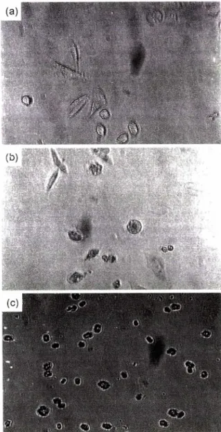
각시료를 50|ig/m/ 농도로첨가한연속 배양에서,암세포의 증식및형태변화를역상광학현미경으로관찰하였다. 성장하는 세포를 trypsin digestion*^., 각배양병에이식을 행하고배양 하면이식 2일에배양병바닥에붙어자라기 시작한다. MCF-7 세포는 Fig. 5a에서보이는바와같이선명한형태를갖추고, 계 속해서성장을유지한다. Fig. 5b는고추잎의추출물 50 을 첨가하여배양한것으로이식후첫날부터착상이 어렵고,배양
Fig. 5a - The pictures above show morphology of MCF-7 cells. The description of its morphology is as follows: (a) 2nd day, starting to grow on the bottom of cultural bottle, (b) 6th day, continuing to grow with clear form, (c) 10th day, continuing to grow with clear form.
Fig. 5b - The pictures above show morphologic changes of MCF-7 cells that occurred at the concentration of 50 |ig/m/ of C.
annuum extract of leaf. The description of its morphologi
cal changes is as follows: (a) 1st day, cannot grow, (b) 3th day, started to be destroyed, (c) 6th day, almost cells destroyed.
, • J 圖
..ᅵ:ᅵ ■ ■ ' ᄉ ) ■■■ ■■二 엾^ 녔M
爾
:
J i l l■Vol. 47, No. 2’ 2003
3일에서 세포탈락과세포파괴가시작되어, 6일째에 거의 모든 세
포의 형태변화와 파괴 현상을 보였다. 그리고 청고추 추출액을
첨가한것으로서,4일째에 세포변형이 관찰되고, 7일째에 세포가
탈락되고 파괴되기 시작했으며,14일째엔 거의 모든 세포가 파
괴되었다. 홍고추 추출액을 첨가한경우에는 4일째에 세포변형
이보이고, 11일째에 세포가 파괴되기 시작하여 16일째엔 거의
모든 세포가파괴되었다. 홍고추씨와 청고추씨 추출물에서도거
의비슷한결과를보였으나 세포변형은 약간늦게 일어났다.
청고추, 홍고추, 씀바귀 추출액은시료에 따라약간의 차이는 있으나약 4일정도에서 일부 세포를 제외하고는틸락을보이며 세포형태변화를보이기 시작했다. 그리고 14~19일정도에서 거
의모든 세포가파괴되었으며 , 그주변에 파괴된세포 파편들이 관찰되었다(Table I).
이형태관찰 결과는 MTT법에서 얻은결과와조금은상이한
결과로서,현미경관찰에서는홍고추추출물의 첨가시료에서 MTT
법의 결과보다세포의 성장과 착상저해작용이 상대적으로 낮게
관찰되었다. 이점은미토콘드리아탈수소효소의 활성에 의해 측
정된 MTT법,즉관련효소활성과 형태변화와의 차이 일가능성
이시사되는 점이다(실험방법에 따른 결과차이). 비교시료로사
용된씀바귀 추출액을첨가한경우 4일째에 세포가잘자라지 못 하고, 8일째에 파괴되기 시작하여 19일엔 거의 모든세포가 파
Fig. 5c - The pictures above show morphology of hepatoma cells.
The description of its morphology is as follows: (a) 3rd day, starting to grow on the bottom of culture bottle, (b) 4th day. (c) 7th day, continuing to grow with clear form.
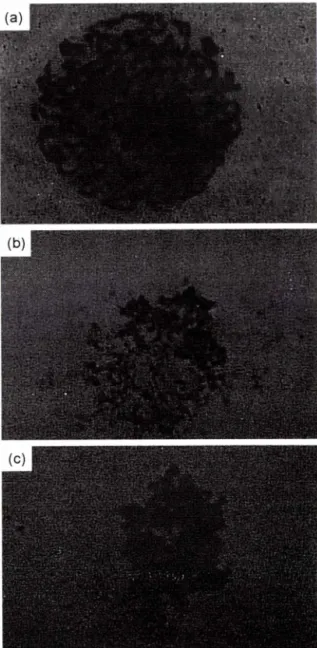
Fig. 5d - The pictures above show morphologic changes of hepa
toma cells that occurred at the concentration of 50 [ig/ml of C. annuurn extract of leaf. The description of its morphological changes is as follows: (a) 2nd day, started to be fragmented, (b) 10th day, s almost cells were destroyed, (c) 18th day, there were a lot of cell debris.
J. Pharm. Soc. Korea
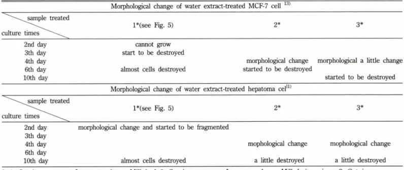
Table I - Morphological changes of MCF-7 cell and hepatoma cell by the microscope
Morphological change of water extract-treated MCF-7 cell 시 sample treated
l*(see Fig. 5) 2* 3*
culture times
2nd day cannot grow
3th day start to be destroyed
4th day morphological change morphological a little change
6th day almost cells destroyed started to be destroyed
10th day started to be destroyed
Morphological change of water extract-treated hepatoma cel11) sample treated
l*(see Fig. 5) 2* 3*
culture times ^시^ ^
2nd day morphological change and started to be fragmented 3th day
4th day mopholᄋgical change mophological change
6th day
10th day almost cells destroyed a little destroyed a little destroyed
*: 1. Capsicum annuum L. var. angulosum M ill, leaf; 2. Capsicum annuum L. var. angulosum M ill, fruit unripen; 3. Capsicum annuum L. var. angulosum M ill, fruit unripen.
괴되었다(씀바귀를대조시료로사용한이유는항암및청혈식 품으로널리 알려져 있고본실험실의관련실험결과1’13)에서도 다른시료들에비해좋은결과를보였으므로계속비교하여실 험하였다.).
간암세포에대한결과에서도고추의모든부위에서 억제작^ >1 높게관찰되었으나, 고추잎의수추출물의 첨가에서는더욱높게 나타나서 배양 2일째에세포의 형태변화와파괴가관찰되었고, 세포중식이거의없는상•태로지속되어 배양 10일에거의모든 세포가파괴되었다. 청고추보다홍고추가증식억제작용이 높이
Day
Fig. 6 - Absorbance on the supernatant of medium.. The cell's treated with 50 [ig/ml of each sample. When the culture medium was changed with new one, the old one was calculated the absorbance by 260 nm spectrophotometer.
They were counted during 9 days.
1. C. annuum L. var. angulosum M ill, Leaf; 2. Lactuca dentata Makino. var. flaviflora Makino; 3. C. annuum L. var.
angulosum M ill, fruit unripen; 4. C. annuum L. var.
angulosum M ill, fruit ripen.
1 2 3 4 5 6
Fig. 7 - Electrophoresis of cells' DNA fragmentation treated with each sample. They were cultured for 24 hr. 1. Marker (DNA 100 base); 2, Marker (DNA 100 base); 3. control; 4.
C. annuum L. var. angulosum M ill, Leaf; 5. C. annuum L.
var. angulosum M ill, fruit unripen; 6. C. annuum L. var.
angulosum M ill, fruit ripen.
Vol. 47, No. 2’ 2003
*: 1. C. annuum L. var. angulosum M ill, leaf; 2. Lactuca dentata M akino. var. flaviflora Makino; 3. C. annuum L. var. angulosum M ill, fruit unripen; 4. C. annuum L. var. angulosum M ill, fruit ripen.
관찰되었고, 배양 4일째에 형태변화가 관찰되고배양 10일에 세
포의 파괴가 관찰되었다. 홍고추씨의 추출물은간암세포에 대한
중식억제 작용이 감소하는 것이 관찰되었다.
염색체의 변화와 DNA 분절에대한관찰
P I 염색법에의한관찰 -Table III는 MCF-7 cell에각시료를
lOOngm/ 농도로 첨가하여 얻은 PI 염색법에 의해 관찰된 결과
이다. 그리고 Fig. 9b는고추잎추출물을 시료로 첨가하여 PI 염
색결과를현광현미경으로 관찰한것이며, 6시간배양에서 세포
핵이 일부파괴되기 시작했으며 24시간에는거의 대부분이 파괴
되었다. 청고추추출물을첨가한경우로서, 6시간과 24시간에서 는세포핵의 누출이 관찰되지 않았으나 4&^ᅵ간후에는많은세포
되지 않았으나 48시간에는 세포핵파괴가관찰되었다. 씀바귀 추 출물을 첨가한경우이며, 6시간과 24시간에서는세포핵 변형이 없었고 48시간후부터 약간 파괴하기 시작하여 72시간후에는 다소파괴되었다.
이실험 결과에서도고추의 모든 부분의 수추출시료가실험
암세포에 대해 증식 억제효과를 보여주었다. 특히 고추잎의 추
출물이 다른 부위추출물에 비해 hepatoma 세포와 MCF-7 mammary gland adenocarcinoma 세포의 세포사를더욱 유도한
다는것이 확인관찰되었다.
염색체의 banded 모형변화- 유방암세포의 염색체는 37시간 경과한후나 40시간뒤가별로차이가 없었다. 그러나시료로고 추잎추출물을 lOO^g/m/ 농도로 첨가한 경우는, 배양 24시간에 염색체의 풀어짐이 관찰되고(Fig. 8b(a)), 배양 27시간•에많은파
괴가 관찰되고, 48시간에는응축과 파괴현상미 함께관찰되었다
(Fig. 8b(c)). 청고추의 추출물 첨가군에서는배양 24시간에 약간
의모형변화와 분산이 관찰되었고, 40시간에 염색체의 많은풀
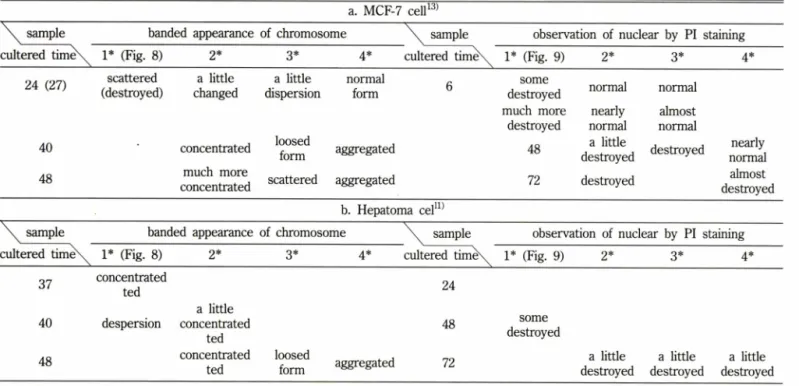
Table III - Morphological changes of chromosomes in MCF-7 and hepatoma cells a. MCF-7 cell13)
sample banded appearance of chromosome \ sam ple observation of nuclear by PI staining cultered time\、1* (Fig. 8) 2* 3* 4* cultered time\^ 1* (Fig. 9) 2* 3* 4*
24 (27)
40 48
scattered (destroyed)
a little changed
concentrated much more concentrated
a little dispersion
loosed form scattered
normal form
aggregated aggregated
6 some
destroyed much more
destroyed 48 72
normal nearly normal a little destroyed destroyed
normal almost normal
destroyed nearly normal almost destroyed b. Hepatoma cel11)
sample banded appearance of chromosome \노 sample observation of nuclear by PI staining cultered time\、 1* (Fig. 8) 2* 3* 4* cultered timex^ 1* (Fig. 9) 2* 3* 4*
37
40
concentrated ted despersion
a little concentrated
ted
24
48 some
destroyed
48 concentrated
ted
loosed
form aggregated 72 a little
destroyed
a little destroyed
a little destroyed
*: 1. C. annuum L. var. angulosum M ill, leaf; 2. Lactuca dentata Makino. var. flaviflora Makino; 3. C. annuum L. var. angulosum M ill, fruit unripen; 4. C. annuum L. var. angulosum M ill, fruit ripen.
Table II - The ratio of the absorbances of DNA in upper layer medium by spectrophotometer 260 nm. MCF-7 cells were cultured with water extract of each sample (50 \i^J ml) for 2, 4, 6,9 days
핵파괴가있었다. 동고추추출물을첨가한것이며, 역시 6시간과
24시간에서는 파괴가관찰되지 않았으며 48시간 후에파괴되기
시작하여 72시간후에는많은파괴가관찰되었다(Fig. 제시생략).
Hepatoma cell에고추잎추출물을시료로 첨가한경우, 3시간
경과에서 핵의 일부누출이 일어나기 시작하였고, 6시간후에는
세포에서 핵의 누출이 현상을보였고 48시간에 핵의 파괴가관
찰되었다(Fig. 9d). 청고추추출물과홍고추추출물을 첨가한경
우로서, 거의 비슷한 6시간과24시간에서 세포핵의 누출이 관찰
A1 1A 7 .2.2 .7 .7 A1 1A 1A IX . 33 X10 1 .1 71
1.3(
.45 1.01 1.5: .05 1.21 1.3(
57
]. Pharm. Soc. Korea
Fig. 8a - The chromosomal states in normal MCF-7cell. The 4 x 10³/m/ cells were cultured for (a) 24 hr (b) 40 hr (c) 48 hr.
어짐이관찰되었으며, 배양 48시간에는풀어짐과흩어짐이관찰 되었다. 홍고추추출물에대한결과는염색체의 농축현상이 배 양 40시간과 48시간에관찰되었다. 간암세포에대한결과는고 추잎추출물을 100 \ig/ml농도로첨가하여,배양 37시간에 염색 체의농축이관찰되다가(Fig. 8d). 배양 40시간이경과하면서 염 색체의 길이가늘어나느슨하게 보였다. 청고추와홍고추는배 양 48시간에풀어짐과응축이 각각관찰되었다.
D N A 분절관찰 - 각시료를 50 \ig/ml농도로첨가하여배양 한세포의 D N A * 분리하여10) 전기영동한결과, 첨가시료에 따
라세포사의 특징인 DNA의저분자로의분절현상이 나타났다.
24시간배양한세포에서분리한 DNA를전기영동법으로관찰한
Fig. 8b - The chromosomal states when the extracts of C. annuum L. var. angulosum M ill, leaf 100 fig/m/ were added to the medium in the MCF-7 cells. Hours from seeding and the morphology's as follows: (a) 24 hr, it seems to be scattered, (b) 27 hr, destroyed, (c) 48 hr, destroyed and aggregated.
결과는 Fig. 7과같으며, 36시간과 48시간배양에서는더 많은 분산과끌림현상이 관찰되었다. Fig. 7에서관찰된결과에서 보 면각고추잎과청고추추출물을시료로첨가한군에서는 DNA
의전기영동 band는흐리고 약하게관찰되었다(대조군에 비해
10% 미만의 약한 band로관찰된배양 12시간이나그이하의배 양후의결과를관찰하지않았으므로확실한결론을지을수는 없으나, 일부는분산되어 끌림을보이고일부는제거된것으로 추정함). 그러나홍고추첨가군에서는 대조군과비슷한수준의 고분자의 band가관찰되었다.
Vol. 47, No. 2’ 2003
Fig. 8c - The chromosomal states in normal hepatoma cells. The 4 x cells were cultured for (a) 37 hr, (b) 40 hr.
Fig. 8d 一 The chromosomal states when the extracts of C. annuum L. var. angulosum M ill,Leaf 100 [ig/ml were added to the medium in the hepatoma cells. Hours from seeding and cell morphology's as follows and above, (a) 37 hr, concentrated, (b) 40 hr, dispersion.
그리고배양액의부유층을 260 nm 흡광도법으로관찰한결과 에서도고추잎추출물을시료로첨가한세포에서는배양초기부터 부유층의흡광도가높게나타났다(Fig. 6). 씀바귀는 24시간배 양한결과에서는부유층의흡광도는대조군과비슷한비교적낮 은값을보여주었으며, 배양후기인 9일에그부유층의흡광도는
Fig. 9a - PI staining in normal MCF-7 cells. Hours from seeding and cell morphology's as follows and above. The MCF-7 cells were added to the medium in 25 cm2 canted neck flask, and then examined by PI staining.
Fig. 9b - PI staining in the MCF-7 cells when the extracts of C.
annuum L. var. angulosum M ill, Leaf 100 (ig/m/ were added to the culture medium. Hours from seeding and cell morphology's as follows and above, (a) 6 hr, some destroyed, (b) 24 hr’ they were cell debris.
J. Pharm. Soc. Korea