INTRODUCTION
Rheumatoid arthritis (RA) is a chronic autoimmune disease characterized by persistent inflammation of the joint synovium and synovial hyperplasia. The ab- errant changes, which occur in the synovium, lead to
joint damage and functional disability. Although the pathogenesis of RA has not been clearly identified, it is clear that a cascading network of pro-inflammatory cytokines plays a major role in the development of RA [1]. Toll-like receptor 4 (TLR4) contributes to the induc- tion of pro-inflammatory cytokines by activating innate
1
Division of Rheumatology, Department of Internal Medicine, Keimyung University Dongsan Medical Center, Daegu;
2
Keimyung University School of Medicine, Daegu;
3Department of Microbiology, Keimyung University School of Medicine, Daegu, Korea
Received : March 4, 2016 Revised : April 8, 2016 Accepted : April 27, 2016 Correspondence to Ji-Min Kim, M.D.
Division of Rheumatology, De- partment of Internal Medicine, Keimyung University Dongsan Medical Center, 56 Dalseong-ro, Jung-gu, Daegu 41931, Korea Tel: +82-53-250-7410
Fax: +82-53-250-7434
E-mail: [email protected]
Background/Aims: Grape seed proanthocyanidin extract (GSPE) has been re- ported to have a beneficial effect on regulating inf lammation. However, the anti-inflammatory mechanism of GSPE remains unclear. The aim of this study was to verify the influence of GSPE on the Toll-like receptor 4 (TLR4)-mediated signaling pathway in the regulation of murine autoimmune arthritis.
Methods: Collagen-induced arthritis (CIA) was induced in dilute brown non-agou- ti (DBA)/1J mice. The mice were treated with GSPE (0 or 100 mg/kg) intraperitone- ally. The severity of arthritis was assessed clinically, biochemically, and histolog- ically. Immunostaining for TLR4 was performed. The expressions of TLR4 and downstream signaling molecules were analyzed by Western blot. The effect of GSPE on lipopolysaccharide (LPS)-induced TLR4 activation was also evaluated using RAW264.7 cells and fibroblast-like synoviocytes (FLSs) from patients with rheumatoid arthritis and from those with osteoarthritis.
Results: GSPE attenuated the clinical severity of arthritis and decreased his- tological damage. GSPE treatment reduced the number of TLR4-stained cells in the synovium of mice with CIA. GSPE also downregulated the expression of TLR4, myeloid differentiation factor 88 (MyD88) and phosphorylated IκBα syno- vial protein in CIA mice. Concurrently, GSPE inhibited the nuclear translocation of nuclear factor-κB (NF-κB) subunits (p65 and p50). LPS-induced TLR4 activa- tion was suppressed by GSPE in human FLS as well as in murine macrophages in vitro.
Conclusions: Our results demonstrated that GSPE ameliorated CIA by regulat- ing the TLR4-MyD88-NF-κB signaling pathway.
Keywords: Grape seed proanthocyanidin extract; Arthritis, experimental; Toll- like receptor 4; Arthritis, rheumatoid
Grape seed proanthocyanidin extract ameliorates murine autoimmune arthritis through regulation of TLR4/MyD88/NF-κB signaling pathway
Sang-Hyon Kim
1, Jihye Bang
2, Chang-Nam Son
1, Won-Ki Baek
3, and Ji-Min Kim
1Korean J Intern Med 2018;33:612-621
https://doi.org/10.3904/kjim.2016.053
immunity under certain circumstances [2]. To date, sev- eral studies have suggested that TLR4 is implicated in the pathogenesis of RA. TLR4 is highly expressed in the inflamed synovium of RA patients [3]. The activation of the TLR4 signaling pathway is involved in persistent in- flammation and joint destruction in RA [4]. Associations between TLR4 single nucleotide polymorphisms and RA disease activity have been reported [5].
Grape seed proanthocyanidin extract (GSPE) is a stan- dardized water-ethanol extract derived from red grape seeds in which various antioxidants including catechins and oligomeric proanthocyanidins are accumulated [6].
GSPE is made up of a combination of ingredients with 15% (+) catechin and (–) epicatechin; 80% (–) epicate- chin 3-O-gallate, dimers, trimers, tetramers, and their gallates; and 5% pentamers, hexamers, heptamers, and their gallates [7]. Proanthocyanidins are the most abun- dant phenolic compounds in grape seeds. They have been reported to have various bio-protective functions such as anti-bacterial, anti-viral, anti-allergic, anti-car- cinogenic, anti-thrombotic, and vasodilatory activities [8]. Proanthocyanidins possess more potent anti-oxi- dative activity than vitamins C, E, and β-carotene [9].
Furthermore, the anti-inflammatory properties of pro- anthocyanidins on experimental inflammation, includ- ing in animal models of ulcerative colitis, skin tumors, and collagen-induced arthritis (CIA), have been shown in previous studies [10-13]. However, little information is available concerning how proanthocyanidins regulate experimental autoimmune arthritis. In this study, we hypothesized that GSPE would influence TLR4 signal- ing pathways, which play a critical role in the pathogen- esis of RA, in the development of murine autoimmune arthritis. We also examined the immunological effect of GSPE on fibroblast-like synoviocytes (FLSs) in patients with RA.
METHODS
Experimental animals
Dilute brown non-agouti (DBA)/1J mice (6 to 8 weeks old; SLC Inc., Hamamatsu, Japan), were housed in poly- carbonate cages and fed with standard mouse chow (Ralston Purina, St. Louis, MO, USA) and water ad libi- tum. All experimental procedures were examined and
approved by the Animal Research Ethics Committee of the Keimyung University (No. KM-2009-06R).
Induction of CIA and GSPE treatment
Male DBA/1J mice were given an intradermal injection of 100 μg of bovine type II collagen emulsified in com- plete Freund’s adjuvant (1:1, w/v; Chondrex, Redmond, WA, USA) into the base of the tail. Three weeks after pri- mary immunization, a booster, which consisted of 100 μg of bovine type II collagen emulsified in incomplete Freund’s adjuvant (IFA; 1:1, v/v; Chondrex), was inject- ed intradermally. Mice were divided into three groups;
control, CIA, and CIA with GSPE treatment. To investi- gate the effects of GSPE on the development of arthri- tis, CIA mice were treated with GSPE (100 mg/kg body weight) five times per week for 3 weeks intraperitoneally, after the booster injection. GSPE was kindly provided by Hanlim Pharmaceutical Company (Seoul, Korea). GSPE dissolved in saline was used for the experiments.
Clinical arthritis severity score
Three observers scored the severity of arthritis inde- pendently from day 24 after primary immunization to day 42. Clinical arthritis scores of the mice were evaluat- ed as follows: 0, no evidence of swelling; 1, mild swelling confined to the ankle or the midfoot (tarsals); 2, mod- erate swelling extending from the ankle to the midfoot (tarsals); and 3, severe swelling and erythema extending from the ankle to the metatarsal joints or ankylosis of the ankle joint. The sum of the scores of four limbs rep- resented the arthritis score of each mouse.
Histological analysis
Joint tissues were fixed with 4% paraformaldehyde.
They were then decalcified for 1 week in hydrochloric
acid before being embedded in paraffin. Tissue sections
(4 μm) were stained with H&E in order to determine the
influx of inflammatory cells. Inflammation was scored
using the following criteria: 0, no inflammation; 1, slight
thickening of lining layer or some infiltrating cells in
sublining layer; 2, slight thickening of lining layer plus
some infiltrating cells in sublining layer; 3, thickening of
lining layer, influx of cells in sublining layer and pres-
ence of cells in the synovial space and synovium high-
ly infiltrated with many inflammatory cells. The tissue
sections were stained with safranin O to evaluate carti-
lage degradation under light microscopic examination.
The extent of cartilage damage scored using the follow- ing criteria: 0, no destruction; 1, minimal erosion limited to single spots; 2, slight to moderate erosion in a limited area; 3, more extensive erosion and destruction. Immu- nohistochemistry was performed using the Vectastain ABC kit and 3,3’-diaminobenzidine as a substrate (both from Vector Laboratories, Burlingame, CA, USA). Non- specific staining was reduced by protein blocking with 20% normal goat serum for 30 minutes, at room tem- perature. Joint tissues were incubated with anti-TLR4 antibody (Santa Cruz Biotechnology, Dallas, TX, USA) overnight at 4ºC.
Enzyme-linked immunosorbent assay
The serum levels of anti-type II collagen specific immu- noglobulin G (IgG; Chondrex), tumor necrosis factor α (TNF-α), interleukin 6 (IL-6), and IL-17 (R&D Systems, Minneapolis, MN, USA) were measured according to the manufacturer’s protocol using an enzyme-linked immu- nosorbent assay (ELISA) kit (R&D). For the determination of cytokine levels, blood samples from the mice were ob- tained on day 42 and stored at –70°C until use. Antibody concentrations for each serum sample were obtained by reference to the standard curve. The absorbance values were determined at 450 nm with an ELISA microplate reader (Vector3, Perkin Elmer, Waltham, MA, USA).
Protein extraction and Western blot analysis
Ankles joints of mice were isolated by disarticulation distal to tibia. Synovial tissues from hind paws were col- lected and frozen until experiments. The frozen synovial tissues were pulverized and synovial protein was extract- ed as follows. Pulverized synovial tissues were lysed in ice-cold lysis buffer (pH 7.4, 50 mM Tris-HCl, 1% Non- idet P-40, 0.25% sodium deoxycholate, 150 mM NaCl, 1 mM Na
3VO
4, and 1 mM NaF) containing protease inhib- itors (2 mM phenylmethylsulfonyl fluoride, 100 μg/mL leupeptin, 10 μg/mL pepstatin, 1 μg/mL aprotinin, and 2 mM ethylenediaminetetraacetic acid). Cytoplasmic and nuclear extracts were prepared from joint tissues using the NE-PER nuclear and cytoplasmic protein extraction reagent kit (Thermo Pierce, Rockford, IL, USA). Samples in each group were subjected to 10% sodium dodecyl sulfate polyacrylamide gel electrophoresis on resolving gels appropriately and transferred to a nitrocellulose
membrane (Millipore, Billerica, MA, USA). Blots were incubated overnight at 4°C with primary antibodies for TLR4, myeloid differentiation factor 88 (MyD88), Toll/IL-1 receptor domain-containing adaptor induc- ing IFN-β (TRIF), IκBα, phosphorylated IκBα (p-IκBα), p65, p50 (all from Santa Cruz Biotechnology), and β-ac- tin (Sigma-Aldrich, St. Louis, MO, USA). Anti-laminB1 (Santa Cruz Biotechnology) was used as an internal control of nucleus protein marker. The membrane was then washed with a mixture of Tris-buffered saline with Tween 20 (TBS-T) and incubated with horseradish per- oxidase-conjugated secondary antibodies (Santa Cruz Biotechnology). Protein bands were detected using en- hanced chemiluminescent reagents (Amersham Phar- macia Biotech, Uppsala, Sweden).
Isolation and culture of FLS
FLS were isolated by enzymatic digestion of synovial tis- sue, provided by the Keimyung Human Bio-Resource Bank, a member of the National Biobank of Korea. The tissue was obtained from patients with osteoarthritis (OA) and from those with RA who underwent total joint replacement surgery or a synovectomy at Keimyung University Dongsan Medical Center. All the patients fulfilled the American College of Rheumatology crite- ria and agreed to give their informed consent (approved by the Institutional Review Board [IRB] of Keimyung University Dongsan Medical Center [IRB No. 2015-02- 005]) [14]. The tissue samples were minced into 2 to 3 mm pieces and treated for 4 hours with 0.5 mg/mL type II collagenase (Worthington Biochemical Corp., Lake- wood, NJ, USA) in Dulbecco�s modified Eagle�s medi- um (DMEM) at 37ºC in a water bath. Synoviocytes were grown in DMEM supplemented with 10% fetal bovine serum, 2 mM L-glutamine, 100 units/mL penicillin, and 100 ng/mL streptomycin. The cells were used for experi- ments from passage 4 to 8, at which time the population was homogeneous.
Statistical analysis
The statistical analysis was performed using the SPSS
version 22 (IBM Co., Armonk, NY, USA). Student t tests
and one-way analysis of variance were carried out. The
accepted level of significance was preset at p < 0.05. Each
experiment was executed at least three times in dupli-
cate. Data are presented as mean ± SD.
RESULTS
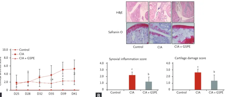
GSPE ameliorates murine autoimmune arthritis We examined the effect of GSPE on the development of experimental arthritis in vivo using a CIA mouse model.
DBA/1J mice were intraperitoneally injected with GSPE five times per week for 3 weeks after the second immu- nization with type II collagen/IFA. The mean arthritis score of GSPE-treated CIA mice was significantly lower than that of CIA mice (Fig. 1A). Representative histolog- ic sections of hind paws of the mice are shown in Fig.
1B. The joints of CIA mice revealed the infiltration of inflammatory cells, synovial proliferation, and cartilage destruction (Fig. 1B). In CIA mice treated with GSPE, there was a reduction in infiltrated inflammatory cells, synovial proliferation, and cartilage damage (Fig. 1B).
GSPE decreases serum levels of pro-inflammatory cytokines in CIA mice
We investigated whether the therapeutic effect of GSPE in CIA mice affects the humoral immune response to type II collagen and serum levels of pro-inflammatory
cytokines. The levels of anti-type II collagen IgG, TNF-α, IL-6, and IL-17 in serum samples of mice were measured using ELISA, 6 weeks after primary immunization. The serum concentrations of anti-type II collagen IgG were significantly reduced in CIA mice following GSPE treat- ment, when compared with CIA mice (Fig. 2A). There was also a decrease in serum levels of TNF-α (Fig. 2B), IL-6 (Fig. 2C), and IL-17 (Fig. 2D) in the GSPE-treated group.
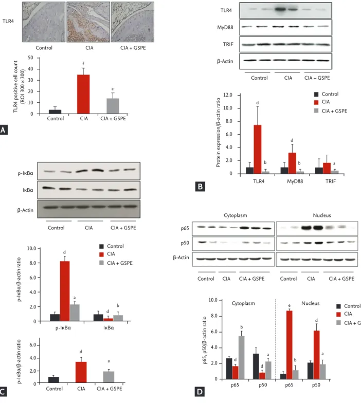
GSPE administration reduces TLR4 expression in the synovium of CIA mice
TLR4 has been reported to play a critical role in the pathogenesis of CIA [15,16]. Therefore, we investigated whether GSPE treatment influenced TLR4 expression in the synovial tissue of CIA using immunohistochem- ical staining. To assess the in vivo effect of GSPE on TLR4 expression, histological sections of the tibiotalar joints were stained in order to visualize the presence of TLR4. As shown in Fig. 3A, less TLR4-positive cells were observed in the joints of GSPE-treated CIA mice than in those of CIA mice. We also investigated the ef-
10.0 8.0 6.0 4.0 2.0 0
4.0 3.0 2.0 1.0 0
4.0 3.0 2.0 1.0 0
Clinical arthritis score
Synovial inflammation score Cartilage damage score
D25 Control CIA CIA + GSPE
D28 D32 D35 D39 D41 Control
Control H&E
Safranin O
CIA
CIA
CIA + GSPE
CIA + GSPE
Control CIA CIA + GSPE
a a a
c
b
c
b
Figure 1. Grape seed proanthocyanidin extract (GSPE) treatment relieves the severity of collagen-induced arthritis (CIA) in mice (n = 8 for each group; control, CIA, GSPE-treated CIA group). (A) For induction of CIA, male dilute brown non-agouti (DBA)/1J mice were given 100 μg of bovine type II collagen emulsified in complete Freund’s adjuvant intradermally into the base of the tail. A booster injection was administered 3 weeks later. GSPE (100 mg/kg) was initiated intraperitoneally after the booster injection. The mice were treated with GSPE (100 mg/kg) five times per week for 3 weeks. The arthritis score for each mouse was calculated as the sum of the scores of both hind paws. Mean values of the arthritis scores are represented on the graph. Error bars indicate standard deviation of the mean. (B) A hind limb of each mouse was stained with H&E (upper panel) and safranin O (lower panel). The joints from CIA mice treated with GSPE demonstrated attenuated inflammation and carti- lage destruction when compared with those from CIA mice (×200). Error bars indicate standard deviation of the mean.
ap < 0.05,
b
p < 0.01, compared with the CIA group,
cp < 0.001 compared with the control group.
A B
fect of GSPE on the intracellular proteins involved in TLR4-mediated signal transduction in the synovium of CIA, by using Western blotting. Synovial protein levels of TLR4, MyD88, and TRIF were analyzed. The expres- sion levels of TLR4 and MyD88 proteins were upregulat- ed in the synovial extracts of CIA mice when compared with those of control mice. TLR4 and MyD88 expres- sion levels were downregulated in GSPE-treated CIA mice when compared with CIA mice (Fig. 3B). The ex- pression level of TRIF showed no increase in CIA mice when compared to the control mice. However, GSPE treatment downregulated the expression level of TRIF in CIA mice when compared to CIA mice (Fig. 3B). In order to explore the effects of GSPE on the activation of nuclear factor-κB (NF-κB) signaling, the levels of IκBα and p-IκBα were measured. GSPE reduced the level of p-IκBα protein in the synovial tissue of CIA mice (Fig.
3C). The ratio of p-IκBα to total IκBα was also lower in GSPE-treated CIA mice than in CIA mice (Fig. 3C). We also examined whether GSPE affected nuclear translo-
cation of the NF-κB subunits, p65 and p50. The nuclear extracts and cytoplasmic lysates were purified from the synovial tissue and subjected to Western blotting. The nuclear localization of p65 and p50 NF-κB was inhibited in the synovial extracts of GSPE-treated CIA mice, while nuclear localization of p65 and p50 NF-κB was promi- nent in those of CIA mice (Fig. 3D).
GSPE suppresses lipopolysaccharide-induced TLR4 activation in RAW264.7 cells
Our data demonstrates that GSPE administration de- creased TLR4 expression in the synovium of CIA mice (Fig. 3A and 3B). We next investigated whether GSPE had an influence on TLR4 expression in vitro by using a mu- rine macrophage cell line, RAW264.7. GSPE downregu- lated the expression of TLR4, which was enhanced by stimulation with lipopolysaccharide (LPS) in RAW264.7 cells (Fig. 4A). We also evaluated the effect of GSPE on the expression of TLR4 in FLS from patients with OA and RA. As shown in Fig. 4B, LPS-induced TLR4 upreg-
d
a 140
120 100 80 60 40 20 0
140 120 100 80 60 40 20 0 700 600 500 400 300 200 100 0
30 25
20 15
10 5
0
collagen antibody (pg/mL)IL-6 (pg/mL) IL-17 (pg/mL)TNF-α (pg/mL)
Control CIA CIA + GSPE
Control CIA CIA + GSPE
Control CIA CIA + GSPE
Control CIA CIA + GSPE
a
d c
c
b
b
Figure 2. Grape seed proanthocyanidin extract (GSPE) treatment suppresses immune response to type II collagen and decreas- es serum levels of pro-inflammatory cytokines. Serum concentrations of (A) type II collagen specific total immunoglobulin G (IgG), (B) tumor necrosis factor α (TNF-α), (C) interleukin 6 (IL-6), and (D) IL-17 were determined using enzyme-linked immu- nosorbent assay in each group of mice (n = 8 for each group). Error bars represent standard deviation of the mean. Each exper- iment was performed three times.
ap < 0.05,
bp < 0.01 compared with the collagen-induced arthritis (CIA) group,
cp < 0.05,
dp <
0.001 compared with the control group.
A
C
B
D
Figure 3. Grape seed proanthocyanidin extract (GSPE) downregulates Toll-like receptor 4 (TLR4) expression and TLR4-medi- ated signal transduction proteins in the synovium of mice with collagen-induced arthritis (CIA; n = 8 for each group). (A) TLR4 expression was assessed by immunohistochemical staining. GSPE treatment led to a reduction in TLR4-positive cells in the synovium of CIA mice, whereas the number of TLR4-stained cells was markedly increased in the synovium of CIA mice, when compared to that of control mice (×200). (B) Western blot analysis of synovial extracts from GSPE-treated CIA mice showed a decreased protein level of TLR4, myeloid differentiation factor 88 (MyD88), and Toll/interleukin 1 receptor domain-containing adaptor inducing IFN-β (TRIF), when compared with CIA mice. (C) The expression level of phosphorylated IκBα (p-IκBα) was also reduced in GSPE-treated CIA mice. (D) GSPE suppressed nuclear translocation of nuclear factor-κB subunits (p65 and p50) in the synovium of CIA mice. Error bars reflect standard deviation of the mean. Each experiment was performed three times.
ROI, region of interest.
ap < 0.05,
bp < 0.01,
cp < 0.001 compared with the CIA group,
dp < 0.05,
ep < 0.01,
fp < 0.001 compared with the control group.
50 40 30 20 10 0
12.0 10.0 8.0 6.0 4.0 2.0 0
10.0
8.0
6.0
4.0
2.0
0
6.0 4.0 2.0 0
TLR4 positive cell count (ROI 300 × 300) Protein expression/β-actin ratio
p-IκBα/β-actin ratio p-IκBα/β-actin ratio
Control
p-IκBα
β-Actin IκBα
CIA CIA + GSPE
Control CIA CIA + GSPE
Control CIA CIA + GSPE
Control
p-IκBα IκBα
CIA CIA + GSPE
Cytoplasm Nucleus
Control CIA CIA + GSPE Control
TLR4
TLR4
MyD88
TRIF
β-Actin
p65
p50
β-Actin CIA CIA + GSPE
Control
TLR4 MyD88
Cytoplasm Nucleus
TRIF Control CIA
CIA CIA + GSPE
CIA + GSPE c
d d
f
b b a
10.0
8.0
6.0
4.0
2.0
0
p65, p50/β-actin ratio
p65 p50 p65 p50
d
d
d e
b b
b
a a
a a
d
d
d Control
CIA CIA + GSPE
Control CIA CIA + GSPE
A
B
C D
ulation was suppressed by GSPE in FLS from patients with RA and OA.
DISCUSSION
Proanthocyanidins, which are the main constituents of GSPE, belong to the category of condensed tannins [17].
The sources of proanthocyanidins include fruits, veg- etables, nuts, seeds, flowers, and bark [18]. Proanthocy- anidins are well-known as naturally occurring anti-oxi- dants. They have been reported to have beneficial effects on modulating inflammation in human cells, such as differentiated adipocytes and human pulmonary epi- thelial cells in vitro, in addition to animal inflammato- ry models as mentioned above [19,20]. Some previous studies have shown that GSPE has an anti-inflammatory effect in an animal model of RA. Cho et al. [13] first re- ported about the therapeutic effect of GSPE on CIA and showed the effects of GSPE on oxidative stress and os- teoclastogenesis in vitro. Another study using a murine model of RA has demonstrated the effect of GSPE focus- ing on inflammation-associated bone destruction [21].
GSPE attenuated both arthritis and obesity in obese CIA mice [22]. However, little is known about the mechanism by which GSPE regulate the inflammatory response. Ox- idative stress is reduced by GSPE, and has been reported
to be involved in the pathogenesis of murine autoim- mune arthritis. However, there has been controversy as to whether oxidative stress instigates, or suppresses the inflammation in a model of murine autoimmune ar- thritis [23-25]. One previous study reported that GSPE influences murine autoimmune arthritis by regulating Foxp3
+regulatory and IL-17-producing T cells, recipro- cal control of which is important in managing autoim- mune arthritis [26]. There was another report that GSPE has an anti-arthritic effect in adjuvant-induced arthritis model by modifying T cell subsets [27]. In this study, we focused on revealing the mechanism of action of GSPE in suppressing murine autoimmune arthritis. Here, we demonstrated that GSPE has an anti-arthritic effect through the regulation of TLR4-mediated signaling for the first time.
TLRs are innate receptors, which play an essential role in the innate immune system [28]. Among diverse TLRs, TLR4 was the first to be characterized, and has been re- ported to play a significant role in the pathogenesis of autoimmune diseases [29]. In particular, a large amount of research provides evidence that TLR4 is implicated in the pathogenesis of RA. TLR4-deficient mice showed lower degree of severity and incidence of CIA compared to the wild-type mice [15]. LPS-induced TLR4 activation was greatly enhanced in peripheral blood mononuclear cells (PBMCs) from patients with RA than in those from
5.0 4.0 3.0 2.0 1.0 0
4.0 3.0 2.0 1.0 0
2.0 1.5 1.0 0.5 0 LPS
GSPE TLR4
β-Actin
TLR4 β-Actin
TLR4 β-Actin
OA-FLS RA-FLS
TLR4/β-actin TLR4/β-actin TLR4/β-actin
– + + –
– – + +
LPS GSPE
– + + –
– – + +
LPS GSPE
– + + –
– – + +
c d
d
a
a a
a
b b
Figure 4. Grape seed proanthocyanidin extract (GSPE) inhibits lipopolysaccharide (LPS)-induced Toll-like receptor 4 (TLR4) expression in vitro. (A) RAW264.7 macrophages were co-incubated with or without LPS (1 μg/mL) for 8 hours after pretreatment with or without GSPE (25 μg/mL) for 16 hours. LPS-induced TLR4 expression was downregulated in RAW264.7 cells after treat- ment with GSPE. (B) Fibroblast-like synoviocyte (FLS) from patients with osteoarthritis (OA; n = 5) and rheumatoid arthritis (RA;
n = 3) were incubated in the same conditions as panel A. GSPE treatment reduced LPS-induced TLR4 expression in FLS from patients with RA as well as in those from patients with OA. Data are presented as the mean ± SD of three independent experi- ments. Each experiment was performed three times.
ap < 0.05,
bp < 0.01 compared with the LPS only-stimulated group,
cp < 0.05,
d