Extract of Rubus coreanus Fruits Increases Expression and Activity of Endothelial Nitric Oxide Synthase in the Human Umbilical Vein Endothelial Cells
Hyun Joong Yoon
1, Soo Young Park
2, Sung-Tack Oh
2, Kee Young Lee
1and Sung Yeul Yang
1*
1
Department of Biochemistry and
2Department of Obstetrics and Gynecology, Medical School and Research Institute of Medical Science, Chonnam National University, Gwangju 501-746, Korea
Received November 17, 2010 /Accepted January 4, 2010
This study aimed to investigate the effects of water extract of Rubus coreanus (RCE) on the expression and activity of endothelial nitric oxide synthase (eNOS), as well as its signal transduction pathways in human umbilical vein endothelial cells (HUVECs). The specific inhibitors of NOS show RCE treat- ment increases NO production in HUVECs due to the up-regulation of eNOS rather than iNOS. The real-time expression level of eNOS mRNA was also increased upon RCE treatment in HUVECs. While a PKC-specific inhibitor, RO-317549, did not alter RCE-induced NO production in HUVECs, tamoxifen (estrogen receptor-specific inhibitor), PD98059 (ERK-specific inhibitor) and LY-294002 (PI3K/Akt-spe- cific inhibitor) did have suppressive effects. Increased NO production by RCE seems to result from a higher level of active eNOS (pSer1177). Specifically, inhibition of ERK not only decreased the level of active eNOS, but also increased the inactive form of the enzyme (pThr495) in HUVECs. This study suggests that RCE treatment increases NO production in HUVECs due to the increased expression and activity of eNOS. It is also shown that RCE-induced eNOS activation occurs partly through the binding of RCE to the estrogen receptor, along with ERK and PI3K/Akt-dependent signal trans- duction pathways. In addition, the regulatory binding proteins of eNOS including Hsp90 and cav- eolin-1 were related to these effects of RCE on eNOS activity in HUVECs.
Key words : Human umbilical vein endothelial cell, NO, eNOS, Rubus coreanus, estrogen receptor
*Corresponding author
*Tel:+82-62-220-4104, Fax:+82-62-223-8321
*E-mail : [email protected]
Introduction
Nitric oxide (NO) plays various roles in regulation, medi- ating many physiological and pathophysiological functions such as vasodilation, neurotransmission/ neuromodulation and cytotoxic activity [2,24,42]. Vasodilation is primarily controlled by the endothelium-dependent production of NO, which acts to induce vascular smooth muscle relaxation in a cGMP-dependent manner [63].
In numerous mammalian cells and tissues, NO is synthe- sized by NO synthase (NOS), which oxidizes the terminal guanidinium residue of L-arginine (L-Arg) to NO and citrul- line [53,54]. There are three NOS isozymes, Ca
2+-dependent endothelial and neuronal NOS (eNOS and nNOS, re- spectively) and Ca
2+-independent inducible NOS (iNOS).
The classic activity of estrogen is mediated by transcrip- tional activation of estrogen-responsive genes involving in- tracellular estrogen receptors (ER). Once estrogen binds to ER, the receptor-hormone complex then binds to a specific estrogen responsive element in the promoter region in the
target genes, leading to transcriptional activation [28,34]. In addition to transcriptional regulation of estrogen-responsive genes, the rapid, membrane-initiated increase in NO release by estrogen, occurring within minutes and intact in the pres- ence of transcriptional inhibitors, were observed [7,21].
Recent evidence implicates cell surface receptors in the rapid response to estrogen in a number of different cell types, in- cluding vascular endothelial cells [8,14,20,29,58].
eNOS exists in at least two distinct subcellular locations:
within caveolae of the plasma membrane and in the Golgi/perinuclear region of the cell [41]. Caveolin-1, a major scaffolding protein in endothelial caveolae, significantly in- hibits eNOS activity as it supplies anchorage and prevents calmodulin binding [16,41]. Conversely, the binding of Ca
2+/calmodulin to eNOS disrupts the eNOS/caveolin-1 complex, leading to enzyme activation [39]. The binding of Hsp90 (90 kDa heat shock protein) to eNOS triggers the tran- sition from the Ca
2+-dependent activation to phosphor- ylation-mediated activation by recruiting Akt to the complex [4]. In fact, active eNOS can be co-immunoprecipitated with Hsp90 and kinases such as Akt [11,17].
Most adult-onset cardiovascular diseases are associated
with age-related vascular dysfunction [10,37]. Age-related
vascular dysfunction due mainly to reduced NO production by eNOS and reduced eNOS expression, which might make endothelial cells more vulnerable to apoptotic stimuli [3,12,23].
Phytoestrogens are biologically active phenolic com- pounds from plants that mimic mammalian estrogens, either structurally or functionally, and therefore play important roles in the prevention of cancer, heart disease, menopausal symptoms and osteoporosis [1,32,48,56,61]. Estrogens influ- ence the growth and function of female and male re- productive tissues, maintain the skeletal and central nervous system, protect the cardiovascular system and prevent colon cancer and skin aging [19,51]. Considering the numerous ef- fects estrogens have on the human body, it is not surprising to consider the potential of phytoestrogens for human health.
Phytoestrogens are naturally occurring molecules that in- clude isoflavonoids, lignans, coumestanes, stilbens and the flavonoids quercetin and kaempherol. These phytochemicals are usually found in fruits, vegetables, legumes and tea and are generally concentrated in the fruit skin, bark and flowers of plants [43]. Resveratrol, daidzein, quercetin and genistein represent four of the most commonly ingested and most in- tensely studied phytoestrogens [38]. Moreover, they may act as “natural”, selective ER modulators because their effects are mediated via the estrogen receptor (ER) in the cell membrane. Recently, the possible application of phytoes- trogens in estrogen replacement therapy for postmenopausal women has been investigated. If successful, there is potential in preventing osteoporosis and cardiovascular diseases with- out adverse effects such as increased risk of breast and endo- metrial cancer, as well as irregular bleeding [5,13,38,64-66].
However, controversy over data requires further studies in order to confirm the health effects of phytoestrogens in vivo [44,61].
Rubus species have been cultivated as fruits for centuries and are used in various countries as natural remedies to sev- eral diseases such as diabetes, many types of infection, colic and burns [49]. Rubus coreanus Miq., a deciduous broadleaf shrub of the family Rosaceae, is popularly known as Bokbunja in South Korea and used in traditional folk medi- cines for treating spermatorrhea, enuresis, asthma and aller- gic diseases [57]. Bokbunja has been used to improve male reproductive function by treating symptoms like impotence, premature ejaculation, seminal emission, spermatorrhea and enuresis in combination with other herbal medicines in tra-
ditional Korean medicine [30].
Bokbunja has been reported to possess anti-oxidant, an- ti-nociceptive, anti-inflammatory, anti-cancer and anti-ana- phylactic effects [9,33,57,67]. The fruits may also improve male fertility by enhancing spermatogenesis [46]. Many re- cent studies have investigated the scientific basis of these purported therapeutic effects on biological systems.
However, the effect of Bokbunja on NO production in endo- thelial cells has not yet been reported. Therefore, in this study we investigated the effect the water extract of Rubus coreanus (RCE) on NO production, the expression and activ- ity of eNOS and the intracellular signal pathway in cultured primary human endothelial cells.
Materials and Methods
Preparation of water extracts from Rubus coreanus fruits
Aqueous extract of Rubus coreanus Miq. fruits collected from Gochang county was prepared by the following methods. The fruits were frozen for overnight at -80
oC and were lyophilized (Hanil Research & Development Co, Korea). The fruits once dried (5 g) were homogenized in distilled water (200 ml) for 5 min using a Polytron homoge- nizer (Model T-25B, IKA Co, Japan), followed by shaking for 20 min at 80
oC in a water bath. Centrifugation was per- formed at 20,000 rpm for 20 min at 4
oC. The resulting super- natant was frozen for overnight at -80
oC and completely lyophilized thereafter. The weight of RCE was about 1.85 g, which was dissolved by distilled water into 10 mg/ml and stored at 4
oC.
Quantification of phenol groups
The phenol content of RCE was measured with Folin- Ciocalteu Reagent (FCR, Sigma, St. Louis, MO, USA) [59].
An aliquot of diluted RCE (200 μl) was mixed with 10% FCR (1 ml) in test tube, which was reacted for 3 min at room temperature. The mixture was next mixed with 7.5% Na
2CO
3(0.8 ml) and incubated for 2 hr in a shaking water bath. After
incubation the absorbance was measured at 760 nm using
UV-spectrophotometer (Model UV-1650PC, Shimadzu,
Japan). The amount of phenol groups was quantified from
the standard curve plotted with authentic catechin. The phe-
nol content of RCE used in this study was ≤200 nM CE
(catechin equivalent).
Cell culture
Human umbilical vein endothelial cells (HUVECs, PDL 21-23) were purchased from Modern Cell & Tissue Technologies (Seoul, Korea) and cultured in Cambrex micro- vascular endothelial cells medium-2 (EGM-2, Cambrex, MD, USA). The cells were incubated at 37°C in a humidified at- mosphere under 5% CO
2.
Measurement of NO production
Production of NO was determined spectrophotometrically using Griess reagent (0.8% sulfanilamide, 0.75% N- (naphthyethylene)diamine in 0.5 N HCl, Sigma, MO, USA).
An aliquot of the incubated supernatant (100 μl) was trans- ferred to 96-well plates and mixed with 100 μl of Griess reagent. After 15 min incubation at room temperature, the nitrite concentration was measured at 540 nm using a micro- plate reader. Sodium nitrate (0.5 to 100 μM) was used as the nitrite standard and the absorbance was found to be line- ar over this concentrations range.
Western blot analysis
The cells were lysed in RIPA buffer (Santa Cruz Biotechnology, CA, USA, 50 mM Tris–HCl, 150 mM NaCl, 1% NP-40, 0.5% sodium deoxycholate, 0.1% SDS, 0.004% so- dium azide, 1 mM PMSF, and 0.5 mM sodium orthovana- date) containing a cocktail of protease inhibitors (10-20 μl per 1 ml RIPA buffer) by incubating for 30 min at 4°C. The mixtures were transferred to microtubes and centrifuged at 12,000× g for 15 min at 4°C, and the supernatants were stor- ed at -80°C before use. Proteins (40 μg) present in the cell lysates were separated by 8% SDS-PAGE and transferred to PVDF membrane. The membrane was blocked with 5% skim milk in TBS-T (Tris buffered saline-Tween-20: 25 mM Tris-base, 137 mM NaCl, 2.7 mM KCl, 0.1% Tween-20, pH 7.4) for 1 hr at room temperature and incubated with an- ti-human eNOS antibody (BD biosciences, Franklin Lakes, NJ, USA), anti-human pS1177 eNOS (BD biosciences) or an- ti-human pThr495 eNOS (BD biosciences) overnight at 4°C.
After washing with TBS-T 3 times, the blot was incubated with secondary antibody (horseradish peroxidase-con- jugated anti-goat antiserum) for 2 hr at room temperature. The antibody-specific proteins were detected using West-ZOL
TM(plus) (iNtRON Biotechnology, Seongnam, Korea).
ELISA
Levels of eNOS protein were also measured using a com-
mercially available ELISA kit (R&D System, MN, USA) fol- lowing the manufacturer’s instruction manual. Absorbances of the samples at 450 nm were converted to protein concen- tration (pg/ml) using standard curves generated with the recombinant human eNOS supplied with the kit.
RNA isolation
Total RNA was isolated from HUVECs using TRI reagent (Molecular Research Center, Cincinnati, OH, USA). The cell lysate was then stored for 5 min at room temperature to permit the complete dissociation of nucleoprotein complexes. 0.2 ml chloroform per 1 ml of TRI reagent and shaken vigorously for 15 sec. The resulting mixture was stor- ed at room temperature for 3 min and centrifuged at 12,000×
g for 15 min at 4°C. Following centrifugation, the mixture was separated into a lower red phenol-chloroform phase, the interphase and the colorless upper aqueous phase. RNA remains exclusively in the aqueous phase, which was trans- fered to a fresh tube, whereas DNA and proteins are sepa- rate into the interphase and organic phase. The aqueous phase was transfered to a fresh tube. The supernatant was removed and the RNA pellet was washed by the addition of 75% ethanol and centrifugation at 7,500× g for 5 min at 4°C. After washing, at least 1 ml of 75% ethanol per 1 ml TRI reagent was added to the RNA pellet, which was briefly air-dried for 3~5 min. The RNA pellet was dissolved in di- ethyl pyrocarbonate (DEPC) water by passing the solution a few times through a pipette tip and incubating for 10~15 min at 55~60°C. Total RNA was quantified spectrophoto- metrically at 260 nm.
Real-time PCR
One microgram of total RNA was reverse transcribed us-
ing a maxime
TMRT premix kit (iNtRON Biotechnology,
Seongnam, Korea) at 42°C for 1 hr. Oligo (dT)
15was used
as primer. The cDNA was amplified by real-time PCR in
the presence of AccuPower
TMGreenStar qPCR premix
(Bioneer, Daejeon, Korea). Specific primer pairs for eNOS
were 5'-CCAGCTAGCCAAAGTCACCAT-3' and 5'-GTCT
CGGAGCCATACAGGATT-3' for sense and antisense,
respectively. For 28S, the sense primer was 5'-TTGAAAAT
CCGGGGGAGAG-3' and antisense primer was 5'-ACATT
GTTCCAACATGCCAG-3'. Real-time RT-PCR was con-
ducted for 45 cycles under the following conditions: 95°C
denaturation step for 15 sec, annealing at 54°C for 20 sec
(eNOS) or 52°C (28S), and extension at 72°C for 20 sec in
a real-time rotary analyzer (Rotor-Gene
TM6000, Corbett Life
Science, San Francisco, CA, USA). Final extension was per- formed at 72°C for variable times depending on product size (eNOS, 354 bp and 28S, 100 bp). The green fluorescent prod- uct was detected at the end of each cycle.
Measurement of protein content
The total protein concentrations of the conditioned media and cell extracts were determined using BCA
TMprotein as- say kit (Pierce Biotechnology, Rockford, IL). Bovine serum albumin was used as a protein standard.
Immunoprecipitations
Immunoprecipitation (IP) was carried out using an Immunoprecipitation Starter Pack (GE Healthcare Bio-Sciences AB, Uppsala, Sweden). The IP procedure for de- tecting the protein level was performed according to the manufacturer’s instructions. nProtein A Sepharose 4 Fast Flow and Protein G Sepharose 4 Fast Flow are supplied pre- swollen in 20% ethanol. It was washed three times by RIPA buffer. It was centrifuged at 12,000× g for 20 sec and dis- carded the supernatant. And, it was mixed by equal volumes of media and RIPA buffer (50% slurry). It was stored at 4
oC.
Cell lysates (100 μl) were transferred to new tubes and added anti-human eNOS antibody (1 μg). These mixtures were gen- tly mixed for 1 hr at 4
oC. These mixtures were added with the 50 μl nProtein A Sepharose 4 Fast Flow or Protein G Sepharose 4 Fast Flow suspension (50% slurry) and gently mixed for 1 hr at 4
oC. After mixing, these mixtures were centrifuged at 12,000× g for 20 sec and save the pellet. These pellets were washed three times with 1 ml RIPA buffer and once with wash buffer (50 mM Tris, pH 8). The final pellets were suspended in 30 μl sample buffer (1% SDS, 100 mM DTT, 50 mM Tris, pH 7.5) and heated to 95
oC for 3 min.
It was centrifuged at 12,000× g for 20 sec to remove the beads. The supernatants were carefully transferred in new tubes. These supernatants were analyzed by western blot.
The following primary antibodies were used: anti-human eNOS from BD Biosciences (Franklin Lakes, USA), and an- ti-human Akt, anti-human Hsp90, and anti-caveolin-1 from Cell Signaling Technology (Danvers, USA).
Statistic analysis
For in vitro data analysis, the Students t-test was per- formed using the Sigma-plot computerized program and a P-value less than 0.05 was deemed significant. The compar- ison was carried out with control or reagent-treated data.
Results
NO production is increased by RCE in HUVECs
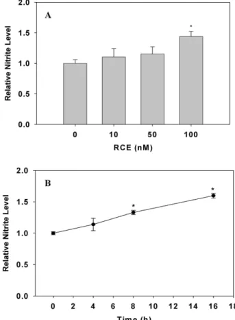
The effect of RCE on the bioavailability of NO in cultured HUVECs (from PDL 21 to 25) was analyzed spectrophoto- metrically by measuring nitrite concentration in culture me- dia with Griess reagent. As shown in Fig. 1, NO production in HUVECs was significantly increased in a dose- (with in- creasing RCE concentrations from 0 to 100 nM) and a time-(with increasing time of incubation from 0 to 16 hr in 100 nM RCE) dependent manner at 37
oC.
The relative contribution of NOS isozymes on RCE-induced NO production in HUVECs
Specific enzyme inhibitors (L-NIO, eNOS-specific in- hibitor; 1400W, iNOS-specific inhibitor) were used to eval-
Fig. 1. Effect of RCE on NO production in HUVECs. HUVECs were treated with RCE in a dose-dependent manner for 16 hr (A) and with RCE (100 nM) in a time-dependent manner (B) at 37oC. Nitrite was measured by the Griess reagent method. Each result represents mean±SD (n=6~
9). The significance was expressed as a comparison with NO production of the untreated group (*
p
<0.05).Fig. 2. Effect of NO synthase-specific inhibitors on RCE-induced NO production in HUVECs. HUVECs were treated with L-Arg (A) or RCE (B) in the presence or absence of NOS-specific inhibitors (L-NIO: eNOS inhibitor and 1400W: iNOS inhibitor) for 16 hr at 37oC. Nitrite was measured by the Griess reagent method. Each result represents mean±SD (n=6~9,
p
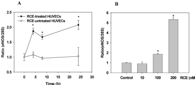
<0.05).Fig. 3. Effect of RCE on content of endothelial NO synthase in HUVECs. HUVECs were treated with RCE in a time (0~16 hr, RCE 100 nM; A and B) and dose- (RCE 0~200 nM, 16 hr; C and D) dependent manner at 37oC under a humidified atmosphere of 5% CO2. Western blot (A and C) and ELISA analysis (B and D) were performed as described in ‘materials and methods’.
Each ELISA result is represented as mean±SD (n=5). The significance was expressed as a comparison with eNOS expression of the untreated group (*
p
<0.05).Fig. 4. Effect of RCE on mRNA expression of endothelial NO synthase in HUVECs. HUVECs were treated with RCE (100 nM) in a time-(A) and dose-(B) dependent manner for 4 hr at 37oC under a humidified atmosphere of 5% CO2. Real-time PCR was performed as described in ‘materials and methods’. Each result represents mean±SD (n=4). The significance was expressed as a comparison with mRNA expression of thhe untreated group (*
p
<0.05).uate the relative contribution of NOS isozymes on RCE-in- duced NO production in human endothelial cells.
The increased NO production in HUVECs by both L-Arg- and RCE treatment was significantly suppressed by accom- panying treatment of the eNOS-specific inhibitor, L-NIO.
However, simultaneous treatment with the iNOS-specific in- hibitor 1400W did not produce any effect on RCE-induced NO production in the cells (Fig. 2).
Expression of eNOS is increased by RCE in HUVECs
In this experiment, the effect of RCE on eNOS expression in HUVECs was evaluated by western blot and ELISA.
HUVECs were treated with RCE in a time-(with increasing time of incubation from 0 to 16 hr in 100 nM RCE, Fig. 3A and 3B) and dose-(with increasing RCE concentrations from 0 to 200 nM for 16 hr, Fig. 3C and 3D) dependent manner at 37
oC. Western blot analysis of HUVEC lysates was per- formed with anti-eNOS antibody. The content of eNOS pro- tein is directly proportional to both RCE concentration and treatment time, as gradual increases in eNOS protein content were observed throughout. ELISA analysis again confirmed that eNOS expression is increased by RCE treatment.
To investigate real-time expression of eNOS, the ex- pression of eNOS mRNA in RCE-treated HUVECs was ana- lyzed using real-time PCR. HUVECs were treated with RCE in a time-(with increasing time from 0 to 24 hr in 100 nM RCE, Fig. 4A) and dose-(with increasing RCE from 0 to 200
nM for 4 hr, Fig. 4B) dependent manner at 37
oC. As shown in Fig. 4B, any increase in eNOS mRNA expression caused by RCE treatment was not observed with a concentration of 10 nM. However, RCE at higher concentrations did effec- tively increase the expression of eNOS mRNA by 1.87-fold (100 nM) and 5.32-fold (200 nM). Treatment with 100 nM of RCE for 4~24 hr maintained a high level (1.87~2.02 fold) of eNOS mRNA expression (Fig. 4A). These results show RCE increases the expression of eNOS in a dose- and time-dependent manner in cultured HUVECs.
Effect of signal-specific inhibitors on RCE-induced NO production and eNOS activation in HUVECs
The inhibitory effect of inhibitors specific to the signal pathways of RCE-induced NO production was analyzed in cultured HUVECs (Fig. 5). Inhibitors used were Tamoxifen (ER-specific inhibitor, 10 μM), RO-317549 (PKC specific in- hibitor, 10 μM), PD98059 (ERK specific inhibitor, 10 μM) and LY-294002 (PI3K/Akt specific inhibitor, 10 μM). HUVECs were treated with signal specific inhibitors in the absence or presence of RCE and western blots were performed with anti-pSer1177-specific eNOS antibody and anti-pThr495-spe- cific eNOS antibody. As shown in Fig. 5, among the four inhibitors, Tamoxifen, PD98059 and LY-294002 suppressed RCE-induced NO production in HUVECs. However, RO-317549 did not have any effect.
The inhibitory effect of inhibitors specific to the signal
pathways of RCE-induced eNOS activation was analyzed in
Fig. 5. Effect of signal transduction inhibitors on NO pro- duction induced by RCE in HUVECs. HUVECs were treated with 100 nM RCE and one signal transduction inhibitor (Tamoxifen, ER inhibitor;
RO-317549, PKC inhibitor; PD98059, ERK in- hibitor; LY-294002, PI3K inhibitor; each concen- tration, 10 μM) for 16 hr at 37oC. Nitrite was meas- ured by the Griess reagent method. Each result represents mean±SD (n=4). The significance was expressed as a comparison with NO production of the control group (*
p
<0.05).Fig. 6. Effect of signal transduction inhibitors on activation of endothelial NO synthase induced by RCE in HUVECs. HUVECs were treated with signal-specific inhibitors (RO-317549, PKC inhibitor; PD98059, ERK inhibitor; LY-294002, PI3K inhibitor;
each concentration, 10 μM) in the absence (A) or presence (B) of RCE for 16 hr at 37oC. The phosphorylation of eNOS was detected by western blot using antibodies phospho-specific to the various eNOS forms (pThr495 and pSer1177). Western blot analysis was performed as described in ‘materials and methods’.
Fig. 7. Alterations in the directly coupling level of the regulatory binding proteins on eNOS in aged HUVECs. The young HUVECs were treated with RCE (100 nM) and incubated for 16 hr at 37oC in a humidified atmosphere of 5% CO2. IP and Immunoblot analysis were performed as de- scribed in the materials and methods (IP: anti-hu- man eNOS, IB: anti-human eNOS, anti-caveolin-1, anti-Akt, and anti-hsp90).
cultured HUVECs (Fig. 6). The amount of pSer1177 band indicating the active form of eNOS was decreased by PD98059 and LY-294002 treatment with or without RCE.
In contrast, the amount of pThr495 band indicating the
inactive form of eNOS was increased by PD98059
treatment with or without RCE. In addition, RO-317549
combined with RCE specifically increased the pSer1177 band and decreased pThr495 band.
Effects of RCE on the regulatory binding proteins associated with eNOS in HUVECs
To investigate the directly coupling change of caveolin-1, Akt and Hsp90 on eNOS in RCE-treated HUVECs, IP was carried out. Akt and Hsp90 directly coupled to eNOS were increased in RCE-treated HUVECs. In contrast, caveolin-1 bound to eNOS was decreased in RCE-treated HUVECs (Fig.
7). These results were shown that the regulatory binding proteins of eNOS including Hsp90 and caveolin-1 were re- lated to these effects of RCE on eNOS activity in HUVECs.
Discussion
Both estrogen and NO play critical roles in blood vessel development, function and remodeling. Understanding the interplay between these two powerful, interdependent vas- cular modulators is possibly the first step in designing treat- ments for conditions such as ischemic, diabetic and post- menopausal vascular dysfunction [52].
It is well known that NO contributes to cardiovascular homeostasis by profoundly affecting blood pressure, vas- cular remodeling, platelet aggregation and angiogenesis [24].
Under normal conditions, the endothelial isoform of NO synthase expressed both in endothelial cells (EC) and cardiac myocytes is the major source of NO in the cardiovascular system [4]. This endothelium-dependent production of NO controls vasodilation in arteries and acts to induce vascular smooth muscle relaxation in a cGMP-dependent manner [63]. Furthermore, NO plays a variety of regulatory functions in vivo, mediating many physiological and pathophysio- logical functions including vasodilation, neurotransmission /neuromodulation and cytotoxic activity [2,24,42].
There exist two NOS isozymes, eNOS and iNOS, that gen- erate NO within the vascular wall. It appears estrogen mod- ulates a cell type- and tissue-specific NOS isozyme.
Specifically, NO production by eNOS in cultured aortic en- dothelial cells was reported to be controlled by estrogen [22].
Estrogen increased iNOS mRNA expression through a clas- sic receptor-mediated pathway in rat peritoneal macro- phages [68]. In addition, phytoestrogen genistein and daid- zein up-regulated LPS-induced iNOS activity through an es- trogen-receptor pathway in RAW264.7 cells [45]. Long-term administration of a soy protein diet rich in genistein and
daidzein increases expression of eNOS and key antioxidant defense genes such as MnSOD and cytochrome c oxidase [36].
In the present study, the NOS isozymes, eNOS and iNOS, were evaluated for their relative contribution to the RCE-in- duced increase of NO production in HUVECs (Fig. 2).
Potentiation of NO production by RCE treatment was sig- nificantly suppressed by concomitant treatment of the eNOS-specific inhibitor L-NIO in HUVECs. However, an iNOS-specific inhibitor, 1400W, did not affect RCE-induced NO production. These results suggest NO production in- creased by RCE treatment occurs through the activation of eNOS instead of iNOS in HUVECs.
eNOS is constitutively expressed in endothelial cells and therefore its activity is most likely controlled by post-transla- tional modifications such as protein-protein interactions, subcellular localization to specialized compartments and phosphorylation [25,50].
Caveolin-1 and eNOS are signaling partners that promote pulmonary vasodilation during the fetal-to-neonatal tran- sition [26]. Importantly, the binding of eNOS to caveolin-1 through specific protein-protein interactions targets the en- zyme into caveolae, thereby rendering it inactive. While eNOS in caveolae is held inactive by its association with cav- eolin-1, eNOS activity can be increased through association with Ca
2+/calmodulin and binding to Hsp90 and dynamin-2 [6,18,27,55]. PKC is highly localized to caveolae in lung (and other) endothelial cells and has been shown to interact with caveolin and affect the morphology and function of caveolae outside the cell [35,47,62]. However, it was shown the PKC-specific inhibitor RO-317549 did not affect RCE-in- duced NO production in HUVECs. Therefore, PKC seems not to be the major regulatory factor of eNOS in HUVECs.
Hsp90 facilitates the phosphorylation of eNOS by forming
a ternary complex with eNOS and Akt [4]. Active eNOS lo-
cated at the plasma membrane can be co-immunoprecipitated
with Hsp90 and kinases, especially Akt [4,11,17]. Dynamin-2
regulates eNOS activity through the binding of its pro-
line-rich domain to the FAD domain of eNOS, promoting
electron transfer between the bound flavins of the reductase
domain and increasing NO production [6]. Among the four
inhibitors used in this study, tamoxifen (ER-specific in-
hibitor), PD98059 (ERK specific inhibitor) and LY-294002
(PI3K/Akt specific inhibitor) suppressed the RCE-induced
NO production in HUVECs. However, RO-317549 (PKC in-
hibitor) did not have any effect on RCE-induced NO
production.
Phosphorylation of Ser116 and Thr495 negatively regu- lates eNOS activity, whereas phosphorylation of Ser635 and Ser1177 has the opposite effect of increasing eNOS activity [11,15,40]. eNOS located at the plasma membrane is usually phosphorylated at Ser1177 (human sequence) by Akt [17].
Conversely, inactive eNOS at the plasma membrane is phos- phorylated at Thr495 (human sequence).
Although the active form of eNOS (pSer1177) is increased by RCE treatment, its activation by RCE is also decreased by PD98059 and LY-294002. Moreover, PD98059 effectively increases the inactive form of eNOS (pThr495) in HUVECs as detected by Western blot more than RCE alone.
Although the activation pathway of eNOS by 17β -estradiol and isoflavones varies in different cell types, the main transduction signal in equol-stimulated NO production occurs via PI3K/Akt, ERK1/2 and Hsp90 [60]. The stim- ulation of ERK1/2 and NO production in endothelial cells by trans-resveratrol was inhibited by the ER antagonists, ICI 182,780 and tamoxifen [31]. RCE also increases both the ex- pression and activity of eNOS mainly via ER, ERK and the PI3K/Akt signal transduction pathway. And, RCE altered the eNOS coupling levels of the regulatory binding proteins including Hsp90, Akt, and caveolin-1.
This study suggests RCE treatment increases NO pro- duction in HUVECs, and this effect is due to the in- crease in eNOS mRNA expression and activity. It was al- so shown that RCE-induced eNOS activation occurs part- ly through binding to ER, and later through the ERK and PI3K/Akt-dependent signal transduction pathways.
In addition, the regulatory binding proteins of eNOS in- cluding Hsp90 and caveolin-1 were related to these ef- fects of RCE in HUVECs.
Interestingly, the band indicating the inactive form of eNOS (pThr495) was increased by RCE treatment. Among the three specific inhibitors, only PD98059 effectively increased the pThr495 band in HUVECs more than RCE alone (Fig. 6).
These specific responses have to be studied thoroughly.
Acknowledgement
This study was financially supported by special research fund of Chonnam National University in 2004.
References
1. Adlercreutz, H. 2002. Phyto-oestrogens and cancer.
Lancet
Oncol.
3, 364-373.2. Bredt, D. S. and S. H. Snyder. 1990. Isolation of nitric oxide synthase, a calmodulin-requiring enzyme.
Proc. Natl. Acad.
Sci.
U.S.A. 87, 682-685.3. Briones, A. M., M. Salaices, and E. Vila. 2005. Ageing alters the production of nitric oxide and prostanoids after IL-1β exposure in mesenteric resistance arteries.
Mech. Ageing Dev.
126, 710-721.
4. Brouet, A., P. Sonveaux, C. Dessy, J. L. Balligand, and O.
Feron. 2001. Hsp90 ensures the transition from the early Ca2+-dependent to the late phosphorylation-dependent acti- vation of the endothelial nitric-oxide synthase in vascular endothelial growth factor-exposed endothelial cells.
J. Biol.
Chem.
276, 32663-32669.5. Brzezinski, A. and A. Debi. 1999. Phytoestrogens: the
‘natural’ selective estrogen receptor modulators?
Eur. J.
Obstet. Gynecol. Reprod. Biol.
85, 47-51.6. Cao, S., J. Yao, and V. Shah. 2003. The proline-rich domain of dynamin-2 is responsible for dynamin-dependent in vitro potentiation of endothelial nitric-oxide synthase activity via selective effects on reductase domain function.
J. Biol. Chem.
278, 5894-5901.
7. Caulin-Glaser, T., G. García-Cardeña, P. Sarrel, W. C. Sessa, and J. R. Bender. 1997. 17β-Estradiol regulation of human endothelial cell basal nitric oxide release, independent of cy- tosolic Ca2+ mobilization.
Circ. Res.
81, 885-892.8. Chambliss, K. L. and P. W. Shaul. 2002. Estrogen modu- lation of endothelial nitric oxide synthase.
Endocrine Rev.
23, 665-686.9. Choi, J., K. T. Lee, J. Ha, S. Y. Yun, C. D. Ko, H. J. Jung, and H. J. Park. 2003. Antinociceptive and antiinflammatory effects of Niga-ichigoside F1 and 23-hydroxytormentic acid obtained from
Rubus coreanus
.Biol. Pharm. Bull.
26, 1436-1441.10. Chowienczyk, P. 2002. Vascular aging.
Clin. Sci.
102, 601-602.11. Dimmeler, S., I. Fleming, B. Fisslthaler, C. Hermann, R.
Busse, and A. M. Zeiher. 1999. Activation of nitric oxide synthase in endothelial cells by Akt-dependent phosphorylation.
Nature
399, 601-605.12. Dimmeler, S., J. Haendeler, and A. M. Zeiher, 2002.
Regulation of endothelial cell apoptosis in atherothrombosis.
Curr. Opin. Lipidol.
13, 531-536.13. Duffy, S. J., J. F. Jr. Keaney, M. Holbrook, N. Gokce, P. L.
Swerdloff, B. Frei, and J. A. Vita. 2001. Short- and long-term black tea consumption reverses endothelial dysfunction in patients with coronary artery disease.
Circulation
104, 151-156.14. Figtree, G. A., D. McDonald, H. Watkins, and K. M.
Channon. 2003. Truncated estrogen receptor alpha 46-kDa isoform in human endothelial cells: relationship to acute ac- tivation of nitric oxide synthase.
Circulation
107, 120-126.15. Fleming, I., B. Fissltahler, S. Dimmeler, B. E. Kemp, and R.
Busse. 2001. Phosphorylation of Thr(495) regulates Ca(2+)/calmodulin-dependent endothelial nitric oxide syn- thase activity.
Circ. Res.
88, E68-75.16. Fulton, D., R. Babbitt, S. Zoellner, J. Fontana, L. Acevedo, T. J. McCabe, Y. Iwakiri, and W. C. Sessa. 2004. Targeting of endothelial nitric-oxide synthase to the cytoplasmic face of the Golgi complex or plasma membrane regulates Akt- versus calcium-dependent mechanisms for nitric oxide release.
J. Biol. Chem.
279, 30349-30357.17. Fulton, D., J. P. Gratton, T. J. McCabe, J. Fontana, Y. Fujio, K. Walsh, T. F. Franke, A. Papapetropoulos, and W. C.
Sessa. 1999. Regulation of endothelium-derived nitric oxide production by the protein kinase Akt.
Nature
399, 597-601.18. Garcia-Cardena, G., R. Fan, V. Shah, R. Sorrentino, G.
Cirino, A. Papapetropoulos, and W. C. Sessa. 1998. Dynamic activation of endothelial nitric oxide synthase by Hsp90.
Nature
392, 821-824.19. Gruber, C. J., W. Tschugguel, C. Schneeberger, and J. C.
Huber. 2002. Production and actions of estrogens.
N. Engl.
J. Med.
346, 340-352.20. Haynes, M. P., D. Sinha, K. S. Russell, M. Collinge, D.
Fulton, M. Morales-Ruiz, W. C. Sessa, and J. R. Bender. 2000.
Membrane estrogen receptor engagement activates endothe- lial nitric oxide synthase via the PI3-kinase-Akt pathway in human endothelial cells.
Circ. Res.
87, 677-682.21. Hisamoto, K., M. Ohmichi, H. Kurachi, J. Hayakawa, Y.
Kanda, Y. Nishio, K. Adachi, K. Tasaka, E. Miyoshi, N.
Fujiwara, N. Taniguchi, and Y. Murata. 2001. Estrogen in- duces the Akt-dependent activation of endothelial nitric-ox- ide synthase in vascular endothelial cells.
J. Biol. Chem.
276, 3459-3467.22. Hishikawa, K., T. Nakaki, T. Marumo, H. Suzuki, R. Kato, and T. Saruta. 1995. Up-regulation of nitric oxide synthase by estradiol in human aortic endothelial cell.
FEBS Lett.
360, 291-293.23. Hoffmann, J., J. Haendeler, A. Aicher, L. Rossog, M. Vasa, A. M. Zeiher, and A. Dimmeler. 2001. Aging enhances the sensitivity of endothelial cells toward apoptotic stimuli:
Important role of nitric oxide.
Circ. Rec.
89, 709-715.24. Ignarro, L. J., G. Cirino, A. Casini, and C. Napoli. 1999.
Nitric oxide as signaling molecule in the vascular system:
an overview.
J. Cardiovasc. Pharmacol.
34, 879-886.25. Jagnandan, D., W. C. Sessa, and D. Fulton. 2005.
Intracellular location regulates calcium-calmodulin-depend- ent activation of organelle-restricted eNOS.
Am. J. Physiol.
Cell Physiol.
289, 1024-1033.26. John, T. A., B. O. Ibe, and J. U. Raj. 2006. Oxygen alters caveolin-1 and nitric oxide synthase-3 functions in ovine fe- tal and neonatal lung microvascular endothelial cells.
Am J. Physiol. Lung Cell Mol. Physiol.
291, 1079-1093.27. Ju, H., R. Zou, V. J. Venema, and R. C. Venema. 1997. Direct interaction of endothelial nitric oxide synthase and cav- eolin-1 inhibits synthase activity.
J. Biol. Chem.
272, 18522-18525.28. Katzenellenbogen, B. S. 1996. Estrogen receptors: bio- activities and interactions with cell signaling pathways.
Biol.
Reprod.
54, 287-293.29. Kelly, M. J. and E. R. Levin. 2001. Rapid actions of plasma membrane estrogen receptors.
Trends Endocrinol. Metab.
12,152-156.
30. Kim, E. J., Y. J. Lee, H. K. Shin, and J. H. Park. 2005.
Induction of apoptosis by the aqueous extract of Rubus cor- eanum in HT-29 human colon cancer cells.
Nutrition
21, 1141-1148.31. Klinge, C. M., K. A. Blankenship, K. E. Risinger, S.
Bhatnagar, E. L. Noisin, W. K. Sumanasekera, L. Zhao, D.
M. Brey, and R. S. Keynton. 2005. Resveratrol and estradiol rapidly activate MAPK signaling through estrogen receptors alpha and beta in endothelial cells.
J. Biol. Chem.
280, 7460-7468.32. Kronenberg, F. and A. Fugh-Berman. 2002. Complementary and alternative medicine for menopausal symptoms: a re- view of randomized, controlled trials.
Ann. Intern. Med.
137, 805-813.33. Ku, C. H. and S. P. Mun. 2008. Antioxidant activities of etha- nol extracts from seeds in fresh Bokbunja (Rubus coreanus Miq.) and wine processing waste.
Bioresour. Technol.
99, 4503-4509.34. Kuiper, G. G., J. G. Lemmen, B. Carlsson, J. C. Corton, S.
H. Safe, T. van der Saag, B. van der Burg, and J. A.
Gustafsson. 1998. Interaction of estrogenic chemicals and phytoestrogens with estrogen receptor beta.
Endocrinology
139, 4252-4263.35. Liu, J., P. Oh, T. Horner, R. A. Rogers, and J. E. Schnitzer.
1997. Organized endothelial cell surface signal transduction in caveolae distinct from glycosylphosphatidylinositol-anch- ored protein microdomains.
J. Biol. Chem.
272, 7211-7222.36. Mahn, K., C. Borrás, G. A. Knock, P. Taylor, I. Y. Khan, D. Sugden, L. Poston, J. P. Ward, R. M. Sharpe, J. Viña, P. I. Aaronson, and G. E. Mann. 2005. Dietary soy isoflavone induced increases in antioxidant and eNOS gene expression lead to improved endothelial function and reduced blood pressure in vivo.
FASEB J.
19, 1755-1757.37. Marin, J. 1995. Age-related changes in vascular responses.
Mech. Ageing Dev.
79, 71-114.38. Mense, S. M., T. K. Hei, R. K. Ganju, and H. K. Bhat. 2008.
Phytoestrogens and breast cancer prevention: possible mechanisms of action.
Environ. Health Perspect.
116, 426-433.39. Michel, J. B., O. Feron, D. Sacks, and T. Michel. 1997.
Reciprocal regulation of endothelial nitric-oxide synthase by Ca2+-calmodulin and caveolin.
J. Biol. Chem.
272, 15583- 15586.40. Michell, B. J., M. B. Harris, Z. P. Chen, H. Ju, V. J. Venema, M. A. Blackstone, W. Huang, R. C. Venema, and B. E. Kemp.
2002. Identification of regulatory sites of phosphorylation of the bovine endothelial nitric-oxide synthase at serine 617 and serine 635.
J. Biol. Chem.
277, 42344-42351.41. Minshall, R. D., W. C. Sessa, R. V. Stan, R. G. Anderson, and A. B. Malik. 2003. Caveolin regulation of endothelial function.
Am. J. Physiol. Lung Cell Mol. Physiol.
285, L1179-L1183.42. Moncada, S., R. M. Palmer, and E. A. Higgs. 1990. Nitric oxide: physiology, pathophysiology, and pharmacology.
Pharmacol. Rev.
43, 109-142.43. Moon, Y. J., X. Wang, and M. E. Morris. 2006. Dietary fla-
vonoids: effects on xenobiotic and carcinogen metabolism.
Toxicol. In Vitro
20, 187-210.44. Moutsatsou, P. 2007. The spectrum of phytoestrogens in na- ture: our knowledge is expanding.
Hormones
6, 173-193.45. Nakaya, M., H. Tachibana, and K. Yamada. 2005. Isoflavone genistein and daidzein up-regulate LPS-induced inducible nitric oxide synthase activity through estrogen receptor pathway in RAW264.7 cells.
Biochem. Pharm.
71, 108-114.46. Oh, M. S., W. M. Yang, M. S. Chang, W. Park, D. R. Kim, H. K. Lee, W. N. Kim, and S. K. Park. 2007. Effects of Rubus coreanus on sperm parameters and cAMP-responsive ele- ment modulator (CREM) expression in rat testes.
J.
Ethnopharm.
114, 463-467.47. Oka, N., M. Yamamoto, C. Schwencke, J. Kawabe, T. Ebina, S. Ohno, J. Couet, M. P. Lisanti, and Y. Ishikawa. 1997.
Caveolin interaction with protein kinase C. Isoenzyme-de- pendent regulation of kinase activity by the caveolin scaf- folding domain peptide.
J. Biol. Chem.
272, 33416-33421.48. Ososki, A. L. and E. J. Kennelly. 2003. Phytoestrogens: a review of the present state of research.
Phytother. Res.
17, 845-869.49. Patel, A. V., J. Rojas-Vera, and C. G. Dacke1. 2004.
Therapeutic Constituents and Actions of Rubus Species.
Current Medicinal Chemistry
11, 1501-1512.50. Prabhakar, P., V. Cheng, and T. Michel. 2000. A chimeric transmembrane domain directs endothelial nitric-oxide syn- thase palmitoylation and targeting to plasmalemmal caveolae.
J. Biol. Chem.
275, 19416-19421.51. Ruggiero, R. J. and F. E. Likis. 2002. Estrogen: physiology, pharmacology, and formulations for replacement therapy.
J. Midwifery Womens Health
47, 130-138.52. Russell, K. S., M. P. Haynes, T. Caulin-Glaser, J. Rosneck, W. C. Sessa, and J. R. Bender. 2000. Estrogen stimulates heat shock protein 90 binding to endothelial nitric oxide synthase in human vascular endothelial cells.
J. Biol. Chem.
275, 5026-5030.53. Schmidt, H. H. and F. Murad. 1991. Purification and charac- terization of a human NO synthase.
Biochem. Biophys. Res.
Commun.
181, 1372-1377.54. Schmidt, H. H., R. Seifert, and E. Bohme. 1989. Formation and release of nitric oxide from human neutrophils and HL-60 cells induced by a chemotactic peptide, platelet acti- vating factor and leukotriene B4.
FEBS Lett.
244, 357-360.55. Sessa, W. C. 2005. Regulation of endothelial derived nitric oxide in health and disease.
Mem. Inst. Oswaldo Cruz.
100, 15-18.56. Setchell, K. D. R. 1998. Phytoestrogens: the biochemistry,
physiology, and implications for human health of soy isoflavones.
Am. J. Clin. Nutr.
68, 1333S-1146S.57. Shin, T. Y., S. H. Kim, E. S. Lee, D. O. Eom, and H. M.
Kim. 2002. Action of Rubus coreanus extract on systemic and local anaphylaxis.
Phytother. Res.
16, 508-513.58. Simoncini, T., A. Hafezi-Moghadam, D. P. Brazil, K. Ley, W. W. Chin, and J. K. Liao. 2000. Interaction of oestrogen receptor with the regulatory subunit of phosphatidylinosi- tol-3-OH kinase.
Nature
407, 538-541.59. Singleton, V. L., R. Orthofer, and R. M. Lamuela-Raventos.
1999. Analysis of total phenols and other oxidation sub- strates and antioxidants by means of Folin-Ciocalteu reagent.
Methods Enzymol.
299, 152-178.60. Siow, R. C., F. Y. Li, D. J. Rowlands, P. Winter, and G. E.
Mann. 2007. Cardiovascular targets for estrogens and phy- toestrogens: transcriptional regulation of nitric oxide syn- thase and antioxidant defense genes.
Free Radic. Biol. Med.
42, 909-925.
61. Sirtori, C. R., A. Arnoldi, and S. K. Johnson. 2005.
Phytoestrogens: end of a tale?
Ann. Med.
37, 423-438.62. Smart, E. J., D. C. Foster, Y. S. Ying, B. A. Kamen, and R.
G. W. Anderson. 1994. Protein kinase C activators inhibit receptor-mediated potocytosis by preventing internalization of caveolae.
J. Cell Biol.
124, 307-313.63. Smith, A. R., F. Visioli, and T. M. Hagen. 2006. Plasma mem- brane-associated endothelial nitric oxide synthase and activ- ity in aging rat aortic vascular endothelia markedly decline with age.
Arch. Biochem. Biophy.
454, 100-105.64. Wade, C., F. Kronenberg, A. Kelly, and P. A. Murphy. 1999.
Hormone modulating herbs: implications for women’s health.
J. Am. Med. Womens Assoc.
54, 181-183.65. Wagner, J. D., M. S., Anthony, and J. M. Cline. 2001. Soy phytoestrogens: research on benefits and risks.
Clin. Obstet.
Gynecol.
44, 843-852.66. Wallerath, T., G. Deckert, T. Ternes, H. Anderson, H. Li, K. Witte, and U. Förstermann. 2002. Resveratrol, a poly- phenolic phytoalexin present in red wine, enhances ex- pression and activity of endothelial nitric oxide synthase.
Circulation
106, 1652-1658.67. Yoon, I., J. H. Wee, J. H. Moon, T. H. Ahn, and K. H. Park.
2003. Isolation and identification of quercetin with anti- oxidative activity from the fruits of
Rubus coreanum
Miquel.Korean J. Food Sci. Technol.
35, 499-502.68. You, H. J., J. Y. Kim, and G. H. Jeong. 2003. 17β-Estradiol increases inducible nitric oxide synthase expression in macrophages.