Effects of Meretrix Extracts on the Collagenase Activity and Procollagen Synthesis in HS68 Human Fibroblasts and Tyrosinase Activity
Kang Hyun Leem*
College of Oriental Medicine, Semyung University
This study was designed to investigate the collagen metabolism and tyrosinase activity of Meretrix extracts (ME).
The effect of ME on type I procollagen production and collagenase activity in human normal fibroblasts HS68 after UVB (312 nm) irradiation was measured by ELISA method. The tyrosinase activity after treatment of ME was measured as well. Type I procollagen production was recovered by ME in UVB damaged HS68 cells. The increased collagenase activity after UVB damage was significantly recovered by ME. The tyrosinase activity and L-DOPA oxidation were significantly reduced as well. However, the effects on tyrosinase activity and L-DOPA oxidation were not powerful enough to be used as whitening agents. ME showed the anti-wrinkle effects and some whitening effects in vitro. These results suggest that ME may be a useful drug as an anti-wrinkle treatments.
Key words : Meretrix, Meretrix extracts, type I procollagen, collagenase, tyrosinase
* To whom correspondence should be addressed at : Kang Hyun Leem, College of Oriental Medicine, Semyung University, Chungbuk 390-711, South Korea
․E-mail : [email protected], ․Tel : 043-649-1341
․Received : 2011/03/17 ․Revised : 2011/04/05 ․Accepted : 2011/05/30
Introduction
Meretrix is the seashell of Meretrix meretrix or Cyclina sinensis1). It is used to clear the Lung and dissolve phlegm.
Other activities are softening hardness and dissipates nodules, and neutralization of gastric acid. It has a mild action to promote urination to treat edema and dysuria1,2).
Aging process in skin (extrinsic aging) is generally referred to as photo-aging due to chronic exposure to short wavelength UV light (UVB) and is characterized by severe wrinkling and pigmentary changes, such as solar lentigo and mottled pigmentation on exposed areas such as the face, neck, and forearm3,4). It has been shown that UV irradiation leads to the formation of reactive oxygen species (ROS) that activate the mitogen-activated protein (MAP) kinase pathway, which subsequently induces the expression and activation of matrix metalloproteinases (MMPs) in human skin in vivo5,6). MMPs including collagenase are considered key factors in the photo-aging process. Melanogenesis was induced after UV irradation as well. The key regulator in melanogenesis is well known as a type of enzyme, tyrosinase. Tyrosinase is a copper-containing enzyme present in animal tissues that
catalyzes the production of melanin7).
In the present study, the effect of ME on type I procollagen production and collagenase activity in human normal fibroblasts HS68 after UVB (312 nm) irradiation were investigated . The tyrosinase activity after treatment of ME was measured as well.
Materials and Methods
1. Sample preparation
Meretrix was purchased from Omniherb (Korea). Meretrix extracts (ME) was prepared as follows. 100 g of Alismatis Rhizoma in 2,000 ml 70% ethanol was heated in a heating extractor for 3 hours. The extract was filtered and concentrated by using the rotary evaporator. The extracts were lyophilized by using freeze dryer (0.09 g). The lyophilized extract was dissolved in water and filtered three times with microfilter paper (Whatman no. 2, 0.45-0.2 μm). It was placed in a disinfected vial and sealed for further study.
2. Reagents
All reagents were purchased from Sigma-Aldrich except as mentioned below (St. Louis, MO, USA).
3. Cell culture
HS68 human fibroblasts (Health Protection Agency Culture Collections, UK) were cultured in Dulbecco's Modified
Eagle's medium (Gibco, USA) containing 10% fetal bovine serum, 1% antibiotics at 37℃ in a humidified atmosphere of 5% CO2. When cells reached above confluency, subculture was conducted at a split ratio of 1:3.
4. UVB irradiation
A UVB lamp (Vilber Lourmat, France) was used as a UVB source. In brief, HS68 cells were rinsed twice with phosphate-buffered saline (PBS), and all irradiations were performed under a thin layer of PBS (200 μl/well)8). Immediately after irradiation, fresh serum-free medium was added to the cells. After 24 hours incubation period, responses were measured. Mock-irradiated blanks followed the same schedule of medium changes without UVB irradiation.
5. Cell viability
General viability of cultured cells was determined by reduction of 3-(4,5-dimethylthiazol-2-yl)-2,5-diphenyltetrazolium bromide (MTT) to formazan. The human fibroblast cells (HS68) were seeded in 24-well plates at a density of 2×10⁵/ml per a well and cultured at 37℃ in 5% CO2. Cells were pretreated with the sample at a concentration of 100, 10, 1 μg/ml for 24 hours prior to UVB irradiation. After UVB irradiation, cells were retreated with the sample and incubated for additional 24 hours, before being treated with 0.05 mg/ml (final concentration) of MTT. The blank and control group was cultivated without sample treatment. The cells were then incubated at 37℃ for additional 4 hours. The medium containing MTT was discarded, and MTT formazan that had been produced was extracted with 200 μl of DMSO. The absorbance was read at 595 nm with a reference wavelength of 690 nm. The cell viability was calculated as follows:
Cell viability (%)
= [(OD595 of sample) / (OD595 of control)] × 100
6. Assays of collagen type I synthesis and collagenase inhibition
HS68 human fibroblasts were inoculated into 24-well plate (2×10⁵cells/well) and cultured at 37℃ in 5% CO2. Cells were pretreated with the sample at a concentration of 10, 30, and 100 μg/ml for 24 hours prior to UVB irradiation. After UVB irradiation, cells were retreated with the sample and incubated for additional 24 hours. The blank and control group was cultivated without sample treatment. After culturing, the supernatant was collected from each well, and the amount of pro-collagen type I was measured with a procollagen type I C-peptide assay kit (Takara Bio, Japan). The activity of
collagenase was measured with a matrix metalloproteinase-1 (MMP-1) human biotrak ELISA system (Amersham life science, USA).
7. Tyrosinase inhibition assay
Tyrosinase activity was determined essentially as previously described9). The reaction mixtures were prepared by adding 40 U of mushroom tyrosinase to 20 μl of ME dissolved in 0.1 M sodium phosphate buffer (pH 6.5), and then adding 40 μl of 1.5 mM L-tyrosine and 220 μl of 0.1 M sodium phosphate buffer. The resulting mixture (300 μl) was incubated for 10 min at 37℃ and then absorbance at 490 nm was measured. The same mixture, but without ME extract, was used as a control.
8. Inhibition of L-DOPA oxidation
The inhibitory effect of ME on L-DOPA oxidation was determined according to the method of Joshi with a slight modification10). 50 μl of ME dissolved in 0.1 M sodium phosphate buffer was added to 40 U of mushroom tyrosinase in 900 μl of 0.1 M sodium phosphate buffer (pH 6.5). After 6 min of incubation at 37℃, 3 mM of L-DOPA was added. Then the mixture was incubated at 37℃ for 15 min. Activities were quantified by measuring absorbance at 475 nm. The same mixture, but without ME extract, was used as a control.
9. Statistical analysis
The results were expressed as means ± standard error of the mean (SEM). Significances of changes were determined using the one-way ANOVA with a Dunnett's post hoc test.
Values of p < 0.05 were considered statistically significant.
Results
1. Cytotoxicity on HS68 human fibroblasts
In order to evaluate the cytotoxicity of ME, samples were prepared at various concentrations and used to treat human fibroblasts (HS68). The results of this evaluation were shown in Fig. 1 at concentrations of 10, 30, 100 μg/ml. The cell viability was recalculated into 100% of control group. The cell viabilities of ME 10 μg/ml treated, ME 30 μg/ml treated, ME 100 μg/ml treated were 103.1 ± 3.1%, 100.4 ± 1.1%, and 100.1
± 0.5%, respectively. ME showed no cytotoxicity up to the effective concentration for anti-wrinkle activity (less than 100 μ g/ml).
2. Assay of collagen type I synthesis
To evaluate the amount of collagen type I synthesis that
occurred upon exposure to the sample, collagen type I was quantitatively detected by using the previously described procollagen type I C-peptide assay kit. Collagens are synthesized as precursor molecules, called procollagens. These molecules contain additional peptide sequences, usually referred to as 'propeptides', at both the amino-terminal end and the carboxy-terminal end. These propeptides are cleaved from the collagen triple-helix molecule during its secretion, after which the triple-helix collagens are polymerized into extracellular collagen fibrils. Thus, the amount of free propeptide stoichiometrically reflects the amount of collagen molecules synthesized11). The amounts of type I collagen synthesis of ME were shown in Fig. 2. ME did not increase the expression of type I collagen at all concentrations of 10, 30, and 100 μg/ml (9.2 ± 3.3 ng/ml, 8.5 ± 0.9 ng/ml, and 5.1
± 0.8 ng/ml) compared with control group (15.3 ± 1.6 ng/ml, Fig. 2).
Fig. 1. Cell viability of Meretrix extracts (ME) on HS68 human fibroblasts. B: blank, distilled water treated group without UVB irradiation. C:
control, distilled water treated group with UVB irradiation. 10, 30, and 100: ME 10, 30, and 100 μg/ml treated group. Data were expressed as the mean ± SEM of three experiments.
Fig. 2. Effect of Meretrix extracts (ME) on collagen type I synthesis in human fibroblast cells.B: blank, distilled water treated group without UVB irradiation. C: control, distilled water treated group with UVB irradiation. 10, 30, and 100: ME 10, 30, and 100 μg/ml treated group. Data were expressed as the mean ± SEM of three experiments.
3. Assay of collagenase activity
To evaluate the collagenase activity, matrix
metalloproteinase-1 (MMP-1) activity was quantitatively measured by using the previously described matrix metalloproteinase-1 assay kit. The activities of MMP-1 of ME treatment were recalculated into 100% of control group(Fig. 3).
ME significantly reduced the MMP-1 activity at all concentrations of 10 μg/ml, 30 μg/ml, and 100 μg/ml (33.4 ± 0.6%, 20.3 ± 0.6%, and 25.0 ± 0.3% respectively, Fig. 3).
Fig. 3. Effect of Meretrix extracts (ME) on collagenase activity in human fibroblast cells. B: blank, distilled water treated group without UVB irradiation. C: control, distilled water treated group with UVB irradiation. 10, 30, and 100: ME 10, 30, and 100 μg/ml treated group. Data were expressed as the mean ± SEM of three experiments. *: significantly different from the control (p <
0.05).
4. Tyrosinase activity assay
The activities of ME on tyrosinase activity were recalculated into 100% of control group(Fig. 4). ME significantly reduced the tyrosinase activity at concentrations of 10 mg/ml (58.1 ± 5.6%, p < 0.05). The tyrosinase activity of ME 0.1 and 1 mg/ml treated group did not show any significance (90.1 ± 0.3% and 80.7 ± 10.9%, respectively).
Fig. 4. Effect of Meretrix extracts (ME) on tyrosinase activity. C:
control, distilled water treated group. 0.1, 1, and 10: ME 0.1, 1, and 10 mg/ml treated group. Data were expressed as the mean ± SEM of three experiments.
*: significantly different from the control (p < 0.05).
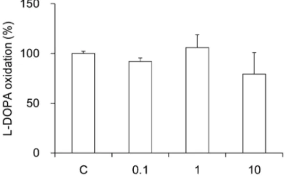
5. L-DOPA oxidation
The activities of ME on L-DOPA oxidation were recalculated into 100% of control group(Fig. 5). ME (0.1, 1, and 10 mg/ml) did not show any significance (92.0 ± 3.5%, 105.9 ±
12.8%, and 79.1 ± 21.7%, respectively).
Fig. 5. Effect of Meretrix extracts (ME) on L-DOPA oxidation. C:
control, distilled water treated group. 0.1, 1, and 10: ME 0.1, 1, and 10 mg/ml treated group. Data were expressed as the mean ± SEM of three experiments.
Discussion and Conclusion
Meretrix is the seashell of Meretrix meretrix or Cyclina sinensis. It is used to clear the Lung heat and dissolve sticky, copious phlegm. It treats accumulation of hot phlegm in the chest that has led to feelings of oppression in the chest, wheezing, dyspnea, cough and copious, yellow sputum. It also treats accumulation of phlegm and fire that has caused chest and hypochondriac pain, cough and wheezing1,2). It softens hardness and dissipates nodules. Clinical applications include treatment of scrofula and thyroid nodules. It neutralizes gastric acid to treat stomach pain, acid reflux, and gastric and duodenal ulcers. This drug has a mild action to promote urination to treat edema and dysuria. When applied topically, it aids healing of wounds, burns and eczema1,2). However, there was no study about the effects on collagenase and tyrosinase.
The skin aging is one of the most obvious evidence of aging. The skin is increasingly exposed to ambient UV-irradiation thus increasing risks for photo-oxidative damage with long-term detrimental effects like photo-aging, characterized by wrinkles, loss of skin tone and resilience.
Photo-aged skin displays alterations in the cellular component and extracellular matrix with accumulation of disorganized elastin and its microfibrillar component fibrilin in the deep dermis and a severe loss of interstitial collagens, the major structural proteins of the dermal connective tissue. MMPs are known to be upexpressed in human fibroblasts within hours after exposure to UV irradiation and are, therefore, considered key factors in the photo-aging process. Therefore, agents with the ability to elevate ECM protein levels or inhibit the major collagen-degrading enzymes like MMPs would prove to be useful in the development of effective anti-aging agents.
Collagen is a group of naturally occurring proteins. In nature,
it is found exclusively in animals, especially in the flesh and connective tissues of mammals12). It is the main component of connective tissue, and is the most abundant protein in mammals, making up about 25% to 35% of the whole-body protein content13). Collagen, in the form of elongated fibrils, is mostly found in fibrous tissues such as tendon, ligament and skin, and is also abundant in cornea, cartilage, bone, blood vessels, the gut, and intervertebral disc. In muscle tissue it serves as a major component of endomysium. Collagen constitutes 1% to 2% of muscle tissue, and accounts for 6% of the weight of strong, tendinous muscles14). Collagen occurs in many places throughout the body. So far, only 29 types of collagen have been identified and described. Over 90% of the collagen in the body, however, is of type I, II, III, and IV.
Among them, collagen type I is placed at skin, tendon, vascular, ligature, organs, and bone (main component of bone).
Collagen-related diseases most commonly arise from genetic defects or nutritional deficiencies that affect the biosynthesis, assembly, postranslational modification, secretion, or other processes involved in normal collagen production .
In this study, we evaluated the cytotoxicity of ME on human fibroblasts (HS68) at various concentrations. There was no cytotoxicity in all ME-treated concentrations. However, the amount of collagen type I was not increased at all concentration of ME treatments. In contrary, the amounts of type I collagen synthesis of ME were a little bit decreased in high dose treatment (100 μg/ml).
To evaluate the collagenase activity, matrix metalloproteinase-1 (MMP-1) activity was quantitatively measured. ME significantly reduced the MMP-1 activity at all concentrations of 10 μg/ml, 30 μg/ml, and 100 μg/ml in a dose-dependent manners. The activity was significantly reduced even in a low concentraion (10 μg/ml). It might suggest the effect of ME is not weak. However, the extracting yield is not economical. Accordingly, further studies on the extracting efficiency of ME and the animal experiments should be undertaken.
Tyrosinase is one of the important enzymes that has a key role in pigmentation process15). L-DOPA oxidation was also undertaken by tyrosinase. The effects of ME on tyrosinase activity was significantly effective at 10 mg/ml treatments.
In conclusion, ME showed the anti-wrinkle and whitening effects. However, the whitening effects were not enough to be used as whitening agents. These results suggest that ME may be a potential drug as an anti-aging treatments such as wrinkle care therapies. I think further studies will be needed to unravel exactly under the effects in vivo and clinical experiments and the molecular mechanisms of the effects
Acknowledgements
This work was supported by a grant from the Ministry of Knowledge Economy of the Republic of Korea(RIC-07-06-01).
References
1. The Boncho-hak textbook committee. Boncho-Hak. Seoul, Young-Lim Press, p 724, 2007.
2. Chen, J.L., Chen, T.T. Chinese medical herbology and pharmacology. CA, Art of Medicine Press. p 713, 2004.
3. Yaar, M., Gilchrest, B.A. Cellular and molecular mechanisms of cutaneous aging. J Dermatol Surg Oncol 16(10):915-922, 1990.
4. Gelse, K., Poschl, E., Aigner, T. Collagens-structure, function, and biosynthesis. Adv Drug Deliv Rev 55(12):1531-1546, 2003.
5. Fisher, G.J., Datta, S.C., Talwar, H.S., Wang, Z.Q., Varani, J., Kang, S., Voorhees, J.J. Molecular basis of sun-induced premature skin ageing and retinoid antagonism. Nature 379(6563):335-339, 1996.
6. Shin, J.Y., Hur, W., Wang, J.S., Jang, J.W., Kim, C.W., Bae, S.H., Jang, S.K., Yang, S.H., Sung, Y.C., Kwon, O.J., Yoon, S.K. HCV core protein promotes liver fibrogenesis via up-regulation of CTGF with TGF-beta1. Exp Mol Med 37(2):138-145, 2005.
7. Wei, X., Liu, Y., Xiao, J., Wang, Y. Protective effects of tea polysaccharides and polyphenols on skin. J Agric Food Chem 57(17):7757-7762, 2009.
8. Moon, H.J., Lee, S.H., Ku, M.J., Yu, B.C., Jeon, M.J., Jeong, S.H., Stonik, V.A., Zvyagintseva, T.N., Ermakova, S.P., Lee,
Y.H. Fucoidan inhibits UVB-induced MMP-1 promoter expression and down regulation of type I procollagen synthesis in human skin fibroblasts. Eur J Dermatol 19(2):129-134, 2009.
9. Vanni, A., Gastaldi, D., Giunata, G. Kinetic investigations on the double enzyme activity of the tyrosinase mushroom. Ann Chim 80: 35-60, 1990.
10. Joshi, P.C., Carraro, C., Pathak, M.A. Involvement of reactive oxygen species in the oxidation of tyrosine and dopa to melanin and in skin tanning. Biochem Biophys Res Commun 142(1):265-274, 1987.
11. Kim, Y.H., Chung, C.B., Kim, J.G., Ko, K.I., Park, S.H., Kim, J.H., Eom, S.Y., Kim, Y.S., Hwang, Y.I., Kim, K.H.
Anti-wrinkle activity of ziyuglycoside I isolated from a Sanguisorba officinalis root extract and its application as a cosmeceutical ingredient. Biosci Biotechnol Biochem 72(2):303-311, 2008.
12. Müller, W.E.G. The origin of metazoan complexity: Porifera as integrated animals. Integr Comp Biol 43(1):3-10, 2003.
13. Di Lullo, G.A., Sweeney, S.M., Korkko, J., Ala-Kokko, L., San Antonio, J.D. Mapping the ligand-binding sites and disease-associated mutations on the most abundant protein in the human, type I collagen. J Biol Chem 277(6):4223- 4231, 2002.
14. Sikorski, Z.E. Chemical and functional properties of food proteins. Boca Raton, CRC Press, p 242, 2005.
15. Lei, T.C., Virador, V.M., Vieira, W.D., Hearing, V.J. A melanocyte-keratinocyte coculture model to assess regulators of pigmentation in vitro. Anal Biochem 305(2):260-268, 2002.