Introduction
Several studies have sought the optimal reconstruction method to improve the mechanical stability of anterior cruciate ligament (ACL) grafts with regard to graft choice, number of bundles,
preservation of the remnant, and tunnel positioning
14). Preserva
tion of the remnant has been shown to provide several advantag
es, including better vascularization and synovial coverage of the graft
5,6), preservation of the mechanoreceptors, and facilitation of mesenchymal stem cell migration
79). However, the clinical ben
efit of remnant preservation has not been clearly established, with recent metaanalyses reporting similar clinical outcomes and mechanical stability after ACL reconstruction performed with and without remnant preservation
10,11). Furthermore, although one study has reported on the importance of preserving a tibial stump as a minimum, if the remnant cannot be fully preserved
12), another study demonstrated that the tunnel position rather than remnant preservation is more important to reproduce the func
tion of the original ACL after reconstruction
13).
Functional stability would further be enhanced after recon
struction by inducing good synovial coverage of the graft and by
Minimal Ablation of the Tibial Stump Using Bony
Landmarks Improved Stability and Synovial Coverage Following DoubleBundle Anterior Cruciate Ligament Reconstruction
Yuya Kodama, MD, PhD, Takayuki Furumatsu, MD, PhD, Tomohito Hino, MD, Yusuke Kamatsuki, MD, and Toshifumi Ozaki, MD, PhD
Department of Orthopaedic Surgery, Okayama University Graduate School of Medicine, Dentistry and Pharmaceutical Sciences, Okayama, Japan
Purpose: To evaluate the clinical effects of using anatomical bony landmarks (Parsons’ knob and the medial intercondylar ridge) and minimal
ablation of the tibial footprint to improve knee anterior instability and synovial graft coverage after doublebundle anterior cruciate ligament reconstruction.
Materials and Methods: We performed a retrospective comparison of outcomes between patients who underwent reconstruction with minimal
ablation of the tibial footprint, using an anatomical tibial bony landmark technique, and those who underwent reconstruction with wide ablation of the tibial footprint. Differences between the two groups were evaluated using secondlook arthroscopy, radiological assessment of the tunnel position, postoperative anterior knee joint laxity, and clinical outcomes.
Results: Use of the anatomical reference and minimal ablation of the tibial footprint resulted in a more anterior positioning of the tibial tunnel, with
greater synovial coverage of the graft postoperatively (p=0.01), and improved anterior stability of the knee on secondlook arthroscopy. Both groups had comparable clinical outcomes.
Conclusions: Use of anatomical tibial bony landmarks that resulted in a more anteromedial tibial tunnel position improved anterior knee laxity, and
minimal ablation improved synovial coverage of the graft; however, it did not significantly improve subjective and functional shortterm outcomes.
Keywords: Knee, Anterior cruciate ligament, Reconstruction, Double-Bundle, Tibial bony landmark pISSN 2234-0726 · eISSN 2234-2451
Knee Surgery & Related Research
Received April 12, 2018; Revised (1st) July 12, 2018;
(2nd) July 26, 2018; Accepted August 9, 2018 Correspondence to: Takayuki Furumatsu, MD, PhD
Department of Orthopaedic Surgery, Okayama University Graduate School of Medicine, Dentistry and Pharmaceutical Sciences, 251 Shikatacho, Kitaku, Okayama 7008558, Japan
Tel: +81862357273, Fax: +81862239727 Email: [email protected]u.ac.jp
348
This is an Open Access article distributed under the terms of the Creative Commons Attribution NonCommercial License (http://creativecommons.org/licenses/bync/4.0/) which permits unrestricted noncommercial use, distribution, and reproduction in any medium, provided the original work is properly cited.
Copyright © 2018 KOREAN KNEE SOCIETY
www.jksrr.org
accurately placing aperture of the tibial tunnel within the tibial footprint of the ACL. We focused on two aspects of the recon
struction technique mainly to acquire good synovial coverage and to create a reproducible anatomical tibial tunnel: preservation of a tibial stump by the use of minimal ablation (MA) of the tibial footprint of the ACL
12)and the use of bony landmarks (Parsons’
knob
14)and the medial intercondylar ridge
15)) on the tibia to de
termine the position of the tibial tunnel. Our aim was to compare the outcomes of our doublebundle ACL reconstruction, using bony anatomical landmarks on the tibia performed with MA of tibial stump, to those of doublebundle reconstruction performed with wide ablation (WA) of the tibial stump of the ACL. We hy
pothesized that MA would improve synovial coverage of the graft and that placement of the tibial tunnel based on the tibial bony landmarks during ACL reconstruction would enhance anterior knee stability postreconstruction.
Materials and Methods
This study was approved by our Institutional Review Board, and all patients provided informed consent prior to participation.
Between January 2012 and March 2015, 83 consecutive patients underwent an outsidein doublebundle ACL reconstruction us
ing hamstring autografts, performed by 2 surgeons. The medical records and intraoperative arthroscopic videos for these patients were reviewed to determine the procedure used: MA versus WA and tunnel positioning with or without reference to the bony landmarks. For procedures performed between January 2012 and March 2014, WA of the ACL stump was used, with creation of anteromedial (AM) and posterolateral (PL) tibial tunnels (WA group). Since April 2014, we modified our surgical technique on the tibial side of the reconstruction, using Parsons’ knob and the medial intercondylar ridge to position the tibial tunnel and at
tempting to preserve as much of the ACL stump as possible (MA
group), for following two reasons: first, we hoped the native ACL fibroblasts would be incorporated as part of the new composite graft to enhance synovial coverage of the graft; and second, pres
ervation of the remaining ACL mechanoreceptors might have a beneficial effect on clinical outcomes
12). Among the 83 patients who underwent ACL reconstruction during the study period, 26 underwent the MA procedure and the remaining 57, the conven
tional WA procedure. From this latter group of 57, we selected 26 patients, matched on demographics to the 26 patients in the MA group.
1. Evaluation of Outcomes
The relevant characteristics of the study group are reported in Table 1, with no preoperative difference between the two groups identified. Outcomes were assessed based on measurement of the position of the AM and PL tibial tunnels on reconstructed com
puted tomography (CT) images, direct examination of the graft during secondlook arthroscopy, quantification of anterior knee stability, and patientreported clinical outcomes measured 2 years after reconstruction, including the Lysholm knee score, the Inter
national Knee Documentation Committee (IKDC) score, and the Knee Injury and Osteoarthritis Outcome Score (KOOS).
2. Surgical Procedure
A doublebundle, outsidein arthroscopic ACL reconstruction was performed in all patients, with the graft formed using the semitendinosus tendon (ST) and, if necessary, the gracilis tendon, as follows. A double bundle was constructed solely from the ST when the harvested ST was >24 cm, with the tendon cut trans
versely into two equal portions. When the harvested ST was <24 cm, additional harvesting of the gracilis tendon was performed to obtain two equal portions (MA group, 3; WA group, 5). The har
vested tendons were doublelooped over an Endobutton fixation device (Smith & Nephew, Andover, MA, USA), with the distal
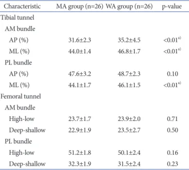
Table 1. Demographic and Clinical Characteristics
Characteristic MA group WA group pvalue
No. of patients 26 26
Age (yr) 26.5±9.2 (16–41) 23.7±5.2 (16–33) 0.60
Sex (male:female) 12:14 13:13
Meniscus injury (rasping/meniscectomy/repair) 14 (7/1/6) 13 (5/2/6)
Body mass index (kg/m
2) 22.0±2.5 (17.7–27.6) 22.4±2.7 (18.0–26.9) 0.46
Interval from injury to ACL surgery (day) 127.6±56.5 (32–280) 119.7±51.7 (35–272) 0.57
Interval from ACL surgery to secondlook arthroscopy (day) 419.7±61.9 (253–417) 411.4±133.8 (273–452) 0.23 Values are presented as mean±standard deviation (range).
MA: minimal ablation, WA: wide ablation, ACL: anterior cruciate ligament.
ends anchored using a Krackow suture, thus recreating the AM and PL bundles of the ACL. To prevent elongation of the grafts, a continuous 30 second loading with 70 N was applied twice to the graft (70 N, 1 min), and then the same loading was repeatedly applied (70 N, 2 min)
16). The femoral tunnel was created using an outsidein technique. The longitudinal linear resident’s ridge
17)and the posterior cartilage, used as landmarks for the ACL femo
ral footprint, were identified. Two 2.4mm guide pins were then inserted, separately, from the outside into the ACL footprint, posterior to the resident’s ridge and just anterior to the articular margin, using an anterolateralentry femoral aimer (Smith &
Nephew). A 5.5mm to 6.5mm tunnel was then created for the AM and the PL grafts by overdrilling of the guide pins. Two En
dobuttonCLs (Smith & Nephew) were connected to the end of each loop graft. The length of the CLs was matched to the length of the femoral tunnel so as to introduce sufficient graft materials (>13 mm) into the bone tunnels. The ACL remnant was resected with a shaver, preserving only the tibial stump.
3. Creation of the Tibial Tunnels for the WA and MA Groups Using the conventional WA procedure, in the WA group, the anterior horn of the lateral meniscus (LM) was identified as a ref
erence landmark
18), and the tunnel was created using WA within the ACL footprint. The AM tunnel was aligned with the anterior horn of LM, with the PL tunnel positioned in the posterior area within the footprint (Fig. 1). This WA approach does not pre
serve the tibial stump.
To preserve the tibial stump in the MA group, the AM tibial bony landmarks (Parsons’ knob and the medial intercondylar ridge) were identified and ablation was limited to the medial side
of the tibial footprint. The AM tunnel was created just lateral to the medial intercondylar ridge and just posterior to the Parsons’
knob. The PL tunnel was created posterior to the AM tunnel and lateral to the medial intercondylar ridge, so as not to overlap with the AM tunnel (Fig. 1).
In all cases, tibial fixation of the graft was performed with the knee flexed at 20°, with an initial tension of 20 N applied to the PL bundle and 30 N to the AM bundle. The tension in each bun
dle was independently measured using a tensiometer
19). 4. CT Image-Based Measurement
CT images were obtained using an Asteion 4 Multislice CT Sys
tem (Toshiba Medical Systems, Tochigi, Japan), at 120 kVp and 150 mA, with 1mm slice thickness. CT reconstruction of the tibial condyles, in the axial plane, was completed using a three
dimensional volumerendering technique (AZE Virtual Place software; AZE Ltd., Tokyo, Japan). The position of the apertures of the tibial and femoral tunnels was obtained using a rectangular measurement grid. On the femoral side, the location of the inser
tion sites was described as a percentage of the distance both par
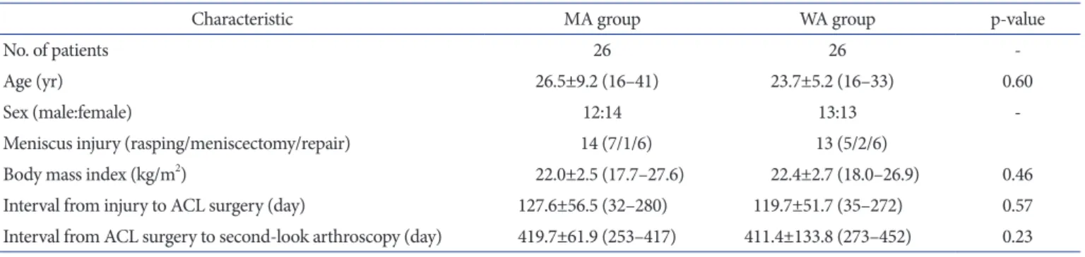
allel and perpendicular to Blumensaat’s line. On the tibial side, the location of the insertion sites was described as a percentage of the anteroposterior and mediolateral dimensions (Fig. 2).
5. Second-Look Arthroscopic Examination
Secondlook arthroscopy was performed, with patients’ con
sent, at approximately 1 year postreconstruction for the removal of the two doublespike plates (Meira, Aichi, Japan), fixed with screws into the tibia, which were used for the tibial fixation of the ACL graft. These data were retrospectively collected from the
MinimalablationWideablation
ACL remnant
ACL remnant
Lateral side
ofthe tibial
stump
LMAH
A B
D E
C
F
LMAH
Fig. 1. Ablation range and tunnel creation
within the anterior cruciate ligament (ACL) tibial footprint in the two groups. Tibial tunnel aperture of the left knee for the min
imal ablation group (A–C), with ablation performed just medial to the footprint (B).
(D–F) Wide ablation of the tibial stump, with no ACL stump retained. Black arrow
heads: Parsons’ knob, white arrowheads:
medial intercondylar ridge, black circle: lat
eral side of the tibial stump, LMAH: lateral
meniscus anterior horn.
records and confirmation from the review of arthroscopic videos.
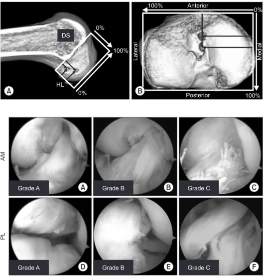
Arthroscopic evaluation was performed using the 3point syno
vial coverage scale as reported by Kondo and Yasuda
5), as follows:
grade A, 3 points; grade B, 2 points, and grade C, 1 point (Fig. 3).
6. Evaluation of Clinical Outcomes
Anterior knee laxity and rotational stability were measured under anesthesia by two experienced surgeons (TF and SM).
Anterior tibial translation was assessed at the primary surgery and secondlook evaluation using a KT2000 arthrometer at maximum manual tension, with the knee in 20° of flexion, as per a previously described procedure
20). Rotational stability was as
sessed using the manual pivot shift test, as previously described
21), at the time of the secondlook arthroscopy. The pivot shift test was subjectively determined by the examiner based on the IKDC criteria: none (−), glide (+), clunk (++), or gross instability (+++).
Functional outcomes were assessed 2 years after surgery using the Lysholm knee score, the IKDC form, and the KOOS, with scores obtained at each visit.
7. Statistical Analyses
Data were presented as means±standard deviations. Differences between the two groups were evaluated using the MannWhitney Utest, with a pvalue <0.05 considered significant. Intra and interobserver repeatability in measurement of the position of the tibial tunnel was assessed by calculating the intraclass correlation coefficient (ICC), with the Kappa value used for grading of syno
vial coverage of the graft.
Results
1. Tunnel Diameter and Position
Comparison of the position of the tibial and femoral tunnels between the two groups is reported in Table 2. Measurement of the tunnel position was consistent, with ICC values of 0.899–0.912 for intraobserver repeatability and 0.902–0.925 for interobserver repeatability. There was no significant difference in the position of the femoral tunnel between the MA and WA groups. However, there was a significant difference between the two groups in the
DS
0%
100%
0%
HL
100% Anterior
0%
Posterior 100%
Lateral
A B
Medial
Fig. 2. Computed tomography imagebased measurement of insertion sites in the femur and tibia. The total sagittal diameter of the lateral femoral condyle measured along Blumensaat’s line. (B) The location of the insertion sites on the tibia was expressed as a percentage of the anteriortoposterior and medialtolateral dimensions. DS:
deeptoshallow distance, HL: hightolow distance.
AMPL
Grade A Grade B Grade C
Grade A Grade B Grade C
A B C
D E F
Fig. 3. Evaluation of synovial coverage.
Arth roscopic grading of synovial coverage of the transplanted anteromedial (AM) (A–
C) and posterolateral (PL) (D–F) bundle grafts. Grade A: synovium covers >75% of the graft (A, D), Grade B: coverage between 50% and 75% (B, E), Grade C: coverage
<50% (C, F).
anteroposterior and mediolateral dimensions of the AM tibial tunnel (31.6%±2.3% and 44.0%±1.4%, respectively, for the MA group, and 35.2%±4.5% and 46.8%±1.7%, respectively, for the WA group; p<0.01) and the mediolateral dimension of the PL tibial tunnel (44.1%±1.7% for the MA group and 46.1%±1.5% for WA group; p<0.01).
2. Synovial Coverage Evaluation
Comparison of the arthroscopic evaluation between the two groups is reported in Table 3. The determination of the grade of synovial coverage was consistent, with a Kappa value of 0.72.
Good synovial coverage (grade A) of the AM graft was identified in 85% of cases (22 knees) in the MA group and 54% of cases (14 knees) in the WA group, with poor coverage (grade C) identified in 8% of cases (2 knees) in the WA group. The synovial cover
age scale of the AM bundle, based on Kondo’s synovial coverage grade, was 2.8±0.4 in the MA group and 2.4±0.7 in the WA group (p=0.03). No difference in the synovial coverage scale was identi
fied between the two groups for the PL bundle (p=0.51).
3. Clinical Outcomes
A comparison of the clinical outcomes at the time of the secondlook arthroscopy examination between the two groups is reported in Table 4. The KT2000 values were significantly
lower for the MA group than for the WA group (0.6±0.5 mm and 1.4±0.8 mm, respectively, p<0.01). With regard to rotation stabil
ity, the pivot test was negative for 50 patients, with a positive “glide (+)” assessed for 2 patients and with no differences between the two groups. Furthermore, there were no differences between the Table 2. Comparison of the Position of the Tibial and Femoral Tunnels
between the Two Groups
Characteristic MA group (n=26) WA group (n=26) pvalue Tibial tunnel
AM bundle
AP (%) 31.6±2.3 35.2±4.5 <0.01
a)ML (%) 44.0±1.4 46.8±1.7 <0.01
a)PL bundle
AP (%) 47.6±3.2 48.7±2.3 0.10
ML (%) 44.1±1.7 46.1±1.5 <0.01
a)Femoral tunnel AM bundle
Highlow 23.7±1.7 23.9±2.0 0.71
Deepshallow 22.9±1.9 23.5±2.7 0.50
PL bundle
Highlow 51.2±1.8 50.1±2.4 0.16
Deepshallow 32.3±1.9 31.5±2.4 0.23
Values are presented as mean±standard deviation.
MA: minimal ablation, WA: wide ablation, AM: anteromedial, AP: ante
ro posterior, ML: mediolateral, PL: posterolateral.
a)
p<0.05.
Table 3. Comparison of Arthroscopic Evaluation Results between the Two Groups
Characteristic MA group
(n=26) WA group (n=26) pvalue AM synovial coverage, no. (%)
Grade A 22 (85) 14 (54)
Grade B 4 (15) 10 (38)
Grade C 0 (0) 2 (8)
Synovial coverage score, mean±SD 2.8±0.4 2.4±0.7 0.03
a)PL synovial coverage, no. (%)
Grade A 18 (69) 13 (50)
Grade B 6 (23) 10 (38)
Grade C 2 (8) 3 (12)
Synovial coverage score, mean±SD 2.5±0.7 2.3±0.7 0.51 Synovial coverage score was evaluated on a 3point scale: grade A, 3 points; grade B, 2 points; and grade C, 1 point.
MA: minimal ablation, WA: wide ablation, AM: anteromedial, SD: standard deviation, PL: posterolateral.
a)
p<0.05.
Table 4. Comparison of the Clinical Outcome at the Final Followup between the Two Groups
Characteristic MA group
(n=26) WA group (n=26) pvalue
Lysholm knee score 95.9±4.3 95.7±6.6 0.87
IKDC evaluation 83.7±9.7 83.8±8.9 0.97
KOOS pain 93.0±6.1 92.7±5.6 0.74
Symptom 92.4±7.9 87.3±8.5 0.06
Activities of daily living 98.1±4.8 97.5±3.4 0.54 Sport/recreation 85.5±12.7 88.1±8.9 0.59 Quality of life 86.7±11.1 80.9±14.9 0.19 KT2000 at secondlook (mm) 0.6±0.5 1.4±0.8 0.003
a)Pivot shift test at secondlook (%) 0.84
None (–) 26 (100) 24 (92)
Glide (+) 0 2 (8)
Values are presented as mean±standard deviation.
MA: minimal ablation, WA: wide ablation, IKDC: International Knee Do cumen tation Committee, KOOS: Knee Injury and Osteoarthritis Out come Score.
a)