Received: 25 April, 2014 Revised: 3 June, 2014 Accepted: 5 June, 2014 Corresponding author: Jerrold S. Petrofsky
Department of Physical Therapy, Loma Linda University, Loma Linda, CA 92350, USA Tel: 1-909-558-7274 Fax: 1-909-558-0481 E-mail: [email protected]
This is an Open-Access article distributed under the terms of the Creative Commons Attribution Non-Commercial License (http://creativecommons.org/licens es/by-nc/3.0) which permits unrestricted non-commercial use, distribution, and reproduction in any medium, provided the original work is properly cited.
Copyright © 2014 Korean Academy of Physical Therapy Rehabilitation Science
http://dx.doi.org/10.14474/ptrs.2014.3.1.20 Phys Ther Rehabil Sci
pISSN 2287-7576 2014, 3 (1), 20-26
eISSN 2287-7584 www.jptrs.org
The effects of low level laser radiation on bacterial growth
Wendy Chung
a, Jerrold S. Petrofsky
b, Michael Laymon
c, Jason Logoluso
a, Joon Park
a, Judy Lee
a, Haneul Lee
baDepartment of Physical Therapy, Azusa Pacific University, Azusa, CA, USA
bDepartment of Physical Therapy, Loma Linda University, Loma Linda, CA, USA
cSchool of Physical Therapy, Touro University, Henderson, NV, USA
Objective: The low level lasers currently in the market vary in wavelength, dosage, and frequency. These devices are used with much different clinical pathology. Most notably, some studies claim that wounds heal faster with low level laser therapy due to the fact that bacteria commonly found in wounds are killed by laser light. Systemic and meta-analysis studies found the difficulty of comparison of numerous research studies because of differences in the intensities and frequencies of low level laser treatment (LLLT). The purpose of this study was to determine the effectiveness of LLLT on controlling bacterial growth.
Design: Cross-sectional study.
Methods: Variables included LLLT dosage and wavelength on 3 bacteria commonly seen in wounds, strains of Staphylococcus aureus, Escherichia coli, and Pseudomonas aeruginosa were used on commercially available 5.0-cm agar plates. Blue, green, and red, ultraviolet (UV) and infrared laser light sources were adjusted to either low or high intensity settings. Five Petri dishes at a time were placed directly beneath laser light sources with the exception of UV which was placed six inches below the suspended light and infrared which was placed directly on top of the Petri dish lid. Each group of five Petri dishes was irradiated for 15 minutes.
Results: The results showed no effect of any of 9 different LLLT intensities or colors on bacteria growth compared to sham light.
Conclusions: At least for claims of bacterial growth inhibition with LLLT, no support for this claim can be found here.
Key Words: Bacteria, Light, Low level laser, Wounds
Introduction
Low level lasers have been used for decades for the treat- ment of a wide range of medical conditions including soft tissue injuries, musculoskeletal disorders, and wound heal- ing [1]. Although low level laser treatment (LLLT) has been widely used, many research studies have shown little or no benefit of LLLT. The physiological mechanism of low level laser is poorly understood, and treatment parameters such as intensity, frequency, wavelength, and dosage are uncertain as well [2]. Even though the effectiveness of LLLT has not been proven, many practitioners continue to use LLLT to treat many conditions such as Carpal Tunnel Syndrome (CTS), musculoskeletal pain, inflammatory disease, venous
leg ulcers, and decubitus ulcers [3].
Naeser et al. [4] performed a randomized, double-blind, cross-over study of LLLT. The study population consisted of eleven patients with mild to moderate CTS. Patients were randomly assigned to receive nine to 12 sessions of active or sham LLLT and transcutaneous electrical nerve stimulation (TENS) treatment. The results showed that seven of the re- maining eight subjects reported pain scores reduced by more than 50% post active LLLT and TENS treatment. All 11 sub- jects reported that they resumed their previous work activ- ities with little to no pain.
The population of this study was small (n=11), and TENS
was used with LLLT and therefore, the results of the study do
not show an effect of LLLT alone [5].
Irvine conducted a double blind randomized controlled trial of LLLT in August 2004. Fifteen CTS patients, 34 to 67 years of age, were randomly assigned to either the control group (n=8) or treatment group (n=7). Both groups were treated three times per week for five weeks. For this study 860 nm lasers were applied at a dosage of 6 J/cm
2to the treat- ment group over the carpal tunnel, and those in the control group were treated with sham laser. After completion of treatment, there was no significant difference in any of the outcome measures between the two groups [6].
Gam et al. [2] conducted a meta-analysis of 23 trials on LLLT for musculoskeletal pain. The mean difference be- tween treatment and placebo on a pain visual analogous scale was 0.3% indicating no effect of LLLT on pain in mus- culoskeletal syndromes. The differences were not weighted by the sample because trials varied in the musculoskeletal diseases treated, LLLT dose, laser type, and wavelength.
Mulcahy et al. [7] conducted a randomized double-blind placebo controlled trial to study the effect of LLLT on lo- calized, painful, soft tissue conditions. Twenty-three pa- tients were randomized to active or placebo groups. LLLT with an intensity of 35 mW was applied two times per week for four weeks to the treatment group. The results showed that 87% of the placebo felt their pain had improved com- pared to 42% of the active group.
Flemming and Cullum [8] reviewed four randomized controlled trials to determine the effect of LLLT on venous leg ulcers. Two studies compared LLLT with placebo treatment. One study compared LLLT with ultraviolet (UV) therapy. The fourth study compared the effect of three treat- ments including LLLT, LLLT plus infrared light therapy, and non-coherent unpolarized red light therapy. Flemming and Cullum [8] did not find any evidence of the benefit of LLLT on venous leg ulcer healing.
Lucas et al. [9] conducted a randomized observer-blind trial in three nursing homes to assess the effect of LLLT on decubitus ulcers. Eighty-four subjects with Stage III decubi- tus ulcers participated in the study. There were 47 subjects in the control group and 39 in LLLT group. The control group received standard wound treatments including patient in- struction, wound cleansing, moist dressings, and frequent alteration of position. Standard wound treatments were ap- plied daily for six weeks. The LLLT group received standard treatment and LLLT. The 904 nm lasers with an average beam power of 12×8 mW were applied five times a week for six weeks. This study did not find any significant differences between the groups.
Saltmarche [10] in 2012, found significant improvement in 61% of wounds with LLLT. But there were several types of wounds and modalities for treatment were mixed. They felt that LLT killed common bacteria and there allowed for faster healing of wounds.
Thus there is confusion in the literature, especially on the healing of wounds with lasers. To start more simply, here we used laser exposure of bacteria to different frequencies to see if the claim as to lasers reducing simple bacterial growth are valid. Further, the bacteria used the most common 3 found in human wounds making this relevant to wound treatment. By using different intensities of light and differ- ent frequencies, we designed the study to see if any of these 3 bacteria would show reduced growth when exposed di- rectly to laser light.
Methods Bacteria culture
Strains of Staphylococcus aureus (ATCC# 49444), Es-
cherichia coli (ATCC# 25922), Pseudomonas aeruginosa(ATCC# 35032), were purchased from Microbiologics (St.
Cloud, MN, USA). Commercially available 5.0-cm agar plates were used in this study for optimal distribution of laser light irradiation over the plate surface [11,12]. The agar plates were prepared and purchased from Hardy Diagno- stics. Nutrient broth was purchased from the Carolina Biological Supply Company. To prepare the nutrient broth, 0.8 g of tryptic soy broth was added to saline to make 100 ml.
One ml of P. aeruginosa culture was transferred to a sterile test tube with nine ml of sterile 0.9 % NaCl saline to create a 10
−6dilution of each bacterial culture. The Petri dishes were gently agitated for uniform distribution over the agar.
This method was repeated for S. aureus and E. coli bacterial cultures.
Laser parameters
An adjustable stand was used to stabilize the lasers di- rectly on top of the Petri dish for an optimal angle of irradiation. The lasers were adjusted one inch over the Petri dish for high level intensity and six inches over the Petri dish for low level intensity. Irradiation of Petri dishes took place in a dark room at room temperature (37
oC). Six different wavelengths of light were used (Unitech Systems Inc./LC LED Inc., Brooklyn, NY, USA). A light meter was used to read the output for each laser light. The lasers utilized were:
red (630 nm), infrared (904 nm), green (525 nm), blue (465
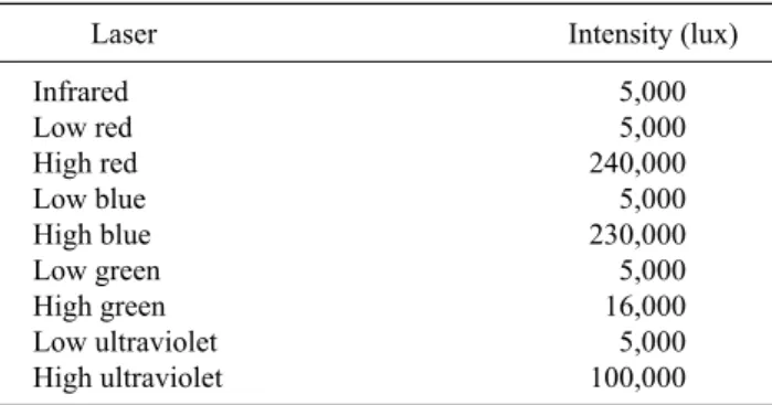
Table 1. Laser types and intensities utilized
Laser Intensity (lux)
Infrared 5,000
Low red 5,000
High red 240,000
Low blue 5,000
High blue 230,000
Low green 5,000
High green 16,000
Low ultraviolet 5,000
High ultraviolet 100,000
Table 2. Irradiation schedule for bacteria exposure to laser light (unit: lux)
IR Low red Low blue Low green Low UV High red High blue High green High UV Escherichia coli (×3 trials) 5,000 5,000 5,000 5,000 5,000 240,000 230,000 16,000 100,000 Pseudomonas aeruginosa (×3 trials) 5,000 5,000 5,000 5,000 5,000 240,000 230,000 16,000 100,000 Staphylococcus aureus (×3 trials) 5,000 5,000 5,000 5,000 5,000 240,000 230,000 16,000 100,000 IR: infrared, UV: ultraviolet.
nm), and UV (350 nm).
Commercially purchased cold lasers commonly use watts as a measure of intensity. A watt is a poor measure to de- termine the brightness of a laser. It refers to how much elec- trical power goes into the laser. However, lux is a measure of how much light is reaching a particular location. Therefore, lux was used as a light measure in this study. Lasers were calibrated to the following intensities (Table 1).
Storage and counting
All bacteria were stored in a Forma Scientific incubator at 37.4
oC. S. aureus and E. coli colonies were counted using a bacteria counter (Hardy Diagnostics) P. aeruginosa was counted as a percentage of growth covering each Petri dish using a grid (Bel-Art Products, Wayne, NJ, USA). One cm
2squares were printed onto transparencies. The 5-cm diame- ter lids were traced onto the grids and were then placed on top of each Petri dish to trace the bacterial growth with a marker. The number of squares covered with growth was counted and divided by the total area of the Petri dish to de- termine percentage growth. The average of four trials was taken and represented in a Table. Reliability of colony count data was performed through using a SD method.
Procedures
Forty four Petri dishes were labeled with the bacterial spe- cies, dilution, date, and the intended laser light source to be
irradiated. Fifty μl of P. aeruginosa at 10
−6dilution was distributed into each of these dishes using a 100-μl pipetteman. Dishes were gently agitated immediately after distribution and covered with their respective lids.
Blue, green, and red laser light sources were adjusted to low intensity settings. Five Petri dishes at a time were placed directly beneath laser light sources with the exception of UV which was placed six inches below the suspended light and infrared which was placed directly on top of the Petri dish lid. Each group of five Petri dishes was irradiated for 15 minutes. After irradiation, the Petri dishes were immediately inverted and placed into an incubator for 24 hours. This pro- cedure was repeated three times.
Blue, green, and red laser light sources were then adjusted to high intensity settings and the UV light was adjusted to one inch above Petri dish height. Groups of four Petri dishes at a time were then irradiated for 15 minute-intervals.
Immediately following irradiation, these Petri dishes were also inverted and placed into an incubator for exactly 24 hours. Four shams were also placed in the incubator for ex- actly 24 hours. These preceding procedural steps were re- peated for S. aureus and E. coli.
After incubation, results were recorded as a percentage of growth of P. aeruginosa over the Petri dishes. For S. aureus and E. coli, single bacterial colonies were counted. These re- sults were entered into an Excel spreadsheet and analyzed for significant data and SD.
See the chart below for all Petri dishes irradiated with their corresponding laser light sources (Table 2).
Data analysis
Data analysis was accomplished by calculating means and SD. ANOVA was conducted to compare mean percent- age of growth among nine different colors of light and sham groups. LSD pairwise comparisons test for multiple com- parisons was used to compare means of variables between any two different groups. The level of significance was p
<0.05.
Figure 1. Staphylococcus aureus percentage of growth per four tri- als exposed to nine different sources of light. UV: ultraviolet, IR:
infrared, (L): low, (H): high.
Table 3. Staphylococcus aureus summary of results
Group Count Sum Average Variance
Low UV 4 46.41 11.60 3.97
Low blue 4 50.87 12.72 6.38
Low green 4 49.53 12.38 7.08
Low red 4 55.2 13.80 13.29
IR 4 47.03 11.76 7.59
High UV 4 40.02 10.01 3.76
High blue 4 44.61 11.15 0.09
High green 4 44.63 11.16 0.97
High red 4 39.24 9.81 5.52
Sham 4 52.51 13.13 0.37
UV: ultraviolet, IR: infrared.
Figure 2. Staphylococcus aureus average percentage of growth among all four trials. UV: ultraviolet, IR: infrared, (L): low, (H):
high.
Figure 3. Pseudomonas aeruginosa percentage of growth per four trials exposed to nine different sources of light. UV: ultraviolet, IR:
infrared, (L): low, (H): high.
Results
S. aureus showed no significant difference in growth
when irradiated with the nine different colors of light and two different laser intensities in comparison to the shams.
Figure 1 shows four trials of S. aureus, and the percentage of growth areas after exposure to nine different light sources including the sham. Figure 2 is an average of the four trials for S. aureus. Statistical differences were not shown among the averages of exposed groups of bacteria when compared to the sham. Overall results are summarized in Table 3.
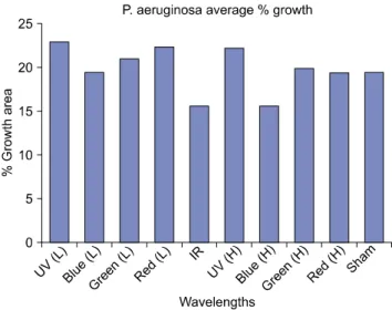
P. aeruginosa also showed no significant difference in
growth when irradiated with the nine different light colors and two different laser intensities in comparison to the shams. Figure 3 shows four trials of P. aeruginosa, and the percentage of growth areas after exposure to nine different light sources including the sham. Figure 4 is an average of the four trials for P. aeruginosa. Although there was a trend for infrared (IR) and Blue (H) lasers to decrease bacterial growth, there were not statistical differences when com- pared to the average growth of the sham plates. Statistical differences were not shown among the averages of exposed groups of bacteria when compared to the shams. Overall re- sults are summarized in Table 4.
Lastly, E. coli showed no significant difference in growth
when irradiated with the nine different light colors and two
Table 5. Escherichia coli summary of results
Group Count Sum Average Variance
Low UV 4 100.83 25.21 1.96
Low blue 4 88.48 22.12 2.25
Low green 4 95.99 24.00 15.32
Low red 4 103.89 25.97 26.53
IR 4 94.92 23.73 6.53
High UV 4 93.12 23.28 2.65
High blue 4 93.14 23.29 3.85
High green 4 90.62 22.66 3.53
High red 4 0.00 0.00 0.00
Sham 4 98.15 24.54 7.64
UV: ultraviolet, IR: infrared.
Figure 4. Pseudomonas aeruginosa average percentage of growth among all four trials. UV: ultraviolet, IR: infrared, (L): low, (H):
high.
Table 4. Pseudomonas aeruginosa summary of results
Group Count Sum Average Variance
Low UV 4 92.5 23.13 13.35
Low blue 4 78.25 19.56 9.43
Low green 4 84.38 21.09 7.68
Low red 4 89.88 22.47 13.82
IR 4 62.63 15.66 15.43
High UV 4 89.38 22.349 30.54
High blue 4 62.75 15.69 1.31
High green 4 80.00 20.00 2.54
High red 4 78.00 19.50 23.83
Sham 4 77.75 19.44 12.56
UV: ultraviolet, IR: infrared.
Figure 5. Escherichia coli percentage of growth per four trials ex- posed to nine different sources of light. UV: ultraviolet, IR: infra- red, (L): low, (H): high.
Figure 6. Escherichia coli average percentage of growth among all four trials. UV: ultraviolet, IR: infrared, (L): low, (H): high.
different laser intensities in comparison to the shams. The following Figure 5 shows four trials of E. coli, and the per- centage of growth areas after exposure to nine different light sources including the sham. Figure 6 is an average of the four trials for E. coli. During the first trial of exposure to the Red (H) laser, the light source malfunctioned, resulting in a lack of data for those four trials. Statistical differences were not indicated among the averages of exposed groups of bac- teria when compared to the shams. Overall results are sum- marized in Table 5.
Discussion
Healthcare professionals have used LLLT for a wide
range of conditions [3,8,13-16]. However, the clinical effec-
tiveness of LLLT has not been proven. Mechanism and treat-
ment parameters of LLLT have not been properly estab-
lished in previous studies. Many research studies show that LLLT has no effect on conditions claimed by manufacturers.
Some show that LLLT is less effective than placebo treatments. Although some claim that LLLT is effective for certain conditions such as CTS, most of these research stud- ies are poorly designed or have no control group [17].
Biostimulation theory is one hypothesis that has ex- plained the mechanism of LLLT in recent studies. This theo- ry states that LLLT will cause a biostimulation effect on tar- get human tissues via non-thermal low intensity irradiation of laser light [18]. However, commercially certified LLLT products in United States are classified as infrared lamps, which are thermal-type devices [13,16,19]. Therefore, sev- eral current indications for LLLT usage contradict this bio- stimulation theory.
Many of LLLT devices emit light that is strong enough to cause thermal effects on human tissues. It was reported that twenty-nine patients had sustained thermal burns from use of the Anodyne Therapy System (890 nm infrared diodes).
The manufacturer of this device received a warning from the Food and Drug Administration in 2005 for promoting laser devices for the treatment of non-certified conditions includ- ing soft tissue injuries, CTS, wounds, neuropathy and lym- phedema [20].
Meta-analysis studies of LLLT have not been able to com- pare studies effectively, often lacking specific treatment pa- rameters including intensity, time, frequency, and irradiated area. Most studies used Watts and Joules to measure how much light is emitted to the target area. The watt refers to how much electric power goes through the device, not how much light it produces. Joules measure how much work has been done per square cm on an area. In our study, lux was used as a light intensity measure because it is more accurate in establishing how many photons of light reach a particular location.
In our study, we irradiated three different bacteria species commonly found in human wounds (S. aureus, E. coli, or P.
aeruginosa) with nine different wavelengths of laser lights.
None of the laser lights caused significant difference in growth on any of the three bacteria species. Therefore, the significance of our study supports the possibility that low-level lasers are not effectively inhibiting or enhancing growth of bacteria when irradiated with the specified parameters.
Biostimulation effects of LLLT are possible when pho- tons of laser light are absorbed into a target human chromophore. However, each chromophore absorbs only a
narrow-range specific wavelength of light. This specific wavelength of light is required to stimulate each chromo- phore in human tissue. For example, an absorption spectrum of lipid peaks at 930 nm and drops low significantly at 970 nm (Conway 1984). Therefore, in order to be effective, laser light must be emitted at a very specific wavelength of light.
An issue of tissue penetration is also a concern. Without the actual occurrence of tissue penetration, the effectiveness of laser light at the cellular level is meaningless. When pho- tons in laser light enter tissues, they can be transmitted, scat- tered, reflected, or absorbed [21,22]. These behaviors of la- ser light are related to the penetration depth capacity of the human tissue [17].
Different human tissues have specific absorption charac- teristics depending on distinct components [22,23]. For ex- ample, infrared light is absorbed primarily by water, while visible and ultraviolet lights are absorbed mainly by hemo- globin and melanin, respectively. Because absorption co- efficients are so specific to tissue components, there lies a tremendous possibility for error when utilizing LLLT to ach- ieve desired cellular effects in the clinical setting.
In conclusion, LLLT in this study had no effect on bacteria growth. Therefore, if there is an effect on wound healing, it cannot be claimed to be due to killing bacteria. Further in- vestigation is warranted to see what LLLT really can do.
Certainly issues with tissue penetration probably limit the effect of LLLT to a placebo effect.
References
1. Gamaleya NF. Laser biomedical research in USSR. Laser Appl Med Biol 1977;1:1-173.
2. Gam AN, Thorsen H, Lønnberg F. The effect of low-level laser therapy on musculoskeletal pain: a meta-analysis. Pain 1993;52:
63-6.
3. Basford JR. Low-energy laser therapy: controversies and new re- search findings. Lasers Surg Med 1989;9:1-5.
4. Naeser MA, Hahn KA, Lieberman BE, Branco KF. Carpal tunnel syndrome pain treated with low-level laser and microamperes transcutaneous electric nerve stimulation: A controlled study.
Arch Phys Med Rehabil 2002;83:978-88.
5. Fontana CR, Kurachi C, Mendonça CR, Bagnato VS.
Temperature variation at soft periodontal and rat bone tissues during a medium-power diode laser exposure. Photomed Laser Surg 2004;22: 519-22.
6. Irvine J, Chong SL, Amirjani N, Chan KM. Double-blind randomized controlled trial of low-level laser therapy in carpal tunnel syndrome. Muscle Nerve 2004;30:182-7.
7. Mulcahy D, McCormack D, McElwain J, Wagstaff S, Conroy C.
Low level laser therapy: a prospective double blind trial of its use in an orthopaedic population. Injury 1995;26:315-7.
8. Flemming K, Cullum N. Laser therapy for the treatment of ve- nous leg ulcers. J Tissue Viability 1999;9:67.
9. Lucas C, van Gemert MJ, de Haan RJ. Efficacy of low-level laser therapy in the management of stage III decubitus ulcers: a pro- spective, observer-blinded multicentre randomised clinical trial.
Lasers Med Sci 2003;18:72-7.
10. Saltmarche AE. Low level laser therapy for healing acute and chronic wounds-the extendicare experience. Int Wound J 2008;5:351-60.
11. Nussbaum EL, Lilge L, Mazzulli T. Effects of 810 nm laser irra- diation on in vitro growth of bacteria: comparison of continuous wave and frequency modulated light. Lasers Surg Med 2002;31:
343-51.
12. Nussbaum EL, Lilge L, Mazzulli T. Effects of 630-, 660-, 810-, and 905-nm laser irradiation delivering radiant exposure of 1-50 J/cm2 on three species of bacteria in vitro. J Clin Laser Med Surg 2002;20:325-33.
13. U.S. Food and Drug Administration. Summary of Safety and Effectiveness. 510(k) Summary Microlight 830 Laser. Silver Spring: U.S. Food and Drug Administration, 2002.
14. U.S. Food and Drug Administration. Summary of Safety and Effectiveness. 510(k) Summary-Acculaser Pro Low Loevel Laser Therapy. Silver Spring: U.S. Food and Drug Administrat- ion, 2002.
15. Summary of Safety and Effectiveness. 510(k) Summary-TUGO Erchonia PL2002. 2002.
16. FDA. Summary of Safety and Effectiveness. 510(k) Summary - AcculaserTM Pro4. 2004.
17. Wang HW, Zhu TC, Putt ME, Solonenko M, Metz J, Dimofte A, et al. Broadband reflectance measurements of light penetration, blood oxygenation, hemoglobin concentration, and drug concen- tration in human intraperitoneal tissues before and after photo- dynamic therapy. J Biomed Opt 2005;10:14004.
18. Kujawa J, Zavodnik L, Zavodnik I, Bryszewska M. Low-in- tensity near-infrared laser radiation-induced changes of ace- tylcholinesterase activity of human erythrocytes. J Clin Laser Med Surg 2003;21:351-5.
19. FDA. Summary of Safety and Effectiveness. 510(k) Summary - BioFlex Professional Therapy Systems. 2003.
20. FDA. Warning Letter: FLA-06-08. Department of Health and Human Services. 2005.
21. van Gemert MJ, Jacques SL, Sterenborg HJ, Star WM. Skin optics. IEEE Trans Biomed Eng 1989;36:1146-54.
22. Sears FW, Young HD. Young sears. In: Sears FW, Zemansky MW, Young HD, editors. University physics. 5th ed. Reading (MA): Addison-Wesley Pub. Co.; 1976.
23. Rosencwaig A, Pines E. A photoacoustic study of newborn rat stratum corneum. Biochim Biophys Acta 1977;493:10-23.