Copyright © 2019, The Korean Society of Veterinary Service. All Rights Reserved. 275
한국가축위생학회지 제42권 제4호 (2019)Korean J Vet Serv, 2019, 42(4), 275-278 ISSN 1225-6552, eISSN 2287-7630 https://doi.org/10.7853/kjvs.2019.42.4.275
< Case Report >
Korean Journal of
Veterinary Service
Available online at http://kjves.org
*Corresponding author: Eun-Jin Choi, Tel. +82-54-912-0460, Fax. +82-54-912-0465, E-mail. choiej@korea.kr
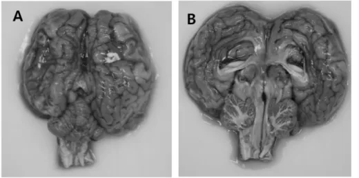
Encephalopathy caused by maternal deficiency of vitamin A in a calf
Kyunghyun Lee
1, Jongho Kim
1, Ujin Jeon
2, Yeon Hee Kim
1, Ha-Young Kim
1, ByungJae So
1, Eun-Jin Choi
1*
1
Animal Disease Diagnostic Division, Animal and Plant Quarantine Agency, Gimcheon 39660, Korea
2