• 검색 결과가 없습니다.

Antioxidant Activities of Various Extracts of Hovenia dulcis Thunb Fruits

N/A
N/A
Protected

Academic year: 2021

Share "Antioxidant Activities of Various Extracts of Hovenia dulcis Thunb Fruits"

Copied!
7
0
0

로드 중.... (전체 텍스트 보기)

전체 글

(1)

Antioxidant Activities of Various Extracts of Hovenia dulcis Thunb Fruits

Weicheng Hu

1,2

, Kabyeon Lee

3

and Myeong-Hyeon Wang

1

*

1

College of Biomedical Science, Kangwon National University, Chuncheon 200-701, Korea

2

College of Biosciences and Biotechnology, Yangzhou University, Yangzhou 225009, China

3

Korea Forest Research Institute, 44-3 Omokchon-dong, Kwonsun-ku, Suwon, 441-847, Korea

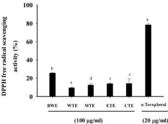
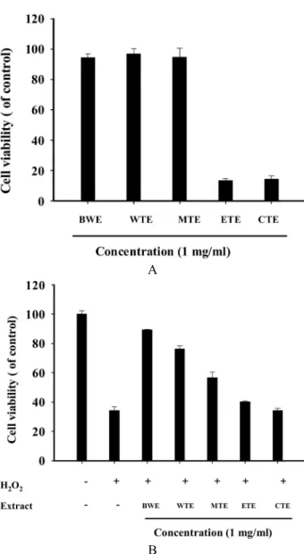
Abstract - Hovenia dulcis Thunb fruits were successively extracted with hot water, water, methanol, ethyl acetate, and

chloroform. The crude extracts were investigated for potential antioxidant by measuring scavenging against DPPH free radicals, reducing power, superoxide radicals, and protection of protein damage and cultured cells from a lethal dose of hydrogen peroxide (H

2

O

2

). In all chemical assays used, the hot water extract of H. dulcis fruits, which contained 61.14 ± 2.57 (Tannic acid mg/g extract, n=3) of total phenolic compounds contents exhibited highest activity in in vitro models of DPPH free radical scavenging activity, reducing power assay, superoxide radical scavenging activity and protection of protein damage. In addition, the hot water extract protected cultured RAW 264.7 macrophages from a lethal dose of H

2

O

2

and reduced reactive oxygen species level in RAW 264.7 cells.

Key words - Hovenia dulcis fruit, Antioxidant, Reactive oxygen species, Protein damage

*Corresponding author. E-mail : [email protected]

Introduction

Reactive oxygen species (ROS) and reactive nitrogen species (RNS) including free radicals such as superoxide ra- dical anion (•O2

-

), hydroxyl radicals (•OH), singlet oxygen (

1

O

2

), hydrogen peroxide (H

2

O

2

) and nitric oxide (NO) are constantly produced continuously in the cells of the human body (Fang et al., 2002). Oxidation is essential in living orga- nisms to obtain the energy needed for biological processes.

However, excessively high levels of free radicals or ROS create oxidative stress, which leads to produce some detri- mental effects, including lipid peroxidation of cellular memb- ranes, alteration of lipid–protein interaction, enzyme inacti- vation and DNA breakage, and in the end, to cause cell injury, necrosis or apoptosis (Lefebvre et al., 2002; Agarwal et al., 2003). Although human body has multiple antioxidant sys- tems to protect the cellular molecules against the oxygen radi- cals induced damage. These defense mechanisms include antioxidative enzymes, such as superoxide dismutase, cata- lase and glutathione peroxidase. The innate defense is not enough for severe oxidative stress, and therefore overpro-

duction of oxidative radicals may cause tissue damages (Blok- hina et al., 2003; Lassen et al., 2008). Consequently, certain amounts of exogenous antioxidants are constantly required to retain an adequate level of antioxidants in order to balance the ROS in human body (Reiter et al., 1997). Therefore, much attention has been focused on natural antioxidants. Plants contain a variety of free radical scavenging molecules such as phenolic compounds, nitrogen compounds, vitamins, terpe- noids, and other endogenous metabolites that are rich in anti- oxidant activity (Su et al., 2009; Huang et al., 2010).

Hovenia dulcis Thunb, belongs to Rhamnaceae family, is

wildly distributed in East Asia. It has been used in traditional

folk remedies for the treatment of liver diseases and detoxi-

fication of alcoholic poisoning. H. dulcis can remarkably re-

duce alcohol concentration in blood; thereby alleviate alco-

holic liver tissue (Hase et al., 1997; Kim, 2001). Some per-

vious literatures showed that H. dulcis possessed antioxi-

dative, antimicrobial, anti-diabetic, and anticancer activities

(Wang et al., 1994; Lee et al., 1999; Cho et al., 2000; Ji et al.,

2002). The fruit of H. dulcis is edible raw or cooked. Some

researchers reported that the fruits of H. dulcis own hepatop-

rotective activity (Ko et al., 2006). Nevertheless, research on

the directly free radical scavenging activity the fruit of H.

(2)

dulcis has not been reported.

To investigate the potential antioxidant properties of fruits of H. dulcis, we utilized a number of in vitro assay systems, including DPPH free radical scavenging activity, reducing power assay, superoxide radical scavenging assay, and pro- tective ability on protein oxidation and H

2

O

2

-injured RAW 264.7 cell death.

Material and Methods

Chemicals and reagents

L-Ascorbic acid, DPPH (1,1-diphenyll-2-2-pricylhydrazyl), ferrous chloride, 2N folin-ciocalteu’s phenol reagent, tannic acid, α-tocopherol, and trichloroacetic acid (TCA) were pur- chased from Sigma (St. Louis, MO, USA). Hydrogen pero- xide, gallic acid, and sodium carbonate were purchased from Junsei (Junsei Chemical Co., Ltd., Tokyo, Japan). Iron (III) chloride hexahydrate was purchased from Cicareagent (Kanto Chemical Co., Ltd., Tokyo, Japan). RPMI medium 1640, fetal bovine serum (FBS), and trypsin-EDTA were acquired from Gibco BRL (Grand Island, NY, USA). The culture supplies (e.g., 96-well plates) were obtained from SPL Brand Products (SPL, Korea). All other unlabelled chemicals and reagents were of analytical grade.

Sample and sample preparation

H. dulcis fruits were harvested in October 2008 from 11- years old plant by Korea forest service. Fruits were dried in the shade at room temperature and then powdered. One hun- dred grams of the fruit powder were extracted separately with distill water, absolute methanol, ethyl acetate, chloroform at 50°C and with hot water for 3 hours. The hot water (BWE), water (WTE), methanolic (MTE), ethyl acetate (ETE) and chloroform extracts (CTE) were filtered (110-mm Whatman filter paper; Maidstone, UK) and evaporated under reduced pressure using a vacuum rotary evaporator (CCA-1110;

Eyela, Tokyo, Japan). The dried samples were weighed and kept in a refrigerator until further analysis.

Determination of the total phenolic compounds content The total phenolic compounds contents were determined using the Folin-Ciocalteu reagent. An aliquot of each sample

(1 ml) was mixed with 1.8 ml of Folin-Ciocalteu reagent, and the solution was allowed to stand at 25°C for 5 min before the addition of 1.2 ml of a 15% sodium carbonate solution.

Tannic acid (Tan) was used as the standard to create a cali- bration curve. The total phenolic content is expressed as mg Tan/g extract.

DPPH free radical scavenging activity

The free radical scavenging activity of text sample was evaluated by the DPPH test. Briefly, 0.5 ml of 0.1 mM DPPH (in methanol) was added to a test tube containing 0.5 ml of the sample. The mixture was then shaken vigorously for 1 min and kept at room temperature for 30 min in the dark. The absorbance of each sample solution was measured at 515 nm using a multiplate spectrophotometer (ELx800TM, BioTek, Winooski, VT, USA)

Reducing power assay

The reducing power was determined as described previ- ously by Hu et al. (2009). One milliliter of sample was mixed with 2.5 ml of sodium phosphate buffer (NaH

2

PO

4

-Na

2

HPO

4

, 0.2 M, pH 6.6) and 2.5 ml of 0.1% potassium ferricyanide, and the mixture was incubated at 50°C for 30 min. After the addition of 2.5 ml of 10% TCA, the mixture was centrifuged at 3,000 ×g for 10 min. The upper layer (2.5 ml) was mixed with 2.5 ml of distilled water and 0.5 ml of 0.1% of ferric chloride, and the absorbance was measured at 700 nm.

Superoxide radical scavenging assay

The superoxide radical scavenging activity was determined by the PMS-NADH generating system which was described by Singh and Rajini. (2004) with minor modifications. Briefly, 100 μl of the extract solution was mixed with 150 μl of NADH (166 μM), 450 μl of NBT (86 μM) and 150 μl of PMS (16.2 μM). After 5 min of incubation at room temperature, the absorbance at 560 nm was measured. Gallic acid at the con- centration of 0.5 mg/ml was used as a positive control.

Protein damage protection assay

The effects of sample on protein oxidation were carried out

according the method of Kızıl et al. (2009). BSA was oxidi-

zed by a H

2

O

2

/Fe

3+

/ascorbic acid system. The reaction mix-

(3)

Table 1. Total phenolic contents of obtained from H. dulcis fruits

Extracts Yield (%)* Phenolic content (Tan mg/g extract)**

BWE 21.6 61.14 ± 2.57

a

WTE 18.7 44.55 ± 3.54

b

MTE 14.3 33.79 ± 0.54

c

ETE 4.3 32.96 ± 2.14

c

CTE 3.5 42.20 ± 1.93

b

*Extraction yield (%) is expressed as: (sample extract weight / sample weight) × 100.

**Tan means tannic acid equivalent. Values with different letters differ significantly (p<0.05).

ture (1.0 ml), containing sample (1 mg/ml), phosphate buffer (20 mM pH 7.4), BSA (1 mg/ml), FeCl

3

(50 µM), H

2

O

2

(1 mM) and ascorbic acid (100 µM) were incubated for 3 hours at 37°C. After incubation, the reaction mixture was analyzed by electrophoresis in 10% SDS polyacrylamide gel. The gel was stained with brilliant blue R staining solution for 2 hours and then destained and digitally photographed. To determine the amount of protein damage, band intensity was estimated using the image analysis software (Quantity One, Bio-Rad, Hercules, CA).

Cytotoxicty and cytoprotective activity of DIM

The cytotoxicity of sample was investigated. RAW 264.7 cells were exposed to 1 mg/ml sample for 24 hours. Then, a total of 20 µl of MTT solution [2 mg/ml in phosphate-buffered saline (PBS)] was added to each well at the time of incuba- tion. After 4 hours of incubation, the supernatant was discar- ded and 200 µl of DMSO was added to each well to terminate the reaction. Absorbance was measured at 550 nm using an enzyme-linked immunosorbent assay (ELISA) plate reader (Bio-Tek, Winooski, VT, USA). Cytoprotective activity of sample on H

2

O

2

induced cell injury was investigated by MTT assay (Hu and Wang, 2009). In total, 2 × 10

4

cells were plated per well in 96-well plates with culture medium for 16 hours and then exposed to test sample for 30 min before exposure to 4 mM H

2

O

2

for 3 hours. The cell viability was measured by MTT method as described previously.

Determination of the ROS level

RAW 264.7 cells (1×10

5

) were treated with 4 mM H

2

O

2

in the presence of BWE for 3 hours at 37°C. The cells were detached and washed with PBS. Cells were treated with 20 µM DHR 123 for 30 min in the dark. Intracellular ROS were measured via flow cytometry (Becton-Dickinson, Franklin Lakes, NJ, USA) at an excitation wavelength of 485 nm and an emission wavelength of 535 nm. For each sample, 10,000 cells were analyzed using three replicates.

Data analysis

All tests were carried out independently in triplicate (n = 3). The data are expressed as the mean ± standard derivation (SD). All analyses were performed using SPSS 16 (SPSS

Institute, Cary, NC, USA); individual comparisons were made using Tukey’s multiple-range test, which was used to determine the differences between the means.

Results and Discussion

Total phenolic compounds content

Yields of H. dulcis fruits obtained from various solvents

were as follows: hot water gave the highest yield (21.6%),

followed by water (18.7%), methanol (14.3%), ethyl acetate

(4.3%), and chloroform (3.5%). Furthermore, the data of ext-

raction yields revealed that water and methanol could solubi-

lise more materials from H. dulcis fruits. This indicated that

most of the soluble compounds in the fruit were high in

polarity. The total phenolic content in each H. dulcis fruits

extract was presented in Table 1. The total phenolic content in

BWE, WTE, MTE, ETE, and CTE was 61.14, 44.55, 33.79,

32.96 and 42.20 mg Tan equivalent/g, respectively. Notably,

BWE had significantly higher total phenolic compounds con-

tents than the other extracts. It is reported that intense heat

from hot water was able to release cell wall phenolics or

bound phenolics due to the breakdown of cellular consti-

tuents, thus causing more polyphenols to be extracted (Lim

and Murtijaya, 2007).

(4)

Fig. 1. DPPH free radical scavenging activity of extracts obta- ined from H. dulcis fruits and α-tocopherol. Concentration of extracts was 100 μg/ml. α-Tocopherol (20 μg/ml) was used as the positive control. Each value is the mean ± SD of triplicate measurements. Values with different letters differ significantly (p<0.05).

Fig. 2. Reducing power activity of extracts obtained from H.

dulcis fruits and L-ascorbic acid. Concentration of extracts was 1 mg/ml. α-Tocopherol (1 mg/ml) was used as the positive control. Each value is the mean ± SD of triplicate measure- ments. Values with different letters differ significantly (p<0.05).

DPPH free radical scavenging activity

Free radicals are known as the major cause of oxidative damage of biological molecules in human body, including coronary heart disease, aging, cancer and dementia (Chen et al., 2006). DPPH is a stable free radical compound, which has been widely used as a substrate to evaluate the antioxidative action of the antioxidants (Leong and Shui, 2002). The

radical-scavenging activity of H. dulcis fruits was shown in Table 1.The values for the DPPH radical scavenging activity of BWE, WTE, MTE, ETE, and CTE were 25.72, 9.56, 12.72, 14.11 and 14.21% at 100 μg/ml, respectively. Based on the DPPH free radical scavenging activity results, BWE showed the highest DPPH scavenging activity. However, scavenging activity of α-tocopherol, known antioxidant, used as positive control, was relatively more pronounced than that of samples.

Alcohol-induced liver injury model was made by Ji and Li (2000) to investigate hepaprotective effect of H. dulcis fruits on alcohol-induced liver injury. The biochemical indexes such as AST and ALT in blood serum and MDA, GSH, cholesterol, and TG from mice liver remarkably reduced after treatment with H. dulcis fruits.

Reducing power assay

There’s a positive correlation between antioxidant activity and reducing power. The reducing activity is associated with the presence of reductones, which act as primary and secon- dary antioxidants; break the free radical chain by donating a hydrogen atom (Duh, 1998; Tanaka et al., 1998). For the measurement of the reductive activity, we investigated the Fe

3+

-Fe

2+

transformation in presence of sample. As shown in Fig. 2, the extracts H. dulcis fruits can react with free radials to covert them to stable products, leading to termination of radical chain reaction. The higher reducing ability of BWE may be due to the high phenolic content which may act as reductones by donating electors to free radicals.

Superoxide radical scavenging activity

Superoxide radical is very harmful to cellular components

as a precursor of ROS, such as single oxygen and hydroxyl

radicals. In cellular oxidation reactions, superoxide radicals

are normally formed first, and their effects can be magnified

because they produce other kinds of cell-damaging free radi-

cals and oxidizing agents. Scavenging of superoxide radicals

is one of the most important ways of clarifying the mecha-

nism of antioxidant activity (Chang et al., 2001). Hence, a

NBT assay was carried out to test studying the scavenging

effects of samples on superoxide radicals. As shown in Fig. 3,

the scavenging effects of test samples (1 mg/ml) on supero-

xide radical were found to be 74.17, 55.28, 46.97, 17.16 and

(5)

Fig. 3. Superoxide radical scavenging activity of extracts obta- ined from H. dulcis fruits and gallic acid. Concentration of extracts was 1 mg/ml. Gallic acid (0.5 mg/ml) was used as the positive control. Each value is the mean ± SD of triplicate measurements. Values with different letters differ significantly (p<0.05).

Fig.4. Visualization of the damage induced by oxidative stress on protein in the presence of extracts obtained from H. dulcis fruits by SDS-PAGE. Line 1, protein incubated without free radicals; Line 2, protein incubated with free radicals; Lines 3-7, protein incubated with free radicals in the presence of extracts obtained from H. dulcis fruits.

14.38% for BWE, WTE, MTE, ETE and CTE, respectively.

Gallic acid was used as the positive control showed 78.68%

superoxide scavenging activity at the concentration of 0.5 mg/ml. These results indicated that BWE had a notable effect on scavenging of superoxide radicals, but a bit weaker than that of gallic acid.

Protein damage assay

Because of their abundance in biological systems, proteins are a major target for oxidants. The protein damages induced by free radicals have been demonstrated to play a significant role in aging and other pathological events (Du and Gebicki,

2004). Radical-mediated damages to proteins might be initia- ted by electron leakage, metal-ion dependent reactions, and autoxidation of lipids and sugars (Bahramikia et al., 2009).

Electrophoretic patterns of BSA after incubation with Fe

3+

/ H

2

O

2

/ascorbic acid system in the presence of the samples was shown in Fig. 4. The density of BSA band decreased to 31.2%

of control after incubated with Fe

3+

/H

2

O

2

/ascorbic acid. A 1 mg/ml of BWE, WTE, MTE, ETE and CTE restored BSA band intensity to 81.6, 69.6, 55.9, 11.1 and 17.6% of the control levels, respectively. These results suggest BWE is more potent in Fe

3+

/H

2

O

2

/ascorbic acid induced protein da- mage.

Cytotoxicity and cytoprotective assay

H

2

O

2

, one of the main ROS, is generated during the redox process and is reported to function as messenger in cell sig- naling pathway (Rhee, 1999). In particular, it is well known that H

2

O

2

could be produced from nearly all sources of oxi- dative stress and can enhance apoptosis in many different cell types through lipid peroxidation and DNA damage (Li et al., 2003). We examined the effect of sample on the viability of RAW 264.7 cells. H

2

O

2

-induced cytotoxic injury to RAW 264.7 cells were quantified by MTT assay. As shown in Fig.

5A, there was no apparent cytotoxic or inhibitory effect on the growth of RAW 264.7 cells incubated with BWE, WTE, or MTE. As illustrated in Fig. 5B, exposure to 4 mM H2O

2

for 3 hours decreased cell viability to 34.23% of the control group, while pretreatment of BWE, WTE, and MTE preven- ted cells from H

2

O

2

- induced damage, restoring cell survival to 89.32, 76.24, and 56.60%, respectively.

ROS level

As it has been stated that ROS in moderate concentrations

exerts some functions is necessary for cell homeostasis main-

tenance. However, when produced in excess they readily

damage biological moleculars and ultimately lead to apop-

totic or necrotic cell death (Jang et al., 2009). Due to BWE

showed higher cytoprotecitve activity on H

2

O

2

induced RAW

264.7 cell death; we determine whether BWE could prevent

H

2

O

2

-induced ROS generation. RAW 264.7 cells exposed to

H

2

O

2

for 3 hours displayed a significant increase in the intra-

cellular level of ROS compared with untreated cells (more

(6)

A

B

Fig. 5. Cytotoxicty and cytoprotective activity of extracts obta- ined from H. dulcis fruits. RAW 264.7 cells was incubated with different extracts for 24 hours (Fig. 5A). RAW 264.7 cells were pretreated with different extracts before exposure to 4 mM H

2

O

2

for 3 hours (Fig. 5B). Then, cell viability was measured by MTT assay.

Fig. 6. Effect of BWE on H

2

O

2

-induced intracellular accumu- lation of ROS. Intracellular ROS levels were measured based on the hydrogen peroxide-sensitive DHR 123 fluorescence.

RAW 264.7 cells were pretreated with 1 mg/ml of BWE before exposure to 4 mM H

2

O

2

for 3 hours. Data were expressed as the mean fluorescent intensity (MFI).

than 2 fold). RAW 264.7 cells pretreated with 1 mg/ml BWE before H

2

O

2

exposure markedly reduced the ROS levels in RAW 264.7 cells (Fig. 6). However, 1 mg/ml of BWE has no significant effect on the ROS levels of the RAW 264.7 cells (data not shown).

The present study clearly demonstrated that hot water ext- ract had good effect on DPPH free radical scavenging activity, reducing power assay, superoxide radical scavenging assay,

and protective ability on protein oxidation and H

2

O

2

-injured RAW 264.7 cell death. An activity-directed methods leads to identification of hot water extract as the most potent radical scavenging extraction way. Additional studies are currently underway to assess the in vivo antioxidant and to identify the active compounds in this plant.

Literature Cited

Agarwal, A., R.A. Saleh and M.A. Bedaiwy. 2003. Role of reactive oxygen species in the pathophysiology of human reproduction. Fertil. Steril. 79: 829-843.

Bahramikia, S., A. Ardestani and R. Yazdanparast. 2009. Pro- tective effects of four Iranian medicinal plants against free radical-mediated protein oxidation. Food Chem. 115: 37-42.

Blokhina, O., E. Virolainen and K.V. Fagerstedt. 2003. Antio- xidants, oxidative damage and oxygen deprivation stress: a review. Ann. Bot. 91: 179-194.

Chang, S.T., J.H. Wu, S.Y. Wang, P.L. Kang, N.S. Yang and L.F. Shyur. 2001. Antioxidant activity of extracts from Aca- cia confuse bark and heartwood. J. Agric. Food Chem. 49:

3420-3424.

Chen, F.A., A.B. Wu, P. Shieh, D.H. Kuo and C.Y. Hsieh CY.

2006. Evaluation of the antioxidant activity of Ruellia tube- rose. Food Chem. 94: 14-18.

Cho, J.Y., J.H. Moon and K.H. Park. 2000. Isolation and identi-

fication of 3-methoxy-4-hydroxybenzoic acid and 3-methoxy-

4-hydroxy-cinnamic acid from hot water extracts of Hovenia

(7)

dulcis Thunb and confirmation of their antioxidantive and antimicrobial activity. Korean J. Food Sci. Technol. 32:

1403-1408.

Du, J. and J.M. Gebicki. 2004. Proteins are major initial cell targets of hydroxyl free radicals. Int. J. Biochem. Cell Biol.

36: 2334-2343.

Duh P.D. 1998. Antioxidant activity of burdock (Arctium lappa Linne): its scavenging effect on free-radical and active oxy- gen. J Am Oil Chem Soc. 75: 455-461.

Fang, Y.Z., S. Yang, and G.Y. Wu. 2002. Free radicals, antioxi- dants and nutrition. Nutrition. 18: 872-879.

Hase, K., M. Ohsugi, Q. Xiong, P. Basnet, S. Kadota and T.

Namba. 1997. Hepatoprotective effect of Hovenia dulcis Thunb on experimental liver injuries induced by carbon- tetrachloride or D-galactosamine/lipopolysaccharide. Biol.

Pharm. Bull. 20: 381-385.

Hu, W. and M.H. Wang. Antioxidant and antiproliferative activities of methanolic extract from celandine. Food Sci.

Biotechnol. 18: 207-212, 2009.

Hu, W., S.I. Heo and M.H. Wang. 2009. Antioxidant and anti- inflammatory activity of Kalopanax pictus leaf. J. Korean Soc. Appl. Biol. Chem. 52: 360-366.

Huang, B., X. Ban, J. He, J. Tong, J. Tong, J. Tian and Y. Wang.

2010. Hepatoprotective and antioxidant activity of ethanolic extracts of edible lotus (Nelumbo nucifera Gaertn.) leaves.

Food Chem. 120: 873-878.

Jang, M.H., H.Y. Kim, K.S. Kang, T. Yokozawa and J.H. Park.

2009. Hydroxyl radical scavenging activities of isoquinoline alkaloids isolated from Coptis chinensis. Arch. Pharm. Res.

32: 341-345.

Ji, Y., S. Chen, K. Zhang and W. Wang. 2002. Effect of Ho- venia dulcis Thunb on blood sugar and hepatic glycogen in diabetic mice. Zhong Yao Cai. 25: 190-191.

Ji, Y., P. Yang and J. Li. 2000. Preventive effect of Hovenia dulcis Thunb on alcohol-induced liver injury. Pharmacology and Clinics of Chinese Material Medica. 16: 19-20.

Kim, O.K. 2001. Protective effects of extracts of Hovenia dulcis Thunb on hepatotoxicity in carbon tetrachloride intoxicated rats. J. Korean Soc. Food Sci. Nutr. 30: 1260-1265.

Kızıl, G., M. Kızıl and B. Ceken. 2009. Protective ability of ethanol extracts of Hypericum scabroides Robson & Poulter and Hypericum tiquetrifolium Turra against protein oxidation and DNA damage. Food Sci. Biotechnol. 18: 130-136.

Ko, B.S., J.S. Jang, S.M. Hong, D.W. Kim, S.R. Sung, H.R.

Park, J.E. Lee, W.K. Jeon and S. Park. 2006. Effect of new

remedies mainly comprised of Hovenia dulcis Thunb on alcohol degradation and liver protection in sprague dawley male rats. J. Korean Soc. Food Sci. Nutr. 35: 828-834.

Lassen, N., W.J. Black, T. Estey and V. Vasiliou. 2008. The role of corneal crystallins in the cellular defense mechanisms against oxidative stress. Semin. Cell Dev. Biol. 19: 100-112.

Lee, M.K., Y.G. Kim, S.W. An, M.H. Kim, J.H. Lee and H.Y.

Lee. 1999. Biological activities of Hovenia dulcis Thunb.

Korean J. Medicinal Crop Sci. 7: 185-192.

Lefebvre, P.P., B. Malgrange, F. Lallemend, H. Staecker, G.

Moonen and T.R. Van De Water. 2002. Mechanisms of cell death in the injured auditory system: otoprotective strategies.

Audiol. Neurootol. 7: 165-170.

Leong, L.P. and G. Shui. 2002. An investigation of antioxidant capacity of fruits in Singapore markets. Food Chem. 76:

69-75.

Li, J., W. Li, J. Su, W. Liu, B.T. Altura and B.M. Altura. 2003.

Hydrogen peroxide induces apoptosis in cerebral vascular smooth muscle cells: possible relation to neurodegenerative diseases and strokes. Brain Res. Bull. 62: 101-106.

Lim, Y.Y. and J. Murtijaya. 2007. Antioxidant properties of Phyllanthus amarus extracts as affected by different drying methods. LWT-Food Sci Technol. 40: 1664-1669.

Reiter, R., L. Tang, J.J. Garcia and A. Munoz-Hoyos. 1997.

Pharmacological actions of melatonin in oxygen radical pathophysiology. Life Sci. 60: 2255-2271.

Rhee, S.G. 1999. Redox signaling: hydrogen peroxide as intracellular messenger. Exp. Mol. Med. 31: 53-59.

Singh, N. and P.S. Rajini. 2004. Free radical scavenging acti- vity of an aqueous extract of potato peel. Food Chem. 85:

611-616.

Su, X.Y., Z.Y. Wang and J.R. Liu. 2009. In vitro and in vivo antioxidant activity of Pinus koraiensis seed extract conta- ining phenolic compounds. Food Chem. 117: 681-686.

Tanaka, M., C.W. Kuie, Y. Nagashima and T. Taguchi. 1988.

Applications of antioxidative Maillard reaction products from histidine and glucose to sadine products. Nippon Suisan Gakkaishi. 54: 1409-1414.

Wang, Y.L., Y. Han and J.P. Qian. 1994. Experimental study on anti-lipoperoxidation of Hovenia dulcis Thunb. Zhong Cao Yao. 25: 306-307.

(Received 2 March 2010 ; Accepted 16 June 2010)

수치

Table 1. Total phenolic contents of obtained from H. dulcis fruits
Fig. 1. DPPH free radical scavenging activity of extracts obta- obta-ined from H. dulcis fruits and α-tocopherol
Fig. 3. Superoxide radical scavenging activity of extracts obta- obta-ined from H. dulcis fruits and gallic acid
Fig. 6. Effect of BWE on H 2 O 2 -induced intracellular accumu- accumu-lation of ROS. Intracellular ROS levels were measured based  on the hydrogen peroxide-sensitive DHR 123 fluorescence

참조

관련 문서

The free radical scavenging activity (1,1-diphenyl-2-picrylhydrazyl, DPPH), total antioxidant capacity by luminol-dependent chemiluminescence assay, and the protective effects

Fraction group의 항산화 활성을 측정하기 위해 DPPH 라디칼 소거 능을 측정하였다(Fig. 그러나 극성인 methanol이 혼합되기 시작한 이 후 용출된 fraction group에서

This study was conducted to determine antioxidant compounds and antioxidative activities of by-products including rice bran and half-crashed rice by rice milling.. DPPH, ABTS

The change of total phenolic compounds, flavonoids, and DPPH radical scavenging activity was dependent of blanching temperature more than time.. The total

Total phenolic content was measured by folin-denis method and antioxidant activity was measured by DPPH free radical scavenging activity (DPPH method) and reducing power

Cosmeceutica activities of different extracts were examined by l.l-diphenyl-2-picrylhydrazyl (DPPH) radical generation, the ABTS+ cation decolorization, tyrosinase activity,

Antioxidant activities were evaluated by assessing total phenolic contents, 2,2-diphenyl-2- picrylhydrazyl (DPPH) radical scavenging activity, chelating effect, reducing power,

We further evaluated β-glucan content, DPPH radical scav- enging activity, ferric ion reducing antioxidant power (FRAP), total phenolic content and total flavonoids content.