Convergence Study on Preparation of Anti-aging Peptides from Fish Collagen Hydrolysates
In Young Bae
1, Yoo Kyung Han
2, Hyun Jeong Je
2, Hyun Jun Lee
3, Hyeon Gyu Lee
4*1
Professor, Food and Nutrition, Far East University
2
Master’s Student, Food and Nutrition, Hanyang University
3
Researcher, Nong Shim Co., Ltd.
4
Professor, Food and Nutrition, Hanyang University
콜라겐 단백가수물을 이용한 항노화 펩타이드 제조에 대한 융합 연구
배인영
1, 한유경
2, 제현정
2, 이현준
3, 이현규
4*1
극동대학교 식품영양학과 교수,
2한양대학교 식품영양학과 석사,
3(주)농심 연구원,
4한양대학교 식품영양학과 교수
Abstract An anti-aging peptide was prepared from fish collagen hydrolysate (FCH) by ultrafiltration (MWCO; 1 kDa) and reversed-phased high performance liquid chromatography (RP-HPLC). Its anti-aging properties were evaluated based on the procollagen-synthesizing and MMP-1-inhibiting activities in Hs68 cells. A potent anti-aging peptide (fraction I-I) increased collagen synthesis by 46% and also inhibited MMP-1 secretion by 77%, compared with unpurified FCH. The amino acid sequence of fraction I-I was identified to be Gly-Arg-Arg-Gly-Asn-Lys (GRRGNK; the repeating Gly-X-Y sequence in collagen), and it had a molecular mass of 686.175 Da. It revealed that the anti-aging activity of GRRGNK was mainly due to skin protective effects. These results demonstrated that fish collagen hydrolysate may be a potential source of anti-aging peptides, which could be utilized in various field, including foods, cosmetics, and pharmaceuticals.
Key Words : Fish collagen hydrolysate, Anti-aging peptide, Collagen synthesis, MMP-1 inhibition, GRRGNK
요 약 본 연구에서는 어류 유래 콜라겐 가수분해물로부터 한외여과법(MWCO; 1 kDa)과 역상액체크로마토그래피를 이용하여 항노화 활성 기능성 펩타이드를 분리, 정제하고자 하였다. 펩타이드의 분리, 정제에 따른 항노화 활성 변화는 Hs68 세포를 사용하여 procollagen 합성능과 MMP-1 저해능을 측정하여 확인하였다. 콜라겐 가수분해물과 비교하 여 최종 분리, 정제된 기능성 펩타이드의 procollagen 합성과 MMP-1 저해는 각각 46%와 77% 향상되었다. 또한 기능성 펩타이드의 구조는 Gly-Arg-Arg-Gly-Asn-Lys (GRRGNK; 콜라겐의 기본 구조인 Gly-X-Y sequence와 유 사)의 서열을 보였고, 분자량은 686.175 Da으로 분석되었다. 따라서 본 연구에서는 어류 콜라겐 가수분해물로부터 분리한 펩타이드가 식품, 화장품, 의약품 등의 피부노화 지연 효과를 보이는 기능성 원료로 다양하게 사용할 수 있는 가능성을 확인하였다.
주제어 : 어류 콜라겐 가수분해물, 항노화 펩타이드, GRRGNK, 콜라겐 합성, MMP-1 저해
This work was supported by the 2016 research fund of Nong Shim Co., Ltd.
*Corresponding Author : Hyeon Gyu Lee([email protected]) Received March 12, 2020
Accepted May 20, 2020 Revised April 22, 2020
Published May 28, 2020
1. Introduction
Collagen, which makes up nearly 80% of the dermis, is categorized into two types (Types 1 and 3). Type 1 collagen is a major constituent of the dermis, whereas there is a limited quantity of type 3 collagen. Type 1 and type 3 collagens are precursor molecules, which are referred to as procollagen, and are synthesized by dermal fibroblasts [1]. Collagen develops a right-handed triple helical structure and sustains skin elasticity [2].
Skin aging, which is associated with wrinkle formation, is caused by imbalance between the synthesis and breakdown of collagen. The enzyme matrix metalloproteinase-1 (MMP-1) destroys collagen (Types 1 and 3) in the dermis [3]. MMP-1 production is facilitated for two reasons, the natural aging process and exposure to UV radiation, and is a critical element in skin aging [4]. It is demonstrated that the quantity of procollagen is reduced in photo-aged skin compared with that of normal skin [5].
Anti-aging properties of collagen have been the main concern of various fields, such as food, cosmetics, and pharmaceuticals [6]. Until recently, the majority of collagen utilized has been extracted from cows and pigs. However, the intake of cow’s collagen has been associated with the fear of diseases, such as bovine spongiform encephalopathy (BSE), and the intake of pig’s collagen has been severely restricted due to religious practices [7]. Therefore, by-products, such as scales and skin generated by the processing of marine products, could be an alternative source of collagen. Recent studies have reported that collagen hydrolysates or peptides isolated from jellyfish [8], fish scales [9], and Rhopilema esculentum skin [10], showed anti-aging effects with increased collagen synthesis and reduced MMP-1 secretion.
Although there have been several researches describing the anti-aging effects of collagen
hydrolysates, only a few peptides have been identified. Furthermore, collagen peptides from different sources, parts, and food each have distinct amino acid contents, which relate to their anti-aging activities [11]. Therefore, the aim of this study was to isolate and identify an anti-aging peptide from fish collagen hydrolysates (FCHs). Also, we investigated the relation between distinct amino acid contents and anti-aging activities of the anti-aging peptide from FCHs. To this end, potential anti-aging peptides were isolated using high-performance liquid chromatography (HPLC), and procollagen synthesis and MMP-1 production were analyzed to characterize the anti-aging ability of the separated fractions.
2. Materials and Methods 2.1 Materials
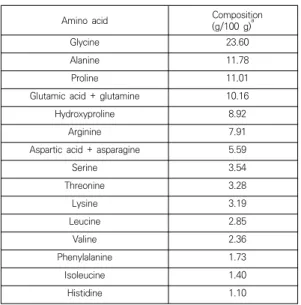
FCHs, produced from tilapia scales hydrolyzed by a neutral protease (Bacillus amyloliquefaciens), were obtained from Nong Shim Co., Ltd (Seoul, Korea). The molecular mass of FCH had an average of 2 kDa, and its amino acid composition is presented in Table 1 [12].
Hs68 cells were obtained from the American Type Culture Collection (Manassas, VA, USA).
Formic acid and 3-(4,5-dimethylthiazol-2-yl)-2,5- diphenyltetrazolium bromide (MTT) were obtained from Sigma-Aldrich Chemical Co. Ltd.
(St. Louis, MO, USA). Phosphate-buffered saline
(PBS) and penicillin-streptomycin were produced
by Lonza (Walkersville, MD, USA). Fetal bovine
serum (FBS) and 0.25% trypsin-EDTA Dulbecco’s
modified Eagle medium (DMEM) were purchased
from Gibco Life Technologies (Grand Island, NY,
USA). Acetonitrile and water for HPLC were
produced by J.T. Baker (Phillipsburg, NJ, USA).
Amino acid Composition (g/100 g)
aGlycine 23.60
Alanine 11.78
Proline 11.01
Glutamic acid + glutamine 10.16
Hydroxyproline 8.92
Arginine 7.91
Aspartic acid + asparagine 5.59
Serine 3.54
Threonine 3.28
Lysine 3.19
Leucine 2.85
Valine 2.36
Phenylalanine 1.73
Isoleucine 1.40
Histidine 1.10
a
Expressed as grams per 100 g of amino acids.
Table 1. Amino acid composition of fish collagen hydrolysate (FCH) (Data citation in the reference of 12)
2.2 Cytotoxicity of FCHs
Hs68 cells were cultivated in DMEM supplemented with 10% FBS and 5% penicillin-streptomycin.
The cells were incubated in an atmosphere of 5%
CO
2at 37°C. To evaluate the cytotoxicity of FCHs, cell metabolic activity was measured by the MTT method that is based on the diversion of yellow MTT to purple formazan when the living cells metabolize [13]. A total of 180 uL of cells were added in a 96-well plate at a concentration of 1 x 10
4cells/well and incubated for 24 hours at 37°C and 5% CO
2. After appropriate incubation time, cells were treated by FCHs at various concentrations and incubated for 24 ho rs. After cultivation, 20 uL of MTT (0.5 mg/mL in PBS) was added to the well and incubated for 4 hours. After incubation, the plate was centrifuged at 1,500 rpm for 5 min to deposit the formed formazan crystals and the supernatant was discarded. Next, to solubilize the formazan salts, 150 uL of dimethylsulfoxide (DMSO) was added.
The absorbance of the formazan salts in each well was measured at 540 nm in a microplatereader (iMark Microplate Reader,
BioRad, Hercules, CA, USA).
2.3 Procollagen synthesis assay
Hs68 cells were added in a 48-well plate at a concentration of 5×10
4cells/well and cultivated at 37°C for 24 hours. After removing the supernatant of each well, the cells were rinsed with PBS solution. All samples for testing were prepared in serum-free DMEM and added to the cells. After cultivation for 24 hours, the supernatant was gathered to determine the quantity of procollagen using a procollagen type I C-peptide assay kit (Takara Bio, Japan).
2.4 UV-induced collagenase inhibition assay Hs68 cells were added in to a 48-well plate at a concentration of 5×10
4cells/well and cultivated at 37°C for 24 hours. After removing the supernatant of each well, the cells were rinsed with PBS. All samples for testing were prepared in serum-free DMEM and were added to the cells.
After 6 hours of cultivation, the cells were rinsed with PBS solution two times. Each well covered with 100 uL of PBS was exposed to 10 J/cm
2UV A with a UVT UV lamp (Dongseo Science Co., Ltd, Korea). The PBS was removed, after which the serum-free medium was added to the cell.
The cells were incubated for 24 hours, and the supernatant was gathered for determining the quantity of collagenase, which was measured using an MMP-1 Human Biotrak ELISA kit (GE Healthcare Life Science, Piscataway, NJ, USA).
2.5 Ultrafiltration
Fractionation of FCHs was performed by an
Amicon model 8400 UF unit (Millipore Corp.,
Billerica, MA) using a Millipore membrane with a
molecular weight cut-off of 1 kDa. Two fractions
with MW <1 kDa and >1 kD were obtained. The
fraction with the highest anti-aging activity was
further separated by reversed-phased high
performance liquid chromatography.
2.6 Reversed-phased high performance liquid chromatography (RP-HPLC)
The FCH fraction with low molecular weight (<1 kDa) was separated by RP-HPLC on a Zorbax Eclipse XDB-C
18column (9.4x250 mm
2, 5 µm, Agilent Technologies, Palo Alto, CA, USA). The sample was eluted with 0.1% formic acid in water (A) and 0.1% formic acid in acetonitrile (B) using 5% A at 0-5 min, 95% A at 10 min, and 5% A at 15 min at a flow rate of 3.0 mL/min. The eluted peaks were monitored by a UV detector at 280 nm. The fraction showing the highest anti-aging activity was collected and concentrated using a rotary evaporator (Eyela N-1000, Tokyo Rikakikai Co., Ltd.). This fraction was then resolved on a Zorbax C
18column (4.6x150 mm, 5um, Agilent Technologies, Palo Alto, CA, USA) at a flow rate of 1 mL/min. A mixture of 0.1% formic acid in water and 0.1% formic acid in acetonitrile (95:5 v/v) was used as the mobile phase. For each separation, the peptide peaks were monitored at 280 nm.
The purification procedures were repeated until the samples were purified to a single peak.
2.7 Amino acid composition
Amino acid was analyzed using a Waters HPLC system equipped with a Waters 510 pump and a Waters Pico-Tag column (3.9 x 300 mm, 4 um).
The mobile phases consisted of 140 mM sodium acetate in 6% acetonitrile (A) and 60%
acetonitrile (B), and the following gradient was used: 86% A at 9 min, 80% A at 9.2 min, 54% A at 17.5 min, and 0% A at 17.7 min. The sample was hydrolyzed with 6N HCl and labeled with phenylisothiocyanate. After labeling, 10 uL of the sample was injected into the column, and peaks were monitored at 250 nm by a UV detector.
2.8 Molecular mass and peptide sequencing The intact mass of the purified peptide was determined by LC/MS analysis using an Agilent 1260 Infinity LC system coupled with an Agilent 6530 Accurate-Mass Q-TOF instrument. The
sample (1 uL, 10 mg/mL) was loaded on a Zorbax Eclipse Plus C18 (2.1 x 50 mm, 1.8 μm) column (Agilent Technologies, Palo Alto, CA, USA). The mobile phases consisted of 0.1% formic acid in water (A) and 0.1% formic acid in acetonitrile (B), and the following gradient was used: 95% A at 0-5 min, 5% A at 10 min, and 95% A at 15 min.
Peptides were detected by full scan mass analysis over the range 0-2,000 (m/z). For peptide sequence analysis, Edman sequencing was performed by standard procedures using an Applied Biosystems Procise Sequencer (Applied Biosystems, Coutaboeuf, France).
2.9 Synthesis of the peptide
The peptide, Gly-Arg-Arg-Gly-Asn-Lys (GRRGNK), was synthesized at AnyGen Ltd (Gwangju, Republic of Korea). The purity of the synthesized peptide was more than 95% as measured by HPLC. The anti-aging properties of the synthesized peptide were evaluated.
2.10 Statistical analysis
SPSS (SPSS Version 21.0, SPSS Inc., Chicago, IL, USA) was used for statistical examination. Data were analyzed by one-way analysis of variance (ANOVA) with Duncan’s test (p<0.05). All experiments were conducted at least in triplicate, and quantitative results are indicated as the mean ± standard deviation (SD).
3. Results and Discussion
3.1 Cytotoxicity and anti-aging properties of FCH The cytotoxicity of FCHs to Hs68 cells was assessed by the MTT assay. As can be seen in Fig.
1 [12], the metabolic activity of Hs68 cells treated
with FCHs (0–1,000 µg/mL) was on average more
than 80% of that of the control; FCHs had no
toxic effect on the Hs68 cells for any of the
tested concentrations.
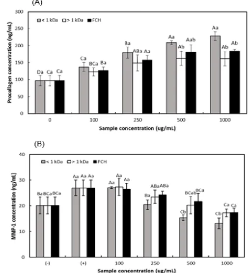
Fig. 2. Procollagen synthesis by Hs68 cells treated with FCHs and two fractions (1> kDa, <1 kDa) obtained from ultrafiltration. (A) Quantitation of MMP-1 production by Hs68 cells treated with fish collagen hydrolysates (FCHs), their <1 kDa fraction, and their >1 kDa fraction for 24 hrs after UVA irradiation (10 J/cm
2). (B) (-) and (+) designate the two control groups (unexposed and exposed to UVA, respectively). Means with capital letters for the same sample and small letters for the same concentration are significantly different ( p <0.05).
Fig. 1. Cytotoxicity of Hs68 cells treated with FCHs (Data citation in the reference of 12).
3.2 Anti-aging properties of the fractions from FCH
The quantities of procollagen and MMP-1 in Hs68 cells treated with ultrafiltration fractions (<1 kDa, >1 kDa) were evaluated by a procollagen type I C-peptide ELISA assay. As exhibited in Fig.
2(A), type 1 procollagen synthesis was increased
in all cells treated with the three types of
samples. The procollagen formation of these
samples might be related to the amino acid composition of FCH (Table 1). The six major amino acids, Gly, Ala, Pro, Glu, Hyp, and Arg, accounted for nearly 73.4% of FCHs. Ala has been found to relate to collagen synthesis [14], and Pro-Hyp functions as a fibroblast growth factor, which facilitates collagen formation [15].
Additionally, Arg is a precursor of proline, which contributes to collagen synthesis [16].
In particular, the <1 kDa fraction significantly increased the amount of synthesized type 1 collagen in a dose-dependent manner at all tested concentrations (100, 250, 500, and 1000 ug/mL;
137.01 ng/mL, 179.18 ng/mL, 208.76 ng/mL, and 228.4 ng/mL), while FCHs and >1 kDa fraction did not show this dose-dependent activity.
Furthermore, at the concentrations of 500 and 1,000 ug/mL, the <1 kDa fraction produced more than twice the amount of collagen synthesis compared to that of the control (97.02 ng/mL) and exhibited a significant difference in the production of collagen compared with FCHs and
>1 kDa fraction (p<0.05).
The amounts of UV-induced MMP-1 in the Hs68 cells treated with FCHs and ultrafiltration fractions (<1 kDa, >1 kDa) are exhibited in Fig.
2(B). All three samples showed a dose-dependent inhibition of MMP-1. At the concentrations of 500–1,000 µg/mL, all samples significantly reduced the production of MMP-1. Several studies have shown that Gly, Glu, Pro, and Hyp consisting of FCH (Table 1) inhibit MMP secretion [8,16]. The MMP-1 inhibition effect of these fish collagen-derived samples may be due to their amino acid composition.
In particular, the <1 kDa fraction was found to inhibit the MMP-1 in Hs68 cells the most effectively among the three samples. In the concentration range of 250–1,000 µg/mL, the <1 kDa fraction significantly inhibited the MMP-1 expression (20.43 ng/mL, 15.33 ng/mL, and 13.18 ng/mL) compared to the UV-irradiated control cells (26.98 ng/mL). Therefore, the <1 kDa
fraction was considered to have the highest anti-aging activity and thus was further isolated into a Zorbax C
18column.
3.3 Anti-aging properties of fractions isolated from HPLC
After ultrafiltration of FCHs, the <1 kDa fraction was separated by HPLC on a C18 semi-prep column, leading to the collection of two fractions (A and B) (Fig. 3(A)). The amounts of procollagen released in fractions A and B were 241.97 ± 5.02 ng/mL and 213.48 ± 3.01 ng/mL, respectively. The collagen-synthesizing and MMP-1-inhibiting abilities of fraction A were 13.35% and 22.15% greater, respectively, compared to those of fraction B. Fraction A was further resolved using a Zorbax analysis C
18column; three fractions (I, II, and III) were collected and lyophilized (Fig. 3(B)). Treatment with fraction I yielded 21.28% higher collagen synthesis and 22.51% lower MMP-1 expression compared with the least bioactive fraction (II).
The fraction with the highest anti-agingpotential, fraction I, was further resolved using an analysis column. Fig. 3(C) shows the two additional fractions (I-I and I-II) isolated from fraction I;
treatment with fraction I–Iyielded 52.67% higher procollagen production and 23.21% lower MMP-1 expression than treatment with fraction I-II.
Therefore, the most bioactive fraction (I-I) was purified into a clear single peak, as shown in Fig.
3(D).
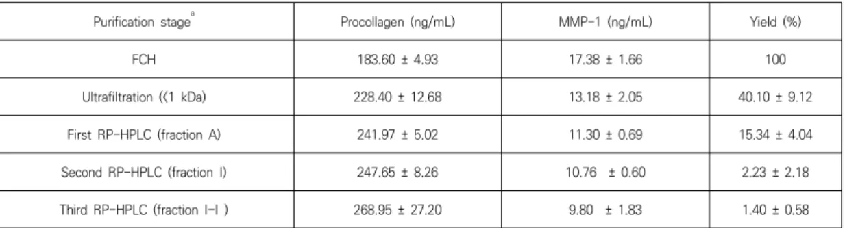
The procollagen synthesis, MMP-1 inhibition, and the yield of the anti-aging peptides at each separation were exhibited in Table 2.
Fig. 3. Purification scheme for obtaining
anti-aging peptides from fish collagen
hydrolysates. (A) First isolation of the <1 kDa
fraction using a semi-preparative column, (B)
second isolation of fraction A using an analytical
column, (C) third isolation of fraction I, and (D)
final purification of fraction I-I.
Purification stage
aProcollagen (ng/mL) MMP-1 (ng/mL) Yield (%)
FCH 183.60 ± 4.93 17.38 ± 1.66 100
Ultrafiltration (<1 kDa) 228.40 ± 12.68 13.18 ± 2.05 40.10 ± 9.12
First RP-HPLC (fraction A) 241.97 ± 5.02 11.30 ± 0.69 15.34 ± 4.04
Second RP-HPLC (fraction I) 247.65 ± 8.26 10.76 ± 0.60 2.23 ± 2.18
Third RP-HPLC (fraction I-I ) 268.95 ± 27.20 9.80 ± 1.83 1.40 ± 0.58
a
The concentration of the sample for each purification stage was 1 mg/mL.
Table 2. Purification of the anti-aging peptide from fish collagen hydrolysate (FCH)
3.4 Identification of the anti-aging peptide The amino acid composition of FCH fraction I-I is presented in Table 3 [12]. The major amino acids in fraction I-I were Gly, Ala, Arg, Lys, Glu;
together, these six amino acids comprised approximately 85.15% of fraction I-I. Gly was the most abundant amino acid in fraction I-I, which may be due to the repeating Gly-X-Y sequence in collagen. Additionally, the high Ala and Lys FCH contents are in agreement with previous studies showing that these two amino acids contribute to collagen formation [14]. Arg is a precursor of proline synthesis [16], which is an important amino acid in collagen synthesis. Thus, the amino composition of fraction I-I is in good agreement with the major components of collagen hydrolysates.
Amino acid Composition
(g/100 g)
aGlycine 32.44
Alanine 23.89
Arginine 15.61
Lysine 8.06
Glutamic acid + glutamine 5.15
Threonine 3.53
Aspartic acid + asparagine 1.71
Phenylalanine 1.58
Hydroxyproline 1.16
Histidine 1.09
a
Expressed as grams per 100 g of amino acids.
Table 3. Amino acid composition of the purified peptide (fraction I-I) from fish collagen hydrolysate (FCH) (Data citation in the reference of 12)
The molecular mass of fraction I-I was determined using LC/MS. The anti-aging peptide was determined to have a molecular mass of 686.175 Da. Through Edman degradation sequencing, the amino acid sequence of fraction I-I was identified to be Gly-Arg-Arg-Gly-Asn-Lys (GRRGNK).
3.5 Anti-aging properties of the synthesized peptide
In order to confirm the anti-aging activity of the isolated peptide, the peptide (GRRGNK) was synthesized. The anti-aging activity of GRRGNK was determined to identify the active component in fraction I-I (Table 4). No significant difference was observed in the amounts of procollagen synthesis and MMP-1 inhibition stimulated by fraction I-I and GRRGNK. Additionally, GRRGNK yielded 81.8% higher collagen synthesis and 63.57% lower MMP-1 expression compared with the UV-irradiated control. Thus, the purified peptide was verified to be actually relevant to the anti-aging effect.
Samples
aProcollagen (ng/mL) MMP-1 (ng/mL) Fraction I-I 268.95 ± 27.20 a 9.80 ± 1.83 b
GRRGNK 267.28 ± 13.75 a 9.50 ± 1.20 b
a