Case Report
원고 접수일 2012년 12월 11일, 원고 수정일 2013년 1월 15일, 게재 확정일 2013년 7월 24일
책임저자 권대근
(700-705) 대구광역시 중구 달구벌대로 2177, 경북대학교 치의학전문대학원 구강악 안면외과학교실
Tel: 053-600-7551, Fax: 053-426-5365, E-mail: kwondk@knu.ac.kr
RECEIVED December 11, 2012, REVISED January 15, 2013, ACCEPTED July 24, 2013
Correspondence to Tae-Geon Kwon
Department of Oral and Maxillofacial Surgery, School of Dentistry, Kyungpook National University
2177 Dalgubeol-daero, Jung-gu, Daegu 700-705, Korea
Tel: 82-53-600-7551, Fax: 82-53-426-5365, E-mail: kwondk@knu.ac.kr
CC This is an open access article distributed under the terms of the Creative Commons Attribution Non-Commercial License (http://creativecommons.org/licenses/
by-nc/3.0) which permits unrestricted non-commercial use, distribution, and reproduction in any medium, provided the original work is properly cited.
상악 측절치 근관치료 중 수산화칼슘 호제근충제 과충전으로 인하여 발생한 신경손상의 치험례
나광명ㆍ김종배1ㆍ진병로2ㆍ김진욱ㆍ김진수ㆍ권대근
경북대학교 치의학전문대학원 구강악안면외과학교실, 1계명대학교 의과대학 치과학교실, 2영남대학교 의과대학 치과학교실
Abstract
Nerve Injury from Overfilled Calcium Hydroxide Root Canal Filling Paste for Maxillary Lateral Incisor Endodontic Treatment
Kwang Myung Na, Jong-Bae Kim
1, Byung-Rho Chin
2, Jin-Wook Kim, Chin-Soo Kim, Tae-Geon Kwon Department of Oral and Maxillofacial Surgery, School of Dentistry, Kyungpook National University,
1Department of Dentistry,
College of Medicine, Keimyung University,
2Department of Dentistry, College of Medicine, Yeungnam University
Calcium hydroxide root canal filing paste (vitapex) is widely used as canal filling paste for infected canal. However, chemical burn is possible because of the high alkali base of calcium hydroxide. A 57-year old woman was admitted to our clinic for consistent dull pain and paresthesia in the left upper lip, zygoma and buccal cheek area, which developed during an endodontic treatment of the left lateral incisor. Radiographic finding showed radiopaque material, which exits from the left incisor root apex, and was within the left canine and first premolar buccal soft tissue. The overfilled Vitapex extended to the soft tissue was surgically curetted. The result of the surgical curettage was favorable. Though slight hypoesthesia on the upper lip was still remained, paresthesia on zygomatic and buccal cheek area was completely recovered. As far as we know, this is the first case report of infraorbital nerve damage from overfilled Vitapex material.
Key words: Vitapex, Calcium hydroxide, Apical leakage, Paresthesia
서 론
Vitapex (Neo Dental Chemical Products Co., Tokyo, Japan)는 수산화칼슘을 주재료로 한 약제로 미완성 치근단공을 지닌 치아의 근첨형성술(apexifacation), 치수절단술(pulpotomy),
치수절제술(pulpectomy) 또는 감염된 근관에서 지속적인 염증으
로 인해 삼출물이 지속적으로 유출되는 감염근관의 치료술에 사용
되는 임시 근관 충전재로로 알려져 있다[1]. Vitapex는 pH 11.3
내외의 높은 알칼리성을 띠는 수산화칼슘을 30.3% 포함하고 있으
며, 요오드포름 40.4%와 실리콘 오일 22.4% 및 기타 성분 6.9%로
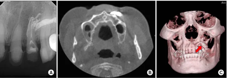
Fig. 1. Preoperative radiographic image: (A) periapical view: the spread of the endodontic paste (Vitapex; Neo Dental Chemical Products Co., Tokyo, Japan) beyond the apex of the right lateral incisor is evident in periapical radiograph. (B) Axial view of computed tomography (CT) image and (C) three dimesional conbeam CT images showing the spread of the endodontic paste (arrow) exit from left incisor root apex to the left canine and first premolar buccal soft tissue.
구성되어 있다.
수산화칼슘은 근관 내 처치 시 염증 부산물에 의해 낮아진 조직의 pH를 높은 알칼리성으로 중화를 시켜주며[2], 치수조직에 대한 NaOCl의 용해효과가 증진되고, 세균의 대사산물이면서 치 근단 골흡수의 주원인인 lipopolysaccharide를 파괴시키는 우수 한 항균작용이 있다[3]. Sundqvist[4]는 감염된 근관 내 수산화칼 슘 적용 시 97%의 근관 내 세균수 감소를 보고하였다. 실리콘 오일은 윤활작용과 단열의 역할을 하며, 조직액, 혈액 등에 의하여 충전제가 용해되는 것을 방지하고, 근관 내벽에 충전재가 잘 밀폐 되도록 하는 기능을 한다. 요오드포름은 감염치료 목적 외에 방사 선 불투과성을 높여주는 역할을 한다. Vitapex는 paste상으로 실린지에 들어 있어 사용이 간편하고 조작성이 용이하며, x-ray 조영성이 있는 등 여러 장점으로 큰 병소를 가진 치근단 염증의 임시충전에서 흔히 사용되고 있다. 이러한 장점들에도 불구하고, 비수용성인 실리콘 오일을 용매로 사용하고 있기 때문에 근관 내 제거가 힘들다는 점, 요오드 성분에 의한 부작용, 수산화칼슘의 높은 알칼리성의 의한 화학적 화상의 부작용 등이 보고되고 있다.
근관 내 임시충전제로 사용되는 수산화칼슘 제제인 Vitapex는 근관치료 중에 일시적으로 사용되어야 하고, 그 후에는 깨끗이 제거되어야 된다. 그럼에도 불구하고, 잘못된 적용방법으로 우발 적으로 치근단공을 넘어 연조직의 감각이상을 초래하는 경우가 종종 보고되고 있다[5-7].
이번 증례에서는 개인 치과의원에서 상악 측절치의 근관치료 도중 우발적으로 Vitapex paste가 상악 좌측 측절치 치근단공을 넘어가, 좌측 윗입술과 관골부의 감각이상을 호소하는 환자를 의뢰 받아 전신마취하 외과적 소파술을 시행 및 경구투약을 통한 경과관찰과정을 통해 만족할 만한 감각이상 해소의 결과를 얻었기 에 문헌고찰과 함께 증례를 소개하고자 한다.
증례보고
57세의 여자 환자가 상악 좌측 안면부의 감각이상을 주소로 경북대학교병원 구강악안면외과로 의뢰되었다. 타 치과의원에서 상악 좌측 측절치의 근관치료를 받고 있었으며, 지속적인 통증과 배농으로 근관형성 후 세정과정을 반복적으로 받고 있었으며 본원 에 내원하기 1일 전 해당 의원에서 임시충전제로 흔히 사용되고 있는 Vitapex를 적용 후 안면부의 감각이상이 시작되었다고 하였 다. 특히 좌측 편측 얼굴의 관골부와 윗 입술부, 협측 뺨 부위의 감각이상을 호소하였으며, 가만히 있을 때도 작열감을 호소하였다.
치근단 방사선사진에서 방사선불투과성 영상이 상악 좌측 측절 치의 근단공을 빠져나와 측절치부터 좌측 제1소구치의 원심면까 지 확장되어 있었다(Fig. 1A). Computed tomography (CT)의 axial 영상과 three dimesional conbeam CT (3D-CBCT)를 통해 방사선 불투과성 영상은 해당치아들의 협측 연조직 내에 위치하고 1.6×1.1×0.6 cm의 크기로 불규칙한 반구형 형태로 관찰되었다(Fig. 1B, 1C).
문진, 임상소견, 방사선 사진 등을 통해 안와하신경(infraorbital
nerve)의 손상으로 판단되었고, 해당부위의 감각이상을 일으킨
방사선 불투과성 물질은 근관 임시 충전제로 사용된 Vitapex라고
진단하고, Vitapex의 주성분인 수산화칼슘에 의한 화학적 화상
및 물리적 압박에 의한 감각이상이라고 판단 후 원인요소의 제거가
필요하다는 판단하에 술 후 10일째에 전신마취하에 외과적 소파술
을 시행하였다. 좌측 상악 측절치부터 수직절개, 전정부 절개
및 판막 거상 후 좌측 상악 측절치 치근단 하방부터 좌측 상악
제1소구치의 원심부의 협측 연조직 사이에 과충전되어 근단공을
빠져나간 Vitapex가 관찰되었다(Fig. 2A). Vitapex 주변부로
골 내 누공을 통해 배농되고 있었으며, 상악 협측골 골흡수가
Fig. 4. (A) Foreign body reaction with granuloma (white arrows). The nerve bundle (black arrow head) is involved and degenerated (black arrow) (H&E, ×200). (B) Birefringent foreign body material under polarized microscope (white arrow) (H&E, ×200).
Fig. 2. (A) After flap elevation, overfilled Vitapex (Neo Dental Chemical Products Co., Tokyo, Japan) was observed. (B) After curettage, bony fistula (white arrow) and bone resorption site (black arrow) was observed.
Fig. 3. Removed overfilled paste (Vitapex; Neo Dental Chemical Products Co., Tokyo, Japan; left) and surrounding soft tissues (right). The size of overfilled paste was similar to those seen in computed tomography image.
관찰되었다(Fig. 2B). 제거된 종물은 방사선사진과 3D-CT image 를 통해 관찰된 크기와 유사하였고, 근관충전제와 함께 인근주위에 있는 감염조직을 함께 제거하였다(Fig. 3). 철저히 소파술 시행 후 활성산소를 만들어 각종 세균의 소독 효과가 있는 H
2O
2를 이용하여 수차례 소독하였다. 수술부위에서 제거된 종물은 헤마톡 실린-에오진으로 염색하였고, 200배의 배율로 광학현미경과 편광 현미경을 이용하여 관찰되었다. 병리조직검사 결과 foreign body 에 의해 histiocyte가 국소적으로 모여 결절성 형태를 띠고 있으며 인근 신경다발이 변성된 소견을 보였다. Foreign body gran- uloma, chronic inflammation으로 최종 진단되었다(Fig. 4).
술 후 CT의 axial 영상과 3D-CBCT를 통해 과충전된 임시충전물
의 완전한 제거가 이루어졌음을 확인하였다(Fig. 5). 수술 후
dexamethason을 2주간 줄여가면서 사용하였으며, Vitamin B12
를 포함한 종합비타민, 아데노신 3 인산 등이 포함된 종합영양제를
복용하였다. 점진적으로 관골부와 협측 뺨 부위의 감각이상은
Fig. 5. (A) Post-operative computed tomography (CT) axial view and (B) three dimesional conbeam CT im- ages showing complete removal of overfilled paste (Vitapex; Neo Dental Chemical Products Co., Tokyo, Japan).
해소되었으며, 현재 수술 후 약 1년 6개월이 경과했고, 아직 상순 부위의 미약한 감각저하를 호소하고 있다. 현재 상악 좌측 측절치 의 근관치료 시행 후 전부도재관으로 보철치료까지 완료하였다.
고 찰
근관치료 중에 나타날 수 있는 신경손상은 신경조직에 물리적, 화학적 손상을 야기할 수 있다. 이러한 손상은 임상과정 중 흔하 지 않은 경우지만, 통증, 감각저하(hypoesthesia), 이상감각 (paresthesia, dysesthesia) 등의 매우 심각한 손상을 가져다 줄 수 있다. International Association for the Study of Pain에서 1994년 정의한 바에 의하면 paresthesia는 자발적으로 일어나는 비정상적인 감각-화끈거림, 얼얼함, 따끔거림, 가려움 같은 불쾌 하지는 않은 감각으로 정의하고 있으며, dysesthesia는 자발적으 로 나타나는 매우 불쾌한 비이상적인 감각으로 설명하고 있다.
하지만 일반적으로 paresthesia terminology가 dysesthesia를 포함하여 일반적인 이상감각을 나타내는 용어로 통용되어 사용 되고 있다[8].
근관치료 중 감각이상이 올 수 있는 요인들을 살펴보면, 1) 지속적인 exudate와 그로 인한 부종과 허혈을 동반하는 근단부 염증, 2) 국소마취 시 오염된 마취제의 이용 또는 신경다발에 직접적인 자극, 3) 근관부 형성 및 세정과정 동안 근관 세척제로 사용하는 NaOCl에 의한 화학적 자극 또는 근단부 하방을 자극하 는 과기구조작(over-instrumentation)등의 물리적 자극, 그리고 이번 환자의 경우에서처럼 4) Vitapex, 수산화칼슘, AH-26 등의 근관약제에 의한 과잉충전에 의해 신경손상이 올 수 있다[9].
근관충전에 의한 신경손상을 설명하는 두 가지 mechanism으 로는 근관충전제에 포함된 성분에 의한 신경독성에 의한 화학적 손상과, 근단공을 통해 유출된 근관충전제에 의한 물리적 압박에 의한 물리적 손상에 의해 설명되고 있다[7,10]. 화학적 손상 기전 에 대한 설명은 다음과 같다. Paresthesia는 수산화칼슘이나 포름 알데하이드 등이 포함된 근관충전제나 근관 내 약제를 신경다발이
가까이에 있는 root canal로 적용 시 과잉충전 및 근단부 유출 등에 의해 나타날 수 있다[11,12]. 특히 Vitapex의 주성분인 수산 화칼슘은 치수조직에 접했을 때 접촉계면에서 새롭게 석화화 층을 형성하여 괴사층을 형성한다고 알려져 있다. 이러한 수산화칼슘 을 포함한 약제가 신경조직에 직접적으로 노출되어 손상을 가할 경우 국소적으로 감각이상을 일으켰다는 있다는 수차례의 보고가 있다. 물리적 손상에 의한 신경손상 기전은 유출된 근관 충전제가 신경말단 조직압박을 일으키고, 그에 따른 신경압박에 의해 동맥 혈액공급의 부족을 일으킨다. 급성 혈액투과도를 증진시키고, hematoma의 생성과 edema에 의해 신경에 산소공급을 줄어들 게 된다. 말단 신경조직은 상대적으로 허혈상태에 저항성이 있지 만, 오랫동안 지속된 신장 또는 압축력은 섬유아세포유입, 반흔조 직의 형성, 신경섬유의 변성 등의 비가역적인 변화를 일으킬 수 있다[13,14].
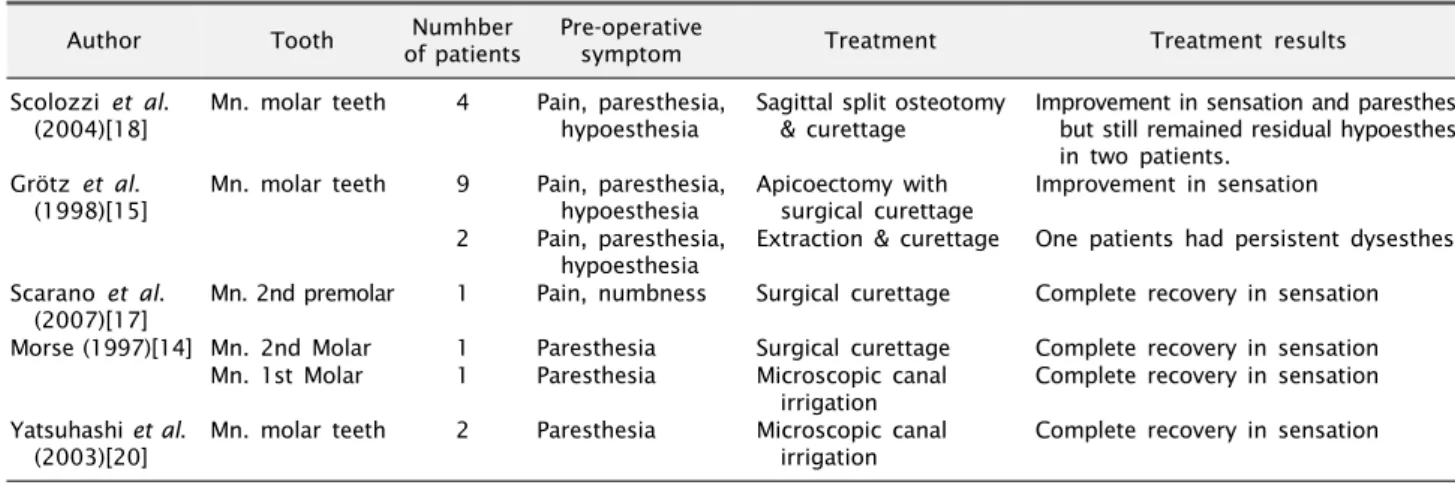
이전의 다른 논문에서는 반복적인 근관세정과 비타민, 아데노 신 3 인산 등의 약물을 이용한 비수술적인 방법으로 이상감각이 해소되었다고 보고하는 증례도 있다[14,15]. 그 외에 decortica- tion, sagittal osteotomy, apicoectomy, curettage 등의 근관충 전제의 제거의 수술적인 방법이 보고된 적이 있으며[15-18], 각각 의 신경손상의 정도는 신경손상 후 경과된 시간에 따라 증가된 양상을 보인다고 하였다[19].
본 증례에서는 술 전 방사선 사진과 3D-CBCT를 통해 유출된 Vitapex의 위치와 양을 정확히 판단 후 최대한 빠른 시일에 전신 마취하 외과적 소파술을 시행하여 화학적 독성 및 물리적 압박을 해소해주었으며, 국소적인 원인인자 제거 외에 dexamethasone 을 감량해가면서 2주간 사용하였고, 전기자극 치료와 온찜질을 포함한 물리치료를 2개월간 시행하고, 보조적으로 비타민 B12가 포함된 종합비타민과 아데노신 3 인산이 포함된 영양제를 복용 후 1년 6개월의 경과 관찰 동안 대부분의 이상감각이 해소되었다.
지금까지 하악구치부 근관치료 중 과잉충전으로 인한 하치조신
경의 이상감각이 생겨 다양한 치료방법에 의해 치유된 증례는
수차례 있었으나(Table 1)[14,15,17,18,20], 상악치아의 근관치
Table 1. Pre-operative symptom, treatment and treatment result due to overfilled endodontic treatment
Author Tooth Numhber
of patients
Pre-operative
symptom Treatment Treatment results
Scolozzi et al.
(2004)[18] Mn. molar teeth 4 Pain, paresthesia,
hypoesthesia Sagittal split osteotomy
& curettage Improvement in sensation and paresthesia but still remained residual hypoesthesia in two patients.
Grötz et al.
(1998)[15]
Mn. molar teeth 9 Pain, paresthesia, hypoesthesia
Apicoectomy with surgical curettage
Improvement in sensation 2 Pain, paresthesia,
hypoesthesia Extraction & curettage One patients had persistent dysesthesia Scarano et al.
(2007)[17] Mn. 2nd premolar 1 Pain, numbness Surgical curettage Complete recovery in sensation Morse (1997)[14] Mn. 2nd Molar 1 Paresthesia Surgical curettage Complete recovery in sensation
Mn. 1st Molar 1 Paresthesia Microscopic canal
irrigation Complete recovery in sensation Yatsuhashi et al.
(2003)[20]
Mn. molar teeth 2 Paresthesia Microscopic canal irrigation
Complete recovery in sensation
Mn., mandible.
료 도중 안와하신경의 손상은 최초로 보고하는 것으로 생각한다.
본 증례를 통하여 임상가들은 paste type의 임시충전재료를 사용 시 강한 압력으로 짜서는 안되며, 천천히 근관 입구에서 빼주면서 충전을 하는 기본을 잘 지켜야 할 것으로 생각한다.
References