Veterinary Science
*Corresponding author
Tel: +82-62-530-2831; Fax: 82-62-530-2809 E-mail: [email protected]
†First two authors contributed equally to this study.
Comparison of cardiac function and coronary angiography between conventional pigs and micropigs as measured by multidetector row computed tomography
Young Keun Ahn
2,3,4,†, Jung Min Ryu
1,†, Hea Chang Jeong
2, Yun Hyeon Kim
5, Myung Ho Jeong
2,3,4, Min Young Lee
1, Sang Hun Lee
1, Jae Hong Park
1, Seung Pil Yun
1, Ho Jae Han
1,*
1
College of Veterinary Medicine, Biotherapy Human Resources Center, Chonnam National University, Gwangju 500-757, Korea
2
The Heart Center,
3Cardiovascular Research Institute,
4Clinical Trial Center, and
5Department of Radiology, Chonnam National University Hospital, Gwangju 501-757, Korea
Pigs are the most likely source animals for cardiac xeno- transplantation. However, an appropriate method for estimating the cardiac function of micropigs had not been established. Computed tomography (CT) analysis aimed at estimating cardiac function and assessing the coronary arteries has not been carried out in micropigs. This study determined the feasibility of evaluating cardiac function in a micropig model using multidetector row computed tomography (MDCT) and compared the cardiac function values with those of conventional pigs. The mean age of the conventional pigs and micropigs was approximately 80 days and approximately 360 days, respectively. The mean body weight in the conventional pigs and micropigs was 29.70 ± 0.73 and 34.10 ± 0.98 kg, respectively. Cardiac MDCT detected ejection fractions of 52.93 ± 3.10% and 59.00 ± 5.56% and cardiac outputs of 1.46 ± 0.64 l/min and 1.21 ± 0.24 l/min in conventional pigs and micropigs, respectively. There were no significant differences in cardiac function between conventional pigs and micropigs in the reconstructed CT images. There were also no differences in the coronary angiographic images obtained by MDCT. It is expected that the results of this study will help improve understanding of cardiac function in micro- pigs. The data presented in this study suggest that MDCT is a feasible method for evaluating cardiac function in micropigs.
Keywords: cardiac function, coronary angiography, MDCT, micropig, multidetector row computed tomography
Introduction
Transplantation is often used to treat fulminant organ failure. However, severe shortages in the availability of suitable human donors have limited the volume of heart transplants [15]. This shortage of donors has stimulated in- terest in the possibility of using animal organs for trans- plantation into humans. Animal-to-human transplantation (or xenotransplantation) would offer an unlimited supply of organs and tissue for transplantation. Both non-human primates and non-primate mammals have been used as sources for heart transplants. Non-human primates such as chimpanzees and baboons are closely related to humans phylogenetically and share many immunological proper- ties with humans [3,5,25]. However, non-human primates are unlikely to be a fruitful source of organs in the future for the following reasons: slow growth rate, limited offspring, difficulty breeding in captivity, and smaller size [27]. Pigs are now considered to be the most likely source animals for human xenotransplantation in the future. They have sev- eral advantages over non-human primates. The heart size of miniature swine including the micropig is compatible with the human heart, and cardiovascular function and he- modynamic parameters are also similar [2]. In addition, the reproduction-related features of pigs, such as early sexual maturity, short gestation time, and generation of large lit- ters, allow for a potentially large pool of animal donors for xenotransplantation [37].
One essential question in xenotransplantation is whether
the animal organ can be an effective physiological proxy
for the human organ. In order to answer to this question, the
functional and anatomical compatibility of a pig heart and
its human counterpart has been investigated [35]. The car-
diac output of porcine and human hearts of similar size was
found to be comparable [19], and their action potentials
exhibit slight genetically-derived metabolic differences [13]. These differences have the potential to create prob- lems for support of the human cardiovascular system after xenotransplantation.
It is clinically important to measure cardiac function in in- dividual micropigs used for heart transplantation for pur- poses of diagnosis and prognosis. The indices of a healthy heart are verified through assessment of ejection fraction (EF), end systolic volume (ESV), end diastolic volume (EDV), cardiac output (CO), and coronary artery angiog- raphy [32,36]. However, there is no reliable method for evaluating these parameters in micropigs. Therefore, we studied the feasibility of evaluating cardiac function in mi- cropigs using multidetector row computed tomography (MDCT) and compared the parameters with those of con- ventional pigs.
In recent years, major technological improvements have been achieved in computed tomography (CT). The most significant development has been the introduction of MDCT, which has brought about substantial improve- ments in spatial and temporal resolution [12,17]. This study is the first to show MDCT to be a reliable method for assessing cardiac function in micropigs and for selecting suitable donor pigs for heart xenotransplantation.
Materials and Methods Animals
All experimental procedures were approved by the Ethics Committee of Chonnam National University Hospital.
Studies were performed using mixed-breed, conditioned Yucatan micropigs and Landrace breed conventional pigs, all of which were provided by the animal breeding house of Chonnam National University Research Institute of Medi- cal Sciences. The pigs were housed individually indoors in cages, fed dry pig food, and provided with water. The mean age was approximately 80 days for conventional pigs and approximately 360 days for micropigs. The mean body weight for the conventional pigs and micropigs was 29.70
± 0.73 kg and 34.10 ± 0.98 kg, respectively.
Radiological assessment of cardiac function
After premedication with azaperone (0.5 mg/kg, intra- muscular) and xylazine (8 mg/kg, intramuscular), normal saline with midazolam (0.2 mg/kg) was infused through a 20-G venous access line placed in an ear vein. CT examina- tions were performed using a two-phase, contrast-en- hanced, ECG-gated, MDCT scanner (SOMATOM Sensa- tion Cardiac 64; Siemens, Germany) set at a 0.75-mm sec- tion thickness, with a gantry rotation time of 330 msec and
CT scanning in the axial plane, together with an ECG-trig- gered examination, was performed from the level of the left ventricular apex after a bolus injection of 60 ml of non-ion- ic contrast media (Ultravist 370; Schering, Germany) fol- lowed by a 60 ml saline bolus injection through the ear vein. Both were injected at a flow rate of 4 ml/sec. Axial images were reconstructed at multiple phases that covered the cardiac cycle in 10% increments of the RR interval be- tween 5% and 95%. Multiphase reconstruction was per- formed with commercially available software (Argus;
Siemens, Germany) by using short axis slices from the base of the heart to the apex. The end-diastole and end-sys- tole were defined as the maximal and minimal left ven- tricular volume, respectively. The EDV, ESV, LVEF, stroke volume (SV), CO, and myocardial mass were compared between the two groups. A 7 Fr. arterial sheath was placed in the left carotid artery after achieving local anesthesia with 2% lidocaine, and a cutdown was made. After infus- ing 10,000 units of heparin, a 7 Fr. coronary artery guiding catheter was placed within the ostia of the left and right cor- onary arteries under fluoroscopic guidance using a mobile C-arm (Phillips BV-25 Gold; Phillips, Netherlands). Coro- nary angiography demonstrated left and right coronary ar- teries with branches. The coronary angiograms for the left and right coronary arteries and their branches were com- pared with the images of the CT angiogram. During the ex- periment, oxygen and normal saline were supplied con- tinuously, and anesthesia was maintained with an addi- tional infusion of midazolam. Continuous ECG monitor- ing was performed in order to confirm that the pigs had a normal ST segment at baseline.
Statistical analysis
Statistical analysis was carried out with Statistical Package for Social Sciences software (SPSS 12.0 for Windows; SPSS, USA). Continuous variables with normal distributions were compared using an unpaired Student's t-test and are expressed as mean ± SD. Where appropriate, categorical variables were compared using a Wilcoxon test. A p-value < 0.05 was considered significant.
Results
The end-systolic and end-diastolic phase images were
displayed (Figs. 1 and 2), and EDV, ESV, EF, SV, CO, and
myocardial mass were calculated automatically (Tables 1
and 2). The EDV and ESV were 48.80 ± 23.30 ml and
22.97 ± 11.30 ml in conventional pigs and 36.70 ± 9.36 ml
and 13.30 ± 7.43 ml in micropigs under a pre-medicated
condition, respectively. There were no significant differ-
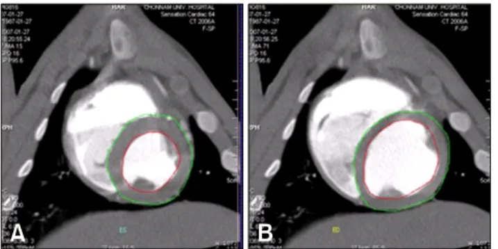
Fig. 1. Left ventriculogram by computerized tomography (A:
end-systolic phase, B: end-diastolic phase) in a conventional pig.
Fig. 2. Left ventriculogram by computerized tomography (A:
end-systolic phase, B: end-diastolic phase) in a micropig.
Table 1. Cardiac parameters in conventional pigs and micropigs as measured by multidetector row computed tomography
Conventional Micropig pig (n = 3) (n = 3)
pEnd diastolic
48.80 ± 23.30 36.70 ± 9.36 0.451 volume (ml)
End systolic
22.97 ± 11.30 13.30 ± 7.43 0.283 volume (ml)
Stroke volume
25.83 ± 12.11 23.40 ± 2.26 0.750 (ml)
Cardiac
1.46 ± 0.64 1.21 ± 0.24 0.567 output (l/min)
Myocardial
41.83 ± 19.47 37.03 ± 6.89 0.602 mass (g)
Table 2. Comparison of ejection fraction (EF) detected by multi- detector row computed tomography (MDCT) and echocardio- graphy in conventional pigs and micropigs
EF (%)
MDCT Echocardiography
(n = 3) (n = 5)*
Conventional pig 52.93 ± 3.10 65.47 ± 5.17
Micropig 59.00 ± 5.56 58.40 ± 8.18
*Cited from Lee et al. [22].