INTRODUCTION
Although the conventional treatment for de novo coronary le- sions is drug-eluting stent (DES) implantation, plain old bal-
loon angioplasty (POBA) is still useful for patients unable to tolerate prolonged dual antiplatelet therapy or anatomically difficult lesions in small sized coronary vessels where stenting is impossible.1,2 Unfortunately, POBA has important limita- tions, including poor vessel patency, high restenosis rates due to elastic recoil, and late negative remodeling.3 The application of a balloon with anti-proliferative coating can overcome some of these deficiencies by preventing restenosis caused by neointimal hyperplasia. In this regard, paclitaxel-coated bal- loon (PCB) treatment is an attractive therapeutic option and may have benefits over POBA.4 The advantages of PCB include a homogeneous drug delivery to the vessel wall, an immedi- ate drug release without the use of a polymer, the potential of reducing the intensity and duration of antiplatelet therapy and the freedom of leaving no foreign object behind in the vessel.5
Comparison of Paclitaxel-Coated Balloon Treatment and Plain Old Balloon Angioplasty for De Novo
Coronary Lesions
Ae-Young Her1, Soe Hee Ann2, Gillian Balbir Singh2, Yong Hoon Kim1, Sang-Yong Yoo3, Scot Garg4, Bon-Kwon Koo5, and Eun-Seok Shin2
1Division of Cardiology, Department of Internal Medicine, Kangwon National University School of Medicine, Chuncheon, Korea;
2Department of Cardiology, Ulsan University Hospital, University of Ulsan College of Medicine, Ulsan, Korea;
3Division of Cardiology, Department of Internal Medicine, University of Ulsan College of Medicine, Gangneung Asan Hospital, Gangneung, Korea;
4East Lancashire Hospitals NHS Trust, Blackburn, Lancashire, UK;
5Department of Internal Medicine and Cardiovascular Center, Seoul National University Hospital, Seoul, Korea.
Purpose: This study compared the angiographic outcomes of paclitaxel-coated balloon (PCB) versus plain old balloon angio- plasty (POBA) treatment for de novo coronary artery lesions. At present, there is no available data comparing the efficacy of PCB versus POBA for the treatment of de novo coronary lesions.
Materials and Methods: This multicenter retrospective observational study enrolled patients with de novo coronary lesions with a reference vessel diameter between 2.5 mm and 3.0 mm and lesion length ≤24 mm who were successfully treated with PCB or POBA. Angiographic measurements and quantitative coronary analysis were performed before and after the procedure, and at 9 months follow-up.
Results: A total of 72 patients (49 receiving PCB and 23 receiving POBA) were enrolled in this study. Late luminal loss was -0.12±0.30 mm in the PCB group and 0.25±0.50 mm in the POBA group (p<0.001). There was a higher percentage of binary reste- nosis (diameter stenosis ≥50%) in POBA, compared to PCB (30.4%, n=7 vs. 4.1%, n=2, p<0.001). Target vessel revascularization was higher in the POBA group (13.0%, n=3 vs. 0%, p=0.033).
Conclusion: PCB treatment of de novo coronary lesions showed better 9-month angiographic outcomes than POBA treatment alone.
Key Words: Paclitaxel-coated balloon, de novo coronary lesion, plain old balloon angioplasty, restenosis, revascularization Yonsei Med J 2016 Mar;57(2):337-341
http://dx.doi.org/10.3349/ymj.2016.57.2.337 pISSN: 0513-5796 · eISSN: 1976-2437
Received: April 10, 2015 Revised: June 7, 2015 Accepted: June 11, 2015
Corresponding author: Dr. Eun-Seok Shin, Department of Cardiology, Ulsan Uni- versity Hospital, University of Ulsan College of Medicine, 877 Bangeojinsunhwan- do-ro, Dong-gu, Ulsan 44033, Korea.
Tel: 82-52-250-7056, Fax: 82-52-250-7058, E-mail: [email protected]
•The authors have no financial conflicts of interest.
© Copyright: Yonsei University College of Medicine 2016
This is an Open Access article distributed under the terms of the Creative Com- mons Attribution Non-Commercial License (http://creativecommons.org/licenses/
by-nc/3.0) which permits unrestricted non-commercial use, distribution, and repro- duction in any medium, provided the original work is properly cited.
The effects of treatment of de novo coronary lesions with PCB in comparison to POBA have not been previously inves- tigated. Accordingly, the aim of this study was to compare an- giographic outcomes between PCB treatment and POBA in de novo coronary lesions using quantitative coronary analysis (QCA).
MATERIALS AND METHODS
This multicenter retrospective observational study enrolled patients treated successfully with PCB and POBA between June 2010 and December 2013 from three teaching hospitals in South Korea. Patients with stable or unstable angina pecto- ris who were scheduled to undergo percutaneous coronary in- tervention (PCI) for de novo coronary lesions were enrolled if they had lesions with a ≥70% diameter stenosis, a reference vessel diameter of between 2.5 mm and 3.0 mm, and a lesion length of ≤24 mm. We retrospectively reviewed angiographic and clinical outcomes of enrolled patients at 9 months follow- up. Successful PCB and POBA treatments of de novo coronary lesions were defined by angiographic, procedural, and clinical criteria.6 Angiographic success of the procedure was consid- ered as residual luminal narrowing in the dilated segment of
<50% immediately after the procedure in the presence of throm- bolysis in myocardial infarction flow grade 3.7 Procedural suc- cess was defined as angiographic success without major clini- cal complications (e.g., death, myocardial infarction, emergency coronary artery bypass surgery) during hospitalization.8 A clinically successful procedure was defined as anatomic and procedural success with relief of signs and/or symptoms of myocardial ischemia after the patient recovered from the pro- cedure until discharge.6 Exclusion criteria included left ven- tricular ejection fraction of <30%, left main disease, heavily calcified or thrombotic lesions, life expectancy <1 year, and known chronic kidney disease (creatinine >2 mg/dL). Target lesion revascularization (TLR) was defined as any clinically driven repeat revascularization caused by a >50% stenosis within the POBA or PCB site or within a 5-mm border proxi- mal or distal to the POBA or PCB site. Target vessel revascular- ization (TVR) was defined as any clinically driven repeat PCI of any segment within the entire epicardial coronary artery containing the target lesion. This study was carried out accord- ing to the Declaration of Helsinki guidelines and was approved by the Institutional Review Board at Ulsan University Hospi- tal. All enrolled patients provided written informed consent.
Interventional procedure, data acquisition and analysis All patients were treated with acetylsalicylic acid 200 mg and a loading dose of clopidogrel 300 mg before the procedure, followed by maintenance clopidogrel 75 mg daily for 6 weeks and for extended periods thereafter at the physician’s discre- tion. After obtaining coronary angiograms, patients underwent
sequential pre-dilation with standard compliant or non-com- pliant balloons with a 1:1 balloon-to-vessel ratio and inflation at nominal pressure. For PCB treatment, the standard balloon was shorter than the intended PCB size, and the PCB (SeQuent Please®, PCB catheter, B. Braun, Melsungen, Germany) was inflated at nominal pressure for 60 seconds. Post-dilation was not performed in PCB or POBA cases. Coronary angiographies before and after the procedure and at 9 months follow-up were analyzed using the Cardiovascular Angiography Analysis Sys- tem (CAAS 5.10, Pie Medical Imaging B.V., Maastricht, the Netherlands) by an independent investigator, who was blind- ed to clinical presentations.
Statistical analysis
All statistical analyses were conducted using SPSS version 18.0 (SPSS Inc., Chicago, IL, USA). Descriptive statistical methods were used to describe the data. Results are presented as mean±standard deviation for continuous variables and fre- quency (percentages) for categorical variables. Comparisons between the two groups were performed using an unpaired t- test for continuous variables and Pearson χ2 test for categori- cal variables. All tests were two-sided, and a p-value <0.05 was considered statistically significant.
RESULTS
Patient characteristics
In total, 72 patients (74 de novo lesions) were successfully treated with PCB (49 patients, 49 lesions) and POBA (23 pa- tients, 25 lesions). Baseline clinical and procedural character- istics of the patients are shown in Table 1. A larger balloon di- ameter was used in the PCB group, compared to the POBA group (2.73±0.47 mm vs. 2.37±0.51 mm, p=0.021); however, there were no group differences in balloon to artery ratio (PCB group, 1.15±0.13 vs. POBA group, 1.18±0.23, p=0.627).
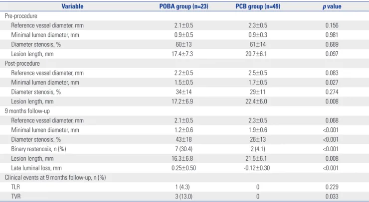
Angiographic follow-up and clinical events at 9 months The QCA and clinical outcomes are shown in Table 2. Nega- tive late luminal loss (LLL) was observed in the PCB group at 9 months follow-up. The in-segment LLL at 9 months was sig- nificantly lower in the PCB group than the POBA group (-0.12±
0.30 mm vs. 0.25±0.50 mm, p<0.001). At 9 months, there was a higher percentage of binary restenosis (diameter stenosis
≥50%) in the POBA group (30.4%, n=7 vs. 4.1%, n=2, p<0.001).
The clinical events observed were TLR and TVR, which oc- curred only in the POBA group.
DISCUSSION
The results of this study demonstrate the superiority of PCB treatment for de novo coronary lesions in suppressing neointi-
mal hyperplasia in comparison to POBA. In fact, the use of the PCB resulted in a negative LLL, compared to POBA treatment alone, with an increase in minimal lumen diameter (MLD) in more than half of the patients (35 of 49) during follow-up.
Historically, remodeling and compression of an atheroscle- rotic plaque were thought to constitute the major mechanism of balloon angioplasty.9 However, previous studies have shown that nearly 50% of the theoretically achievable cross-sectional area is lost after balloon angioplasty, because of the elastic properties of the vessel and intimal hyperplasia.9-11 Coronary stents were developed in part to overcome the risk of elastic recoil and restenosis from balloon angioplasty. Despite the clear benefits of coronary stents, alternatives are occasionally required for patients with de novo lesions requiring revascu- larization who are unable to tolerate long-term dual antiplatelet therapy due to high bleeding risk, poor compliance, or pending non-cardiac surgery, or where coronary anatomy prevents stent deployment.1,2 In these scenarios, PCB treatment as an adjunct to POBA provides an effective and safe alternative to coronary stent implantation.
Recent data suggests PCB treatment as an adjunct to POBA is feasible in patients with de novo coronary lesions.12,13 The Valentines II trial demonstrated that PCB achieves high pro-
cedural success rates (99%) with acceptable rates of bail-out stenting (12%) and low adverse cardiac events rates at mid- term follow-up (8.7%), and offered an alternative for revascu- larization in patients unsuitable for DES implantation.12 An- other study showed that PCB treatment in de novo coronary arteries after pre-dilatation without major dissection and recoil led to late lumen increase (1.75±0.55 mm vs. 1.91±0.55 mm, p<0.001).13 In the same context, our study showed that PCB treatment, unlike POBA, achieves lumen increase in de novo coronary lesions at 9 months follow-up, although both PCB and POBA equally achieved procedural success upon com- pletion of the procedure.
Although the exact mechanism of the late lumen increase is not well understood, this is thought to be likely due to the local drug delivery effects of paclitaxel. The sustained pharmacolog- ical effects of paclitaxel are exerted by binding to the subunit of tubulin, resulting in arrest of microtubule function, up-regula- tion of pro-apoptotic factors, and the promotion of prolonged antiproliferation.14,15 Preceding laboratory results have shown that even a short contact between taxane compounds and vas- cular smooth muscle cells can inhibit the proliferation of the cells for a long period.16,17 A previous study showed that the most pronounced lumen enlargement is seen in areas with Table 1. Baseline Clinical and Procedural Characteristics
Variables POBA group (n=23) PCB group (n=49) p value
Age, yrs 65.7±8.7 61.8±9.1 0.105
Male, n (%) 14 (60.9) 35 (71.4) 0.778
LV ejection fraction, % 56.5±12.0 64.3±6.3 0.007
Cardiovascular risk factors, n (%)
Hypertension 13 (56.5) 32 (65.3) 0.981
Diabetes mellitus 3 (13.0) 22 (44.9) 0.019
Dyslipidemia 10 (43.5) 33 (67.3) 0.391
Current smoker 3 (13.0) 18 (36.7) 0.386
Family history of CAD 4 (17.4) 3 (6.1) 0.403
Clinical diagnosis, n (%) 0.224
Stable angina 8 (34.8) 25 (51.0)
Unstable angina 15 (65.2) 24 (49.0)
Procedural findings
Number of diseased vessel, n (%) 2.4±0.7 1.2±0.4 <0.001
Culprit vessel, n (%) 0.004
LAD 5 (21.7) 30 (61.2)
LCX 8 (34.8) 12 (24.5)
RCA 10 (43.5) 7 (14.3)
ACC/AHA B2/C lesion, n (%) 15 (65.2) 30 (61.2) 0.607
POBA balloon diameter, mm 2.37±0.51 2.73±0.47 0.021
POBA balloon to artery ratio 1.18±0.23 1.15±0.13 0.627
POBA inflation pressure, mm Hg 8.40±2.13 10.31±2.56 0.019
PCB balloon diameter, mm NA 2.86±0.44 NA
PCB balloon length, mm NA 23.15±5.15 NA
POBA, plain old balloon angioplasty; PCB, paclitaxel-coated balloon; LV, left ventricular; CAD, coronary artery disease; LAD, left anterior descending artery; LCX, left circumflex artery; RCA, right coronary artery; NA, not available; ACC, American College of Cardiology; AHA, American Heart Association.
Data are mean±standard deviation or number (percentage).
the highest plaque burden.13 However, plaque regression or other healing mechanisms cannot be excluded without as- sessment with optical coherence tomography or intravascular ultrasound.
There are some limitations to our study that need consider- ation. Firstly, this study was a retrospective observational clin- ical study with small numbers. Secondly, the patients selected had relatively small coronary vessels; however, this is the cur- rent indication for PCB reimbursement in Korea. Although a further study is needed to evaluate these findings in larger ar- teries after PCB treatment, ethical considerations may make such a study hard to undertake, because of the clear benefits of coronary stents in large vessels amongst patients with no other contraindications. Finally, post-procedure reference ves- sel diameter and MLD were smaller in the POBA treatment group than the PCB treatment group. These two parameters may have affected the binary restenosis and TLR.
In conclusion, PCB treatment of de novo coronary lesions showed better angiographic outcomes at 9 months after the procedure than POBA treatment alone.
REFERENCES
1. Brilakis ES, Banerjee S, Berger PB. Perioperative management of patients with coronary stents. J Am Coll Cardiol 2007;49:2145-50.
2. Agostoni P, Biondi-Zoccai GG, Gasparini GL, Anselmi M, Moran- do G, Turri M, et al. Is bare-metal stenting superior to balloon an-
gioplasty for small vessel coronary artery disease? Evidence from a meta-analysis of randomized trials. Eur Heart J 2005;26:881-9.
3. Mintz GS. Remodeling and restenosis: observations from serial intravascular ultrasound studies. Curr Interv Cardiol Rep 2000;2:
316-25.
4. Vos NS, Dirksen MT, Vink MA, van Nooijen FC, Amoroso G, Her- rman JP, et al. Safety and feasibility of a PAclitaxel-eluting balloon angioplasty in Primary Percutaneous coronary intervention in Amsterdam (PAPPA): one-year clinical outcome of a pilot study.
EuroIntervention 2014;10:584-90.
5. Kleber FX, Rittger H, Bonaventura K, Zeymer U, Wöhrle J, Jeger R, et al. Drug-coated balloons for treatment of coronary artery dis- ease: updated recommendations from a consensus group. Clin Res Cardiol 2013;102:785-97.
6. Smith SC Jr, Feldman TE, Hirshfeld JW Jr, Jacobs AK, Kern MJ, King SB 3rd, et al. ACC/AHA/SCAI 2005 guideline update for per- cutaneous coronary intervention: a report of the American Col- lege of Cardiology/American Heart Association Task Force on Practice Guidelines (ACC/AHA/SCAI Writing Committee to Up- date the 2001 Guidelines for Percutaneous Coronary Interven- tion). J Am Coll Cardiol 2006;47:e1-121.
7. Smith SC Jr, Dove JT, Jacobs AK, Kennedy JW, Kereiakes D, Kern MJ, et al. ACC/AHA guidelines of percutaneous coronary inter- ventions (revision of the 1993 PTCA guidelines)--executive sum- mary. A report of the American College of Cardiology/American Heart Association Task Force on Practice Guidelines (committee to revise the 1993 guidelines for percutaneous transluminal coro- nary angioplasty). J Am Coll Cardiol 2001;37:2215-39.
8. Kent KM, Bentivoglio LG, Block PC, Cowley MJ, Dorros G, Gosse- lin AJ, et al. Percutaneous transluminal coronary angioplasty: re- port from the Registry of the National Heart, Lung, and Blood In- stitute. Am J Cardiol 1982;49:2011-20.
Table 2. Pre-Procedure, Post-Procedure, and 9-Month Angiographic Follow-Up Quantitative Coronary Analysis and the Clinical Events at 9 Months Follow-Up
Variable POBA group (n=23) PCB group (n=49) p value
Pre-procedure
Reference vessel diameter, mm 2.1±0.5 2.3±0.5 0.156
Minimal lumen diameter, mm 0.9±0.5 0.9±0.3 0.981
Diameter stenosis, % 60±13 61±14 0.689
Lesion length, mm 17.4±7.3 20.7±6.1 0.097
Post-procedure
Reference vessel diameter, mm 2.2±0.5 2.5±0.5 0.083
Minimal lumen diameter, mm 1.5±0.5 1.7±0.5 0.027
Diameter stenosis, % 34±14 29±11 0.274
Lesion length, mm 17.2±6.9 22.4±6.0 0.008
9 months follow-up
Reference vessel diameter, mm 2.1±0.5 2.3±0.5 0.068
Minimal lumen diameter, mm 1.2±0.6 1.9±0.6 <0.001
Diameter stenosis, % 43±18 26±13 <0.001
Binary restenosis, n (%) 7 (30.4) 2 (4.1) <0.001
Lesion length, mm 16.3±6.8 21.5±6.1 0.008
Late luminal loss, mm 0.25±0.50 -0.12±0.30 <0.001
Clinical events at 9 months follow-up, n (%)
TLR 1 (4.3) 0 0.229
TVR 3 (13.0) 0 0.033
POBA, plain old balloon angioplasty; PCB, paclitaxel-coated balloon; TLR, target lesion revascularization; TVR, target vessel revascularization.
Data are mean±standard deviation or number (percentage).
9. Haude M, Erbel R, Issa H, Meyer J. Quantitative analysis of elastic recoil after balloon angioplasty and after intracoronary implanta- tion of balloon-expandable Palmaz-Schatz stents. J Am Coll Car- diol 1993;21:26-34.
10. Rensing BJ, Hermans WR, Strauss BH, Serruys PW. Regional dif- ferences in elastic recoil after percutaneous transluminal coro- nary angioplasty: a quantitative angiographic study. J Am Coll Cardiol 1991;17(6 Suppl B):34B-8B.
11. Liu MW, Roubin GS, King SB 3rd. Restenosis after coronary an- gioplasty. Potential biologic determinants and role of intimal hy- perplasia. Circulation 1989;79:1374-87.
12. Waksman R, Serra A, Loh JP, Malik FT, Torguson R, Stahnke S, et al. Drug-coated balloons for de novo coronary lesions: results from the Valentines II trial. EuroIntervention 2013;9:613-9.
13. Kleber FX, Schulz A, Waliszewski M, Hauschild T, Böhm M, Dietz U, et al. Local paclitaxel induces late lumen enlargement in coro-
nary arteries after balloon angioplasty. Clin Res Cardiol 2015;104:
217-25.
14. Pires NM, Eefting D, de Vries MR, Quax PH, Jukema JW. Sirolimus and paclitaxel provoke different vascular pathological responses after local delivery in a murine model for restenosis on underly- ing atherosclerotic arteries. Heart 2007;93:922-7.
15. Gray WA, Granada JF. Drug-coated balloons for the prevention of vascular restenosis. Circulation 2010;121:2672-80.
16. Axel DI, Kunert W, Göggelmann C, Oberhoff M, Herdeg C, Küt- tner A, et al. Paclitaxel inhibits arterial smooth muscle cell prolif- eration and migration in vitro and in vivo using local drug deliv- ery. Circulation 1997;96:636-45.
17. Scheller B, Speck U, Schmitt A, Böhm M, Nickenig G. Addition of paclitaxel to contrast media prevents restenosis after coronary stent implantation. J Am Coll Cardiol 2003;42:1415-20.