1. Physiology of Testis 2. Male Infertility
3. Male Sexual Dysfunction
ANDROLOGY ( 남성과학 )
고환의 생리
(PHYSIOLOGY OF TESTIS )
Testis: dual function 1. spermatogenesis
2. testosterone formation
학습목적 : 정자를 형성하여 생식에 관여하고 , 남성호르몬을 분비하여
내분비적 중요 기능을 하는 고환의 구조와 생리를 이해한다
.
학습목표 ( 한국의과대학장협의회 )
- 테스토스테론의 기능을 설명한다 .
- 테스토스테론의 분비 조절에 대해 설명한다 .
- 고환의 라이디히세포 (Leydig cell) 의 기능애 대하여 설명한다 .
학습목표 ( 대한비뇨기과학회 )
1. Hypothalamic pituitary gonadal axis 에 대해 설명한다 . 2. 고환의 Leydig cell 의 기능에 대해 설명한다 .
3. 고환의 Sertoli cell 의 역할에 대해 기술한다 .
4. spermatogenesis 의 hormone control 에 대해 설명한다 . 5. 남성호르몬의 생성과 분비에 대해 기술한다 .
6. 남성호르몬의 운반에 대해 기술한다 . 7. 남성호르몬의 기능에 대해 기술한다 . 8. 부고환의 기능에 대해서 설명한다 .
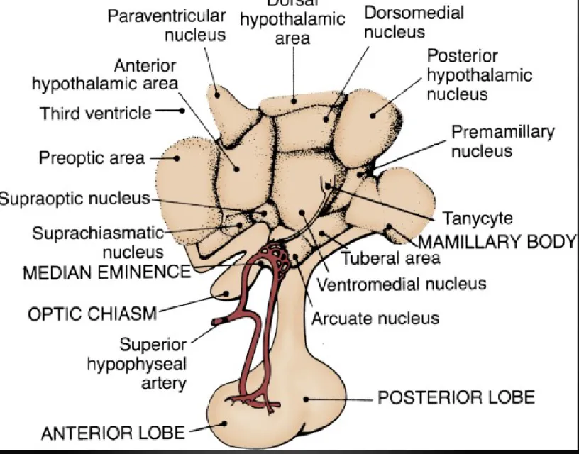
시상하부 뇌하수체 정소 축
Hypothalamic-Pituitary-Gonadal Axis
Figure 18-1 Diagram of the hypothalamic-pituitary-testis axis.
LH, luteinizing hormone; FSH, follicle-stimulating hormone.
1. Extra-hypothalamic central nervous system limbic system
2. Hypothalamus; release single decapeptide GnRH episodically neural message from CNS
neurotransmitter (norepinephrine, dopamine, serotonin, acetylcholine) neuropeptide (endogenous opioid peptide)
humoral message from testis
* Gonadotropin-releasing hormone
enhanced synthesis and release of LH, FSH 3 type of rhythmicity
1. seasonal: peaking in spring
2. circadian: highest during early morning 3. pulsatile every 90 - 120 min
constant exposure to GnRH, result in inhibiting effect prompt release of LH, much less extent of FSH
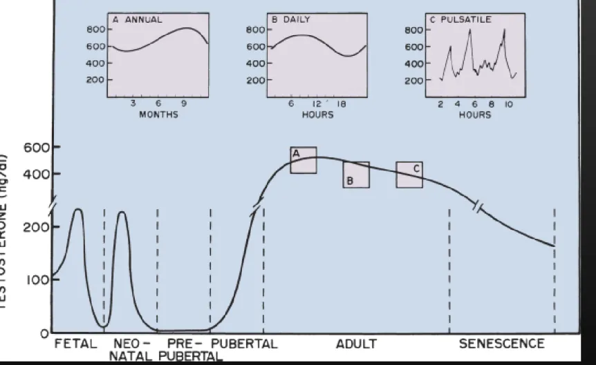
Figure 18-8 Concentration of testosterone in peripheral blood of the human male at different times of the life cycle.
The peak of testosterone in the peripheral blood of the fetus occurs between 12 and 18 weeks of gestation (lower left corner; gestational age not shown). The peak of testosterone in the peripheral blood of the neonate occurs at approximately 2 months of age. Testosterone declines to low levels during the prepubertal period. The pubertal increase in testosterone concentration in peripheral blood occurs between 12 and 17 years of age. Testosterone concentration in the adult reaches its maximum during the second or third decade of life and then declines slowly through the fifth decade. Testosterone concentration in peripheral blood declines dramatically during senescence.
Inset A shows the annual rhythm in testosterone concentration in peripheral blood of the human male. The peak and nadir occur in the fall and spring, respectively. Inset B shows the daily rhythm in testosterone concentration in peripheral blood of the adult human male. The peak and nadir occur in the morning and evening, respectively. Inset C shows the frequent and irregular fluctuations in testosterone concentration in peripheral blood of the human male.
3. Pituitary gland
posterior lobe (neurohypophysis): oxytocin, vasopressin
anterior lobe (adenohypophysis): LH, FSH, ACTH, GH, TSH, prolactin
LH, FSH are synthesized in pituitary gland, secretion into systemic circulation LH: stimulate testosterone production by Leydig cell in interstitium
FSH: stimulation of Sertoli cell, support spermatogenesis in seminiferous tubule
1) Feed back control
testosterone is primary inhibition of LH secretion estradiol (testis, peripheral conversion of androgen);
potent inhibition of LH, FSH
FSH inhibited by inhibin (secreted primarily by Sertoli cell, inhibin B) 2) Prolactin
inhibit production of GnRH
decrease libido and sexual function
Figure 18-2 Organization of the hypothalamus and pituitary. (From Swerdloff RS, Wang C: Physiology of hypothalamic-pituitary function.
In Walsh PC, Wein AJ, Retik AB, Vaughan ED [eds]: Campbell's Urology, 7th ed. Philadelphia, WB Saunders, 1997, pp 1239-1253.)
고 환 (Testis)
15-25 ml in volume, 4.5-5.1 cm in length human testis is an organ of dual function spermatogenesis in seminiferous tubule
secretion of androgen by Leydig cell, in interstitial tissue combined length of 600-1200 tubules is approximately 250 m Rete testis coalesce to form 6~12 efferent duct, conduit to carry testicular fluid and spermatozoa
Figure 18-4 Drawing of the human testis
showing the seminiferous tubules, epididymis, and ductus deferens. (Illustration based on Hirsh AV: The anatomical preparations of the human testis and epididymis in the Glasgow Hunterian Collection. Hum Reprod Update 1995;1:515-521.)
Figure 18-3 Scanning electron micrograph of the cut surface of the human testis. Note the relationship of interstitial tissue to seminiferous tubules. (From Christensen AK: Leydig cells. In Greep RO, Astwood EB [eds]: Handbook of Physiology. Washington, DC, American Physiology Society, 1975, pp 57-94.)
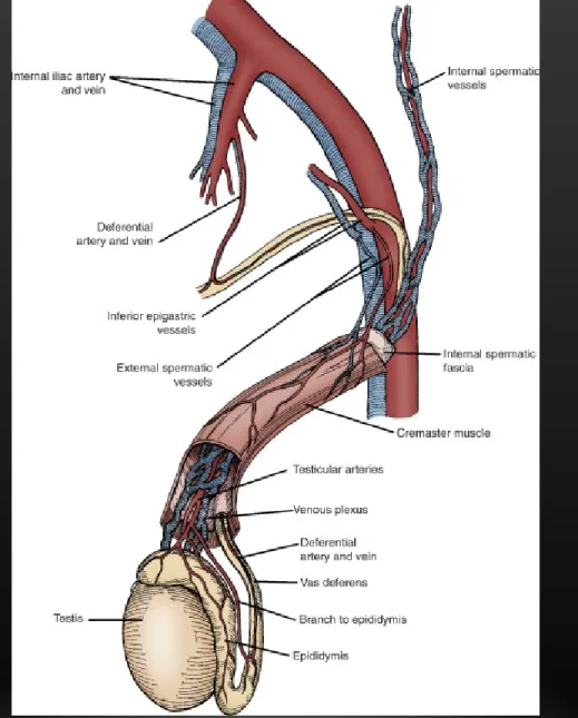
3 arteries; 1. internal spermatic artery 2. deferential artery
3. external spermatic or cremasteric artery Count-current exchange of heat in spermatic cord
Testis is 2~4 o C lower than rectal temperature
Figure 18-5 Schematic illustration of the
interconnections between internal spermatic, external spermatic (cremasteric), and deferential vessels in the peritesticular region and schematic cord.
1. Interstitium : 20-30% of total testicular volume
contain blood vessel, lymphatics, fibroblastic supporting cell, macrophage, mast cell, Leydig cell.
1) Leydig cell
round polygonal cytoplasm prominent mitochondria abundant s-ER
secretion of testosterone response to LH pulse and has diurnal pattern
Figure 18-6 Location and fine structure of human Leydig cells. Leydig cells occur in clusters in the interstitial tissue between the seminiferous tubules (upper left). Interstitial tissue (upper right) contains macrophages and fibroblasts as well as capillaries and lymph vessels. Seminiferous tubules are surrounded by a boundary layer of myoid cells. The most abundant organelle within the cytoplasm of the Leydig cell is the smooth endoplasmic reticulum (lower left). Some of the organelles are seen in greater detail in a selected area of cytoplasm (lower right).
Figure 18-7 Luteinizing hormone (LH) is the primary tropic stimulus for testosterone
production. LH is shown binding to its receptor.
Highlighted in red are the events associated with the acute regulation of steroidogenesis, namely, mobilization of the initial substrate for testosterone biosynthesis, cholesterol. These events occur within minutes of LH binding. The green arrows denote chronic events triggered by LH, which include increased transcription and translation of the genes encoding the
steroidogenic enzymes. LH stimulation also increases the number and size of Leydig cell organelles that are involved in steroidogenesis, such as the mitochondria and smooth
endoplasmic reticulum (SER) membranes. These events are chronic and require several hours, to days, before they become evident. The LH receptor (in yellow) has seven membrane- spanning domains and is coupled to G proteins that modulate its activation of adenylate cyclase. Cyclic adenosine monophosphate (cAMP) stimulates protein kinase A. LH binding also initiates several other events in parallel:
calcium influx leading to calmodulin activation of a calcium/calmodulin kinase; arachidonic acid mobilization from phospholipase A2 activity; and efflux of chloride ions (all shown in red). The net effect of these changes is to make more
substrate cholesterol available for
steroidogenesis. The three main sources of cholesterol in the Leydig cell are (1) externally, from bloodborne lipoprotein and internalization of cholesterol/lipoprotein receptor complexes, (2) from de novo synthesis from acetate, and (3) from stored cholesterol esters in lipid droplets.
Maintenance of cholesterol stores is part of the normal resting function of the Leydig cell; LH stimulation evokes cholesterol mobilization through cholesterol esterase activity. The free cholesterol then associates with steroid acute regulatory protein (StAR) for transport to the inner membrane of the mitochondrion. A key part of the transport mechanism is the signal sequence (depicted as a blue threadlike tail) that enables StAR protein to pass through mitochondrial membranes. Peripheral
benzodiazepine receptor (PBR) forms a channel in the mitochondrial membranes and also facilitates cholesterol entry.
MAJOR EPOCHS IN TESTOSTERONE PRODUCTION
1. Differentiation and development of fetal reproductive tract
2. Neonatal organization or imprinting of androgen dependent target tissue
3. Masculinization of male at puberty
4. Maintenance of growth and function of androgen-dependent organ in adult
2) Transport of androgen
2% free : physiologically active androgen
44% bound to sex hormone binding globulin (SHBG) 54% bound to albumin and other protein
in seminiferous tubule bound to androgen binding protein (ABP)
in target cell convert to more potent androgen dihydrotestosterone
(DHT) by 5 alpha reductase
3) Function of androgen target tissue
a. regulation of gonadotropin secretion by hypothalamic-pituitary axis b. initiation and maintenance of spermatogenesis
c. differentiation of internal and external male genital system during fetal development
d. promotion of sexual maturation at puberty
2.
Seminiferous tubule
600-1200 tubules in testis, approximately 250 m
contain germ cell and Sertoli cell, 85-90% of testicular volume
1) Sertoli cell
linked by tight junction, divide into basal and adluminal compartment close approximation of myoid cell, serve to form blood-testis barrier create unique microenvironment in adluminal compartment
facilitate spermatogenesis
maintain in immunologically privileged location nourishment of developing germ cell
phagocytosis damaged cell
production of androgen binding protein (ABP) production of inhibin
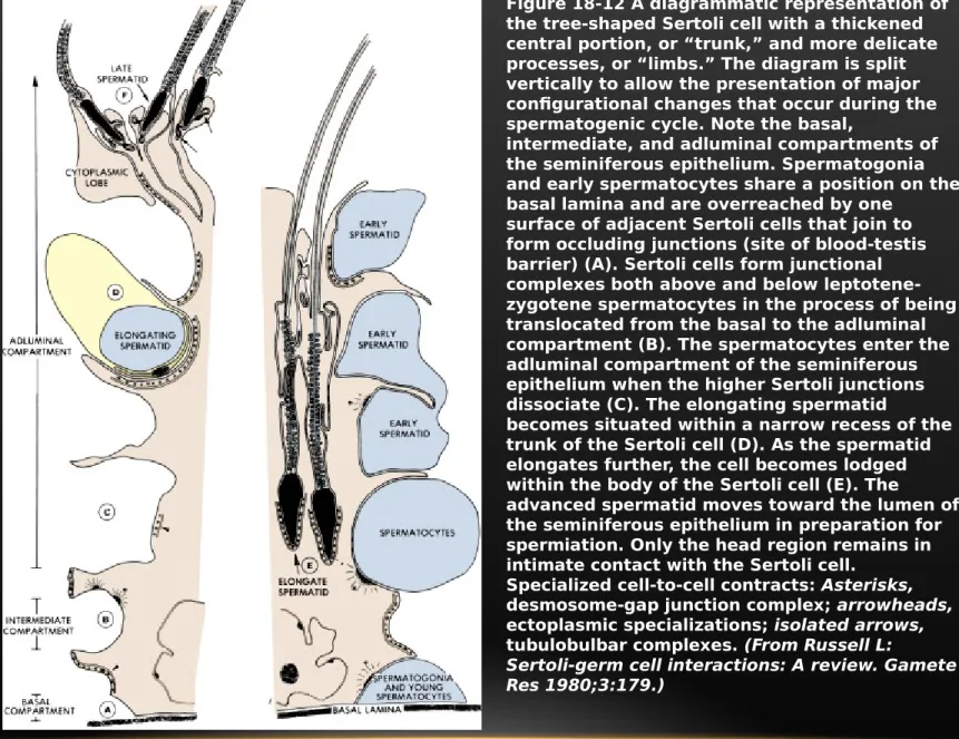
Figure 18-12 A diagrammatic representation of the tree-shaped Sertoli cell with a thickened central portion, or “trunk,” and more delicate processes, or “limbs.” The diagram is split vertically to allow the presentation of major configurational changes that occur during the spermatogenic cycle. Note the basal,
intermediate, and adluminal compartments of the seminiferous epithelium. Spermatogonia and early spermatocytes share a position on the basal lamina and are overreached by one
surface of adjacent Sertoli cells that join to form occluding junctions (site of blood-testis barrier) (A). Sertoli cells form junctional complexes both above and below leptotene- zygotene spermatocytes in the process of being translocated from the basal to the adluminal compartment (B). The spermatocytes enter the adluminal compartment of the seminiferous epithelium when the higher Sertoli junctions dissociate (C). The elongating spermatid
becomes situated within a narrow recess of the trunk of the Sertoli cell (D). As the spermatid elongates further, the cell becomes lodged within the body of the Sertoli cell (E). The
advanced spermatid moves toward the lumen of the seminiferous epithelium in preparation for spermiation. Only the head region remains in intimate contact with the Sertoli cell.
Specialized cell-to-cell contracts: Asterisks, desmosome-gap junction complex; arrowheads, ectoplasmic specializations; isolated arrows, tubulobulbar complexes. (From Russell L:
Sertoli-germ cell interactions: A review. Gamete Res 1980;3:179.)
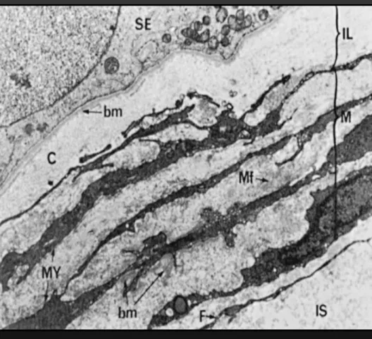
Figure 18-9 Low-power electron micrograph of the
peritubular tissue in a human testis. Peritubular tissue lies between the basement membrane (bm) of the seminiferous epithelium (SE) and the interstitial tissue (IS). Three zones are present in peritubular tissue: the inner lamella (IL); the myoid layer (M), containing myoid cells (MY) with abundant microfibrils (Mf); and an adventitial layer containing
fibroblasts (F). (From Hermo L, Lalli M, Clermont Y:
Arrangement of connective tissue elements in the walls of seminiferous tubules of man and monkey. Am J Anat
1977;148:433-446.)
2)
Germinal epithelium
spermatogonia divide, either stem cell renewal (reproduce their number) or daughter cell (become spermatocyte)
13 germ cell type;
dark type A spermatogonia (Ad) pale type A spermatogonia (Ap)
type B spermatogonia (B) preleptotene primary spermatocyte (R)
leptotene primary spermatocyte (L) zygote primary spermatocyte (Z) pachytene primary spermatocyte (P) secondary spermatocyte (II)
Sa, Sb, Sc, Sd1, Sd2 spermatid result of meiosis is Sa spermatid
6 recognizable cellular association (stage of cycle of seminiferous epithelium) entire spermatogenic process in man require 64 days
spermiogenesis
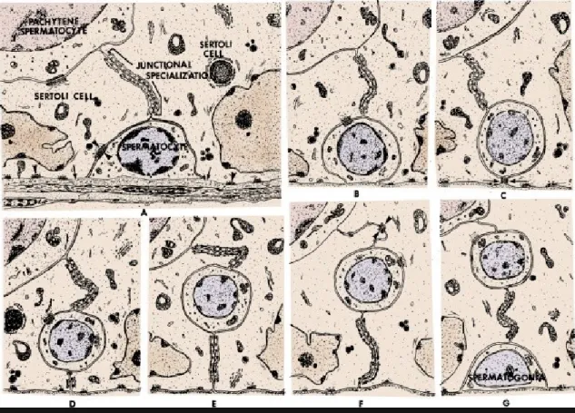
Figure 18-11 Diagram of the steps required to transfer rat primary spermatocytes from the basal to the adluminal compartment of the seminiferous tubule. Initially (A, B) the Sertoli cells are attached to each other above the spermatocytes by tight junctions. Next (C to E), Sertoli cells form new junctions below the spermatocytes, isolating the spermatocytes in an intermediate compartment, above and below which are tight junctions.
The junctions above the spermatocytes then break down (F, G), and the spermatocytes enter the adluminal compartment. (From Russell L:
Desmosome-like junctions between Sertoli cells and germ cells in the rat testis. Am J Anat 1977;148:313.)
Figure 18-13 The steps of
spermatogenesis in man. Ad, dark type A spermatogonium; AP, pale type A spermatogonium; B, type B
spermatogonium; R, resting or
preleptotene primary spermatocyte; L, leptotene spermatocyte; Z, zygotene spermatocyte; P, pachytene
spermatocyte; II, secondary
spermatocyte; Sa(a), Sb1(b1), sb2(b2), Sc(c), sd1(d1), Sd2(d2), spermatids;
Rb, residual body. The table shows the cells that make up the six stages of the cycle of the seminiferous epithelium (I to VI): D1, diakenesis; ID and IID, first and second maturation divisions of spermatocytes. (Modified from
Clermont Y: Renewal of spermatogonia in man. Am J Anat 1966;118:509.)
Figure 18-14 Cellular composition of the six cellular associations (stages I to VI) found in human seminiferous tubules. Ser, Sertoli nuclei; Ad and Ap, dark and pale type A
spermatogonia; B, type B spermatogonia; R, resting (preleptotene) primary spermatocytes; L, leptotene spermatocytes; Z, zygotene spermatocytes; P, pachytene spermatocytes; Di, diplotene spermatocytes; Sptc-Im, primary spermatocytes in division; Sptc-II, secondary spermatocytes in
interphase; Sa, Sb, Sb2, Sc, Sd1, Sd2, spermatids at various steps of spermatogenesis. (Modified from Heller CG, Clermont Y: Kinetics of the germinal epithelium in man. Rec Prog Horm Res 1964;20:545.)
Figure 18-15 Helical configuration of stages of the seminiferous tubule epithelium in man. (From Schulze W, Rehder U: Organization and morphogenesis of the human seminiferous epithelium. Cell Tissue Res 1984;237:395-407.)
부고환 (Epididymis)
caput epididymis consist of 8-12 efferent duct 5 – 6 m in length
testicular spermatozoa are non-motile and incapable of fertilizing ova
spermatozoa gain progressive motility and fertilizing ability after passing epididymis epididymis has contractility due to prominent muscular development
* Function of epididymis 1) sperm transport
2) sperm storage
3) maturation of spermatozoa sperm motility maturation sperm fertility maturation
Figure 18-16 Schematic drawing of the human epididymis showing
regionalization of the ductal epithelium and muscle layer.
Locations of epididymal segments shown in cross sections are identified by number. (From Baumgarten HG, Holstein AF, Rosengren E: Arrangement,
ultrastructure, and
adrenergic innervation of smooth musculature of the ductal efferentes, ductus epididymidis, and ductus deferens in man. Z Zellforsch Mikrosk Anat 1971;120:37.)
Figure 18-18 Patterns of tail movement in human epididymal spermatozoa. A, The pattern shown by spermatozoa taken from proximal regions of the epididymis is characterized by a high-amplitude, low- frequency beat producing little forward movement. B, In contrast, tail movement in a large proportion of
spermatozoa from the cauda epididymis is characterized by low-amplitude, rapid beats that result in forward progression. (From Bedford JM, Calvin HI, Cooper GW:
The maturation of spermatozoa in the human epididymis.
J Reprod Fertil 1973;18:199-213.)
Figure 18-19 Sperm fertility maturation in the human epididymis. Sperm fertilizing ability was assessed using zona pellucida–
free hamster eggs and by changes in motility, which increases in the distal regions of the human epididymis. (From Bedford JM: The bearing of epididymal function in strategies for in vitro fertilization and gamete intrafallopian transfer. Ann N Y Acad Sci 1988;541:284- 291.)
Figure 18-20 Diagram of a typical mammalian spermatozoon. The
plasma membrane is omitted in order to illustrate the major components of the spermatozoon. Cross-sectional insets show the orientation of the internal cell structures. (From Fawcett DW: The mammalian spermatozoon.
Dev Biol 1975;44:394-436.)
정 낭 (SEMINAL VESICLE)
major volume of seminal plasma from seminal vesicle (60%) provide nourishing substrate fructose
prostaglandin, coagulating substance
seminal plasma has buffering effect on acidic vaginal environment
전립선 (PROSTATE)
ejaculated semen liquefy within 20 min as result of prostatic proteolytic enzyme add zinc, phospholipid, spermine, phosphatase to seminal fluid
first portion of ejaculate contain most of spermatozoa and prostatic secretion second portion is composed seminal vesicle secretion and only few spermatozoa