• 검색 결과가 없습니다.

Open Surgical Repair Using the Femoral Vein for a Mycotic Superior Mesenteric Artery Aneurysm

N/A
N/A
Protected

Academic year: 2021

Share "Open Surgical Repair Using the Femoral Vein for a Mycotic Superior Mesenteric Artery Aneurysm"

Copied!
4
0
0

로드 중.... (전체 텍스트 보기)

전체 글

(1)

ISSN: 2233-601X (Print) ISSN: 2093-6516 (Online)

− 209 −

Received: September 25, 2017, Revised: October 25, 2017, Accepted: November 1, 2017, Published online: June 5, 2018

Corresponding author: Keon Hyon Jo, Department of Thoracic and Cardiovascular Surgery, Seoul St. Mary’s Hospital, College of Medicine, The Catholic University of Korea, 222 Banpo-daero, Seocho-gu, Seoul 06591, Korea

(Tel) 82-2-2258-2858 (Fax) 82-2-594-8644 (E-mail) khjo@cmcnu.or.kr

© The Korean Society for Thoracic and Cardiovascular Surgery. 2018. All right reserved.

This is an open access article distributed under the terms of the Creative Commons Attribution Non-Commercial License (http://creativecommons.org/

licenses/by-nc/4.0) which permits unrestricted non-commercial use, distribution, and reproduction in any medium, provided the original work is properly cited.

Open Surgical Repair Using the Femoral Vein for a Mycotic Superior Mesenteric Artery Aneurysm

Min Namkoong, M.D. 1 , Seok Beom Hong, M.D. 1 , Hwan Wook Kim, M.D. 1 , Keon Hyon Jo, M.D. 1 , Jang Yong Kim, M.D. 2

1

Department of Thoracic and Cardiovascular Surgery and

2

Division of Vascular Surgery, Department of Surgery, Seoul St. Mary’s Hospital, College of Medicine, The Catholic University of Korea

Superior mesenteric artery (SMA) aneurysms are rare and often fatal. A 72-year-old man had previously been admitted to the emergency room with epigastric pain and heart murmur. The echocardiographic diag- nosis was vegetation on the aortic and mitral valves, with moderate regurgitation from both valves due to infective endocarditis. No aneurysm was detected on abdominal computed tomography, and emergency dou- ble-valve replacement was performed. On postoperative day 25, the patient experienced abrupt abdominal pain, and computed tomography revealed a mycotic SMA aneurysm. Open surgical repair of the SMA aneur- ysm was performed using the femoral vein, and the patient’s postoperative course was uneventful.

Key words: 1. Superior mesenteric artery aneurysm 2. Endocarditis

3. Femoral vein 4. Sternotomy

Case report

A 72-year-old man visited the emergency room with a 1-month history of poor oral intake and gen- eral weakness, as well as a 2-hour history of peri- umbilical pain. The patient had no medical history except hemorrhoidectomy, which had been per- formed 2 years earlier. Upon admission, the patient’s vital signs were stable. Laboratory testing revealed leukocytosis (11,190 white b lood cells per microliter) and an elevated C-reactive protein level (7.48 mg/L).

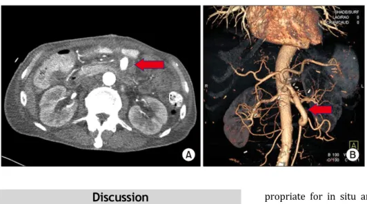
A physical examination demonstrated epigastric ten- derness and heart murmur, and abdominal computed tomography (CT) showed hepatomegaly and splenic infarction, but no signs of an aneurysm (Fig. 1A).

Transthoracic echocardiography revealed 10-mm and

8-mm areas of vegetation on the aortic and mitral valves, with moderate regurgitation in both valves.

Thus, the patient underwent an emergency double- valve replacement (aortic valve: Carpentier-Edwards 19 mm [Edwards Lifesciences, Irvine, CA, USA]; mi- tral valve: Carpentier-Edwards 29 mm [Edwards Lifesciences]). Enterococcus casseliflavus was identi- fied in the b lood culture, although culture of the sur- gical specimen revealed negative findings. The patient received antibiotic treatment in the general ward and did not experience any specific complications.

On the 25th day after surgery, the patient experi- enced abrupt abdominal pain and a pulsatile mass was found in the abdomen. Abdominal CT revealed an unruptured 3.5-×5-cm mycotic superior mesen- teric artery (SMA) aneurysm (Fig. 1B). Emergency

Korean J Thorac Cardiovasc Surg 2018;51:209-212 □ CASE REPORT □

https://doi.org/10.5090/kjtcs.2018.51.3.209

(2)

Min Namkoong, et al

− 210 −

Fig. 1. Abdominal computed tomography. (A) Before cardiac surgery. No signs of aneurysm (arrow). (B) The 25th day after the cardiac surgery. An unruptured 3.5-×5-cm mycotic superior mesenteric artery was observed (arrow).

Fig. 2. Intraoperative images. (A) The mycotic superior mesenteric artery aneurysm was visible, although there was no bowel ischemia or aneurysm rupture. (B) Approximately 10 cm of the femoral vein was harvested. (C) After repair using the femoral vein.

surgery was performed, although bowel ischemia and aneurysm rupture were not observed (Fig. 2A).

Careful dissection of the aneurysm was performed to preserve the intestinal vascular arcades. After clamping the SMA b ase, the aneurysm was opened and the thrombus was removed. Two jejunal branch- es from the SMA were identified inside the aneur- ysm, and color change occurred after ligation of the jejunal branch. We planned to harvest a vein for in- terposition at the branching point from the SMA to the jejunal b ranch. We chose to not use the great sa- phenous vein because of size mismatch (a small di- ameter). Thus, we harvested approximately 10 cm of the femoral vein by sharp dissection and hand-tie (Fig. 2B). End-to-end anastomosis was performed on the SMA and the jejunal branch using a femoral vein

graft. After the femoral vein graft interposition, the

color of the bowel returned to normal (Fig. 2C). SMA

angiography was performed to confirm flow at the

anastomosis site. Slight stenosis was found at the

distal branch and a percutaneous transluminal angio-

plasty balloon was inserted. After confirming that the

flow had improved, the aneurysmal sac was closed

using an omental patch. To prevent postoperative ve-

nous hypertension and leg edema at the donor site, a

compression stocking was applied and early ambula-

tion was performed. The patient was discharged 45

days after the repair without any complications and

with good leg and digestive function. A follow-up CT

after 1 year revealed that the patency of the graft

had been maintained (Fig. 3).

(3)

Open Repair of Mycotic SMA Aneurysm

− 211 −

Fig. 3. Postoperative computed to- mography (CT). (A) The aneurysm had decreased in size compared to the previous CT scan (arrow). (B) Follow-up CT after 1 year revealed that the patency of the graft had been maintained (arrow).

Discussion

Splanchnic artery aneurysms are rare, and SMA aneurysms only account for approximately 5% of all splanchnic artery aneurysms. These cases have a high mortality rate (38%–50%) that is related to in- tractable complications, such as internal ischemia and aneurysm rupture [1,2]. Infectious aneurysm is a complication of infective endocarditis that is caused b y septic emb olism, although it typically involves an intracranial vessel [3]. However, the occurrence of an SMA aneurysm is also often associated with infective endocarditis. In such cases, the patient may present with upper abdominal pain and fever, which is re- lated to the compressive mass effect of the aneurysm. SMA aneurysms must be treated promptly to avoid death or complications, such as expansion and eventual rupture, thrombosis, and distal emboli- zation causing intestinal ischemia [4]. Therefore, ab- dominal pain in a patient with a recent history of in- fective endocarditis should be closely evaluated using CT and/or ultrasonography. In the present case, the correct diagnosis was confirmed using CT.

Although early treatment is important in these cas- es, there are no guidelines for treating infective en- docarditis and SMA aneurysm. The current treatment for a mycotic SMA aneurysm involves resection of the potentially infected aneurysm and autologous tis- sue reconstruction [5].

An autologous graft, usually of the saphenous vein, can be used to reconstruct the SMA [6]. However, the saphenous vein was too small in the present case, so we elected to use the femoral vein. In this context, an autologous superficial femoral vein is ap-

propriate for in situ arterial reconstruction, as it has been associated with advantages in an infected field [7].

Interestingly, most reported cases of mycotic SMA aneurysms have been diagnosed at the same time as infective endocarditis. Furthermore, SMA aneurysm has generally been treated before cardiac surgery [8].

However, the SMA aneurysm in the present case was not detected at the diagnosis of infective endocardi- tis, and appears to have occurred after cardiac surgery.

In conclusion, patients with infective endocarditis can experience mycotic aneurysms in non-intracranial vessels. Thus, these patients should be evaluated for aneurysms before and after cardiac surgery. In addi- tion, the subsequent development of abdominal pain, fever, and a pulsatile mass should elicit suspicion of a mycotic SMA aneurysm. Artery reconstruction using the femoral vein should be considered when the sa- phenous vein is not available for autologous grafting.

Conflict of interest

No potential conflict of interest relevant to this ar- ticle was reported.

References

1. Lai KM, Rosenthal D, Wellons ED, Bikk A, Franklin JS.

Mycotic superior mesenteric aneurysm. J Vasc Surg 2007;45:191.

2. Aerts PD, van Zitteren M, van Kasteren ME, Buiting AG,

Heyligers JM, Vriens PW. Report of two in situ re-

constructions with a saphenous spiral vein graft of

(4)

Min Namkoong, et al

− 212 −

Coxiella burnetii-infected aneurysms of the abdominal aorta. J Vasc Surg 2013;57:234-7.

3. Shiraishi M, Ohki S, Misawa Y. Mycotic superior mesenteric pseudoaneurysm draining into a vein. Interact Cardiovasc Thorac Surg 2011;12:91-3.

4. Lorelli DR, Cambria RA, Seabrook GR, Towne JB. Diagnosis and management of aneurysms involving the superior mes- enteric artery and its branches: a report of four cases.

Vasc Endovascular Surg 2003;37:59-66.

5. Liao WP, Loh CH, Wang HP. Mycotic aneurysm of superior mesenteric artery branch presenting as pulsatile abdomi-

nal mass. Am J Emerg Med 2006;24:128-9.

6. Zhao J. Massive upper gastrointestinal bleeding due to a ruptured superior mesenteric artery aneurysm duodenum fistula. J Vasc Surg 2008;48:735-7.

7. Daenens K, Fourneau I, Nevelsteen A. Ten-year experience in autogenous reconstruction with the femoral vein in the treatment of aortofemoral prosthetic infection. Eur J Vasc Endovasc Surg 2003;25:240-5.

8. Chu PH, She HC, Lim KE, Chu JJ. Mycotic aneurysm of the

superior mesenteric artery in a young woman. Int J Clin

Pract 2005;59:614-6.

수치

Fig. 1. Abdominal computed tomography. (A) Before cardiac surgery. No signs of aneurysm (arrow)
Fig. 3. Postoperative computed to- to-mography (CT). (A) The aneurysm  had decreased in size compared to  the previous CT scan (arrow)

참조

관련 문서

Key Words: Femur, Subtrochanteric fracture, limited open reduction, Proximal femoral nail anti-rotation (PFNA)... 수술 방법 및

In this study we will survey the changes in DNA repair factors during aging in rat, including mismatch repair (MMR), base excision repair (BER), and double-strand break

The clinical outcome and complication for treating proximal femoral shaft fracture were compared and analyzed through the group treated with closed

Kameoka, “Sentinel lymph node biopsy for breast cancer patients using fluorescence navigation with indocyanine green,” World Journal of Surgical Oncology, Vol..

The locations of aneurysms were middle cerebral artery in 15 patients, cerebral artery in 15 patients, cerebral artery in 15 patients, cerebral artery in

16) http://www.macmillandictionary.com/open-dictionary/entries/greycation.htm.. This means using the Outernet will be more like watching a program broadcast on TV,

The average position of the posterior superior alveolar artery, the wall thickness of the lateral wall, and the average volume of the maxillary sinus will

This study attempts to quantify the economic effects of increases in international oil prices on Korea’s energy and biodiesel industry by using a small open computable