• 검색 결과가 없습니다.

Expression of Matrix Metalloproteinase-2 and -13 and Tissue Inhibitor of Metalloproteinase-4 in Varicose Veins

N/A
N/A
Protected

Academic year: 2021

Share "Expression of Matrix Metalloproteinase-2 and -13 and Tissue Inhibitor of Metalloproteinase-4 in Varicose Veins"

Copied!
5
0
0

로드 중.... (전체 텍스트 보기)

전체 글

(1)

http://dx.doi.org/10.5090/kjtcs.2011.44.6.387 ISSN: 2233-601X (Print) ISSN: 2093-6516 (Online)

*Department of Thoracic and Cardiovascular Surgery, Jeju National University Hospital

**Department of Pathology, Jeju National University Hospital

†This study is supported by a clinical research grant of Jeju National University Hospital.

Received: May 11, 2011, Revised: August 22, 2011, Accepted: September 7, 2011

Corresponding author: Young-Hee Maeng, Department of Pathology, Jeju National University Hospital, 1753-3, Ara 1-dong, Jeju 690-716, Korea (Tel) 82-64-717-1411 (Fax) 82-64-717-1131 (E-mail) yhmaeng@jejunu.ac.kr
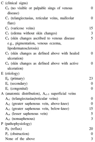
C

The Korean Society for Thoracic and Cardiovascular Surgery. 2011. All right reserved.

CC

This is an open access article distributed under the terms of the Creative Commons Attribution Non-Commercial License (http://creative- commons.org/licenses/by-nc/3.0) which permits unrestricted non-commercial use, distribution, and reproduction in any medium, provided the original work is properly cited.

Expression of Matrix Metalloproteinase-2 and -13 and Tissue Inhibitor of Metalloproteinase-4 in Varicose Veins

Jee Won Chang, M.D.*, Young-Hee Maeng, M.D.**, Su Wan Kim, M.D.*

Background: The relationship between the degree of expression of matrix metalloproteinases or tissue inhibitor of metalloproteinases and venous reflux remains to be investigated. Materials and Methods: Primary varicose vein tissues were obtained from 23 patients, 18 females and 5 males, aged from 19 to 73. Cephalic or basilic veins were obtained for the control group from 10 patients who underwent vascular access for maintenance hemodialysis.

Two operative techniques (high ligation with stripping or endovenous laser coagulation) were used. The expression of matrix metalloproteinase-2 and 13 and tissue inhibitor of metalloproteinase-4 in the varicose vein group and con- trol group was assessed semi-quantitatively by immunohistochemical slides stained with primary antibodies. Results:

Twenty (87%) of the varicose vein group patients had greater or lesser saphenous vein diseases with reflux. The focal weak (+) stain for matrix metalloproteinases-2, and 13, and tissue inhibitor of matrix metalloproteinase-4 was dominant in the varicose vein group; the focal or diffuse strong stain (++ or +++) was prevalent in the con- trol group. The differences were statistically significant (p<0.01). The degree of reflux and the duration of symp- toms were not significantly related to the expression of MMP-13 (p=0.317 and p=0.654, respectively). Conclusion:

Further study should be performed to investigate the relationship between the clinical characteristics related to ve- nous hypertension or reflux and expression of MMPs and TIMP in varicose veins.

Key words: 1. Veins

2. Varicose veins 3. Extracellular matrix

INTRODUCTION

Reflux, or pathologic retrograde flow, caused by an in- competent venous valve has been reported as one of the most important etiologic factors of varicose veins (VV) in the low- er extremities. Furthermore, venous hypertension followed by reflux can cause changes in the physical properties of the ve- nous walls by increased expression of matrix metallopro- teinases (MMPs), which will lead to a dilated and serpentine

morphology of VV [1]. However, the relationship between

venous hypertension or reflux and the expression of various

subtypes of MMPs, or role of each subtype of MMPs, in cre-

ating morphologic changes is still controversial [2]. This

study is focused on comparing the level of expression of

MMP-2, MMP-13, and tissue inhibitor of metalloproteinase-4

(TIMP-4) between VV and non-varicose veins in order to in-

vestigate the relationship between the expression of MMPs

and the clinical characteristics related to reflux in VV.

(2)

Table 1. Patient characteristics based on the classification of chronic venous disease (n=23)

C (clinical signs)

C

0

(no visible or palpable sings of venous disease)

C

1

(telangiectasias, reticular veins, malleolar flare)

C

2

(varicose veins)

C

3

(edema without skin changes)

C

4

(skin changes ascribed to venous disease e.g., pigmentation, venous eczema, lipodermatosclerosis)

C

5

(skin changes as defined above with healed ulceration)

C

6

(skin changes as defined above with active ulceration)

E (etiology) E

p

(primary) E

s

(secondary) E

c

(congenital)

A (anatomic distribution), A

s1-5

superficial veins A

s1

(telangiectasias/reticular veins)

A

s2

(greater saphenous vein, above-knee) A

s3

(greater saphenous vein, below-knee) A

s4

(lesser saphenous vein)

A

s5

(nonsaphenous) P (pathophysiology)

P

R

(reflux) P

o

(obstruction) None of the above

0

0

15 3 5

0

0

23 0 0

0 0 15 5 3

20 0 3

MATERIALS AND METHODS

Twenty-three patients who underwent surgery for primary varicose veins from October 2009 to February 2010 were included. Informed consent was obtained from all the pa- tients, and this study was approved by Institutional Review Board of Jeju National University Hospital. The specimens used from the control group of patients (n=10) who under- went surgery for vascular access formation for maintenance hemodialysis, were cephalic or basilica veins. None of the control group patients had varicose veins in their lower extremities.

The mean age of the patients with VV was 52.5 (range 19

∼73) and 78.3% (n=18) were female. The patients’ charac- teristics based on the classification of chronic venous disease in the “Reporting standards in venous disease” [3] are pre-

sented in Table 1.

Symptoms, cosmetic needs, or concerns about complica- tions were treated by surgical resection. A careful medical history, physical examination, and routine preoperative labo- ratory studies were performed for each patient, and duplex ul- trasonography of deep and superficial veins of the legs was performed in order to rule out secondary varicose veins and detect venous reflux. Two operative techniques for VV were used: high ligation with stripping and endovenous laser coag- ulation with an 810 nm diode laser (Dinona Inc., Daejeon, South Korea) with or without additional individual ligation and excision. In cases of severely tortuous and dilated VV, high ligation and stripping was preferred because the insertion and progression of the laser catheter can be difficult and in- complete obliteration or recurrence should be prevented. In the cases in which no clear differences between the two tech- niques were predicted, one of the techniques was selected based on the patient’s preference.

Immunohistochemical analysis for the expression of the MMPs and TIMP-4 was performed as follows. Ten percent formalin-fixed, paraffin-embedded tissue was cut at 4 μm slices, deparaffinized in xylene, and rehydrated with graded ethanol. A standard immunohistochemical technique was per- formed using a Ventana BenchMark XT immunostainer (Ventana Medical Systems Inc., AZ, USA). Heat epitope re- trieval provided by the immunostainer was performed for 60 minutes. The enzymatic reactivity was visualized with 3,3’-diaminobenzidine. The primary antibodies used were an- ti-MMP-2 (Diagnostic BioSystems, California, USA; clone A-Gel VC2; dilution 1 : 25), anti-MMP-13 (American Diagno- stica inc., Connecticut, USA; polyclonal; 1 : 50), and an- ti-TIMP-4 (Diagnostic BioSystems, California, USA; poly- clonal; dilution 1 : 50). They were diluted with PBS buf- fer-based dilution solution and incubated for 32 minutes at 37

o

C. For the negative controls, the slides were stained by omitting the primary antibody from the protocol and sub- stituting it with commercially available nonimmune mouse immunoglobulin G serum (DAKO, Carpinteria, CA, USA).

The immunohistochemical slides were evaluated and in-

terpreted by a pathologist. The slides were scored semi-quan-

titatively for staining intensity and quantity as follows: - no

signal detectable, + focal weak staining, ++ focal (<50%)

(3)

Fig. 1. Photomicrographs show immunohistochemical findings of MMP2 (A: +, B: +++), MMP13 (C: +, D: +++), and TIMP4 (E: +, F: +++) in the vascular wall (×40). MMP=Matrix metalloproteinase; TIMP=Tissue inhibitor of metalloproteinase.

Table 2. Preoperative clinical characteristics (n=23) Symptoms*

Fatigue of legs Heaviness

Burning sensation or itching Pain

Night cramp Duration of symptoms

Less than 1 year 1 year∼3 years 3 years∼5 years Longer than 5 years

Grade of reflux on duplex ultrasonography

++

+++

++++

13 7 2 8 10

0 12 8 3

3 6 9 2

*Each patient presented more than one symptom.

strong or diffuse (≥50%) weak staining, and +++ diffuse strong staining (Fig. 1).

A Pearson chi squared test (SPSS, version 12.1 for Windows, Chicago, IL, USA) was used for determining the

differences in expression between MMPs and TIMP-4; a p-value <0.05 was considered statistically significant.

RESULTS

The patients’ clinical characteristics including their symp- toms, the duration of symptoms, and grade of reflux on du- plex ultrasonography are presented in Table 2. Twenty (87%) patients had greater or lesser saphenous vein disease with reflux. Ligation of the sapheno-femoral or sapheno-popliteal junction with stripping was performed for 17 patients and en- dovenous laser coagulation with additional individual ex- cisions was performed for 6 patients.

Immunohistochemical analysis showed that focal or diffuse

strong staining for MMP-2 and MMP-13 tended to be prom-

inent in the control group, whereas focal weak staining was

prevalent in the disease group (Table 3). No specimen

showed diffuse strong staining for MMP-2 in the varicose

vein group. Expression of TIMP-4 also showed a similar pat-

tern to that of MMPs. Half (n=5) of the control group

showed diffuse strong staining for TIMP-4, but strong stain-

(4)

Table 3. Differences in expression of MMPs and TIMP between the disease group and the control group

MMP-2 MMP-13 TIMP-4

Disease Control Disease Control Disease Control

+++

++

+ Total

0 (0%) 1 (4.3%) 22 (95.7%)

23 (100%)

5 (50%) 2 (20%) 3 (30%) 10 (100%)

1 (4.4%) 9 (39.1%)

13 (56.5%) 23 (100%)

9 (90%) 1 (10%) 0 (0%) 10 (100%)

0 (0%) 12 (52.2%) 11 (47.8%) 23 (100%)

5 (50%) 2 (20%) 3 (30%) 10 (100%) MMP=Matrix metalloproteinase; TIMP=Tissue inhibitor of metalloproteinase.

Table 4. Expression of MMP-13 according to the duration of symptoms or reflux

MMP-13

++ or

+++ (n=12)

MMP-13

+ (n=11)

Reflux

++ or +++

Reflux

+ Duration

Less than 3 years (n=12) Duration

Longer than 4 years (n=11)

7

5

6

6

6

5

6

5 MMP=Matrix metalloproteinases.

ing for TIMP-4 in the varicose vein group was not indentified. When focal or diffuse strong staining (++ or

+++) is considered to identify a ‘significantly stained group’ and focal weak stain (+) is considered to identify a

‘group that is not significantly stained’, the differences in the expression of MMP-2, MMP-13, and TIMP-4 between the disease and the control groups were all statistically significant (p<0.001).

The relationship between the degree of reflux observed on the duplex ultrasonography or the duration of symptoms and expression of MMP-13 is presented in Table 4, and there is no significant increase in expression of MMP-13 with a rise in the degree of reflux or a longer duration of symptoms (p=0.317 and p=0.654, respectively).

DISCUSSION

The etiologic factors causing structural changes observed in VV remain to be clarified. Although venous hypertension fol-

lowed by valvular incompetence was believed to be the pri- mary cause, the biochemical or molecular mechanisms con- necting venous hypertension and structural changes are still unclear. Vascular wall remodeling is reported to be related to the activity of macrophages, smooth muscle cells, endothelial cells, fibroblasts [4], MMPs, the enzymes secreted by macro- phages, collagen, fibronectin, laminin, and other components of the extracellular matrix.

It was initially hypothesized that this study would reveal

the increased expression of MMP-2 and -13 and TIMP-4 in

VV because most of the studies that have compared the ex-

pressions of MMPs and TIMPs between human VV and

non-varicose vein specimens showed up-regulation of the var-

ious subtypes of MMPs [5]. Those results agree with the ex-

isting hypothesis that morphologic characteristics of VV are

caused by degradation of the extracellular matrix. Further-

more, Raffetto and colleagues [6] reported that the magnitude

and the duration of the venous wall tension in vitro are re-

lated to over-expression of MMP-2. This supported our hy-

pothesis about over-expression of MMPs and TIMP in VV

because the duration of the symptoms or the awareness of

protruded veins was present for at least one year in all of the

patients who were included. It was speculated that this study

would show a significant relationship between the over-ex-

pression of MMPs or TIMP and clinical characteristics such

as duration of symptoms or grade of reflux on duplex

ultrasonography. However, the results of this study did not

show any relationships between the degree of expression of

MMPs and clinical characteristics. The expression patterns of

MMPs and TIMP between the two groups were quite con-

trary to the initial hypothesis (The fact that the control group

consisted of upper extremity veins may not have had an ef-

fect on the results because they had not been exposed to the

(5)

effects of gravity or venous hypertension.). Reasons for such results could be assumed to be as follows. First, observing the degree of reflux on the duplex ultrasonography was not the proper method for measuring the stimulatory effect that causes an increase in expression of MMPs. Ishikawa et al.

[7] reported that levels of expression of MMP-2 and TIMPs in varicose veins were not different from those in the corre- sponding normal vessels and shear stress or increased intra- vascular pressure may lead to vascular wall remodeling. The degree of reflux can be subjective and may not properly rep- resent hemodynamic stress. Second, the effect of prolonged exposure to venous hypertension in vivo on changes in the venous wall remains to be clarified. Raffetto and colleagues [6] suggested that increased venous wall tension causes over-expression of MMPs and consequently promotes degra- dation of the extracellular matrix, but they also mentioned that in vitro tension time was 24 hours in their experiment, and whether or not time-dependent acute venodilatory effects of MMPs have prolonged effect in vivo should be inve- stigated.

The decrease, lack of change, or regional variation of MMP-2 or MMP-13 in varicose veins has also been pre- viously noted [8,9]. MMPs are zinc-dependent endopeptidases that degrade all kinds of extracellular matrix and process bio- active molecules. Grouping or subtyping MMPs is based on the substrate specificity and cellular localization, but it is dif- ficult to clearly classify the subtypes because the substrates or locations of MMPs overlap. Likewise, the precise role of each MMP subtype during the pathogenesis of VV remains to be clarified.

CONCLUSION

MMP-2, 13, and TIMP-4 did not show significantly in- creased expression in VV when compared to the control

group. Clinical characteristics such as degree of reflux on du- plex ultrasonography or duration of symptoms were not sig- nificantly related to the increased expression of MMP-13.

Further studies should be performed to investigate the rela- tionship between the clinical characteristics related to venous hypertension or reflux and the expression of MMPs and TIMP in VV.

REFERENCES

1. Somers P, Knaapen M. The histopathology of varicose vein disease. Angiology 2006;57:546-55.

2. Lim CS, Davies AH. Pathogenesis of primary varicose veins. Br J Surg 2009;96:1231-42.

3. Eklöf B, Rutherford RB, Bergan JJ, et al. Revision of the CEAP classification for chronic venous disorders: consensus statement. J Vasc Surg 2004;40:1248-52.

4. Woodside KJ, Hu M, Burke A, et al. Morphologic charac- teristics of varicose veins: possible role of metalloprote- inases. J Vasc Surg 2003;38:162-9.

5. Sansilvestri-Morel P, Rupin A, Jullien ND, et al. Decreased production of collagen Type III in cultured smooth muscle cells from varicose vein patients is due to a degradation by MMPs: possible implication of MMP-3. J Vasc Res 2005;42:

388-98.

6. Raffetto JD, Qiao X, Koledova VV, Khalil RA. Prolonged increases in vein wall tension increase matrix metal- loproteinases and decrease constriction in rat vena cava: po- tential implications in varicose veins. J Vasc Surg 2008;48:

447-56.

7. Ishikawa Y, Asuwa N, Ishii T, et al. Collagen alteration in vascular remodeling by hemodynamic factors. Virchows Arch 2000;437:138-48.

8. Gillespie DL, Patel A, Fileta B, et al. Varicose veins possess greater quantities of MMP-1 than normal veins and demon- strate regional variation in MMP-1 and MMP-13. J Surg Res 2002;106:233-8.

9. Badier-Commander C, Verbeuren T, Lebard C, Michel JB,

Jacob MP. Increased TIMP/MMP ratio in varicose veins: a

possible explanation for extracellular matrix accumulation. J

Pathol 2000;192:105-12.

수치

Table 1. Patient characteristics based on the classification of  chronic venous disease (n=23)
Fig. 1. Photomicrographs show immunohistochemical findings of MMP2 (A: +, B: +++), MMP13 (C: +, D: +++), and TIMP4 (E: +,  F: +++) in the vascular wall (×40)
Table 3. Differences in expression of MMPs and TIMP between the disease group and the control group

참조

관련 문서

For this study—our third on global workforce trends, follow- ing studies in 2014 and 2018—Boston Consulting Group and The Network surveyed some 209,000 people in 190 countries

Basic aspects of AUTOSAR architecture and methodology Safety mechanisms supported by AUTOSAR.. Technical safety concepts supported by AUTOSAR Relationship to ISO

GDP impact of COVID-19 spread, public health response, and economic policies. Virus spread and public

1 John Owen, Justification by Faith Alone, in The Works of John Owen, ed. John Bolt, trans. Scott Clark, "Do This and Live: Christ's Active Obedience as the

Potential risks and uncertainties that could cause the actual results of operations or financial condition of the Company to differ materially from those expressed or

Micro- and nano-sized pores were formed on the surface of the alloy using PEO and anodization methods, and the pore shape change according to the Zr

In this way, it is possible to shorten the time of accident restoration and maintenance by minimizing the expression error of the high-strength facial

In other words, pomegranate extract has an effect of inhibiting the progression of alveolar bone loss due to periodontal inflammation by reducing the expression of COX-1 and