Osteomyelitis of Mandibular Condyle : A Case Report in 9-year-old Child
Kyung-Eun Lee, D.D.S.,M.S.D., Soon-Jeong Choi, D.D.S.,M.S.D., Bong-Jik Suh, D.D.S.,M.S.D,,Ph.D.
Dept. of Oral medicine, School of dentistry, Chonbuk National University, Institute of Oral Biosciences
Osteomyelitis means inflammation of the bone marrow. It usually begins in the medullary cavity, involving the cancellous bone; then it extends and spreads to the cortical bone and eventually to the periosteum. The cause is usually thought to be microbiological. But there still are factors that predispose to produce a possible bone infection such as injuries, syphilis, actionomycosis, chronic kidney failure, alcoholism, malnutrition, radiotherapy, and chemotherapy.
Treatment of modalities have been directed toward eradicating microbes and improving circulation in the early stage.
In the case presented, surgical debridement and IV antibiotics were the treatment of choice.
Osteomyelitis in children is mainly affected in the mandible. And in childhood, the mandibular condyle is regarded as an important center of mandibular growth. Therefore, in young patients, osteomyelitis involving this region may cause a restraint of mandibular development, resulting in facial asymmetry. So diagnosis in the early stage is important in child with osteomyelitis.
Recently, we have encountered an interesting case of osteomyelitis of the mandibular condyle in 9-year-old boy. So we present the case and review the literature about osteomyelitis.
Key words: Osteomyelitis, Mandibular condyle, Children
1)
I. INTRODUCTION
Osteomyelitis means inflammation of the bone marrow. It usually begins in the medullary cavity, involving the cancellous bone; then it extends and
Corresponding author : Bong-Jik Suh Dept. of Oral medicine, School of Dentisty, Chonbuk National University
Duckjin-dong 664-14, Jeonju, Jeonbuk 561-756, Korea Tel: 82-63-250-2107
Fax: 82-63-250-2206 E-mail: [email protected] Received: 2009-07-23
Revised: 2009-08-21 Accepted: 2009-09-07
* This study was supported by research funds from Chonbuk National University hospital, 2009.
spreads to the cortical bone and eventually to the periosteum. The cause is usually thought to be microbiological. After contemporary availability of antimicrobial therapy has contributed to the declining prevalence and the ultimate control of osteomyelitis.
1)But there still are factors that predispose to produce a possible bone infection such as injuries, syphilis, actinomycosis, chronic kidney failure, alcoholism, malnutrition, radiotherapy, and chemotherapy.
2)Although the maxilla can also become involved in
osteomyelitis, it does so rarely compared with the
mandible.
1)Because the mandible is made up of a
well-developed periosteum, a solid cortex, and
extensive spongiosa in the subapical region of the
body of the jaw. The spongiosa also extends into the
ascending ramus and mental tuberance. The thick cortex is not easily penetrated by a suppurative process, but the infection spreads in the cancelleous part of the bone easily.
3)In the mandible, the tooth bearing segment was the major site of involvement.
4)The parts of the mandible most commonly involved are the anterior region and the body from the mental foramen to the ascending ramus.
3)The ramus is less frequently the seat of osteomyelitis. The condylar process may be infected by an abscess in the adjacent pterygo- mandibular space.
3)Cartilage covers the surface of the mandibular condyle at the temporomandibular joint. Hyperplasia, hypertrophy, and endochondral replacement do occur there although this cartilage is not like the cartilage at an epiphyseal plate or a synchondrosis. As a growth site, the chin is almost inactive. It is translated downward and forward, as the actual growth occurs at the mandibular condyle and along the posterior surface of the ramus. It becomes apparent that mandibular condyle is the principal site of growth of the mandible.
5)And from an anatomical point of view, mandibular structure in childhood is complicated. Because it is also still developing and deciduous and permanent teeth germs come closed each other.
6)Therefore osteomyelitis in children is frequently fulminating and can be very serious
3)and the involvement of the condyle and the mandibular joint may cause serious secondary deformities during the period of skeletal growth. The downward and forward growth of the jaw can be arrested on the affected side, causing asymmetry of the face.
Recently, we have encountered an interesting case of osteomyelitis of the mandibular condyle in 9-year-old boy, so present the case and review the literature about osteomyelitis of children.
Case Report
A 9-year-old boy visited our department of Oral Medicine on February 24, 2009. one week before his
that his pain and limitation of mouth opening had became progressively worse. He stated that ball had hit on his left face slightly 2 week before but had no discomfort at that time.
Review of his history showed no significant medical problems.
The clinical examination showed tenderness and slightly diffuse swelling over the left temporoman- dibular joint area. Interincisal distance was 18mm with pain and there was deflection of the mandible to the right on mouth opening. But his teeth and gingiva appeared non-specific although there were treated teeth. Body temperature was 36.8°C.
At first visit, panoramic view was taken and demonstrated irregular bony destruction of the mandibular condyle on the left side(Fig. 1).
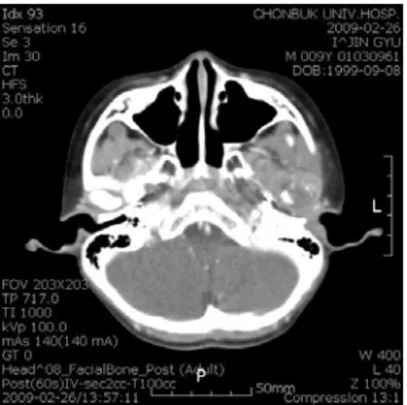
Cone beam CT(Fig. 2a, 2b) and computed tomography(Fig. 3a, 3b) were obtained for further evaluation. They showed osteolysis and bone destruction on the mandibular condyle. Bone scan showed reactive bone lesion in head of the mandibular condyle on the left side(Fig. 4). Based on these findings, osteomyelitis of the mandibular condyle was suspected.
And 3 days after his first visit, he was referred
to the department of Pediatrics for hospitalization
and antibiotic therapy. But his symptom and sign
did not subside. Then he was referred to the
department of Oral Maxillofacial Surgery for bony
curettage and biopsy of left condylar head on March
5, 2009. Biopsy and bony curettage from the
diseased area was performed under general
anesthesia.
Fig. 2a. Coronal view of left mandibular condyle by cone beam CT.
Fig. 2b. Sagittal view of left mandibular condyle by cone beam CT.
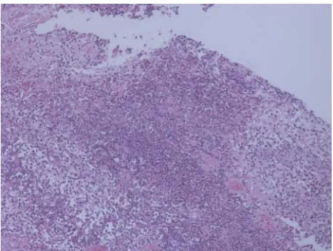
Histological examination revealed chronic osteomyelitis with foreign body reaction and acute suppurative inflammation with granulation tissue formation(Fig. 5a, 5b, 5c). Microbiologic culture of bone fragments was negative.
For 20 days after biopsy and curettage, antibiotics therapy was performed during hospitalization. He was discharged on March 27, 2009. Oral examination showed slightly openbite at the anterior teeth and right posterior teeth. So he was referred to the department of Orthodontics and is being under orthodontic treatment.
At 4 month after discharge, cone beam CT were obtained. It demonstrated bony formation at the left mandibular condyle without the relapsing symptom of disease(Fig. 6a, 6b).
Fig. 3a. Horizontal view of CT
Fig. 3b. Coronal view of CT
Fig. 4. Bone scintigraphy view
Fig. 5a. Microscopic view shows infiltration of infla- mmatory cells( x10, H& E stain).
Fig. 5b. Microscopic view shows infiltration consisting of multiple macrohages, lymphocytes with embedded giant cells( x40, H& E stain).
Fig. 6a. Coronal view of left mandibular condyle at 4 month after discharge by cone beam CT.
Fig. 6b. Sagittal view of left mandibular condyle at 4 month after discharge by cone beam CT.
Ⅱ. DISCUSSION AND CONCLUSION 1. Pathogenesis
The term osteomyelitis literally means inflammation of the bone marrow. It usually begins in the medullary cavity, involving the cancellous bone; then it extends and spreads to the cortical bone and eventually to the periosteum.
1,7)The cause is usually thought to be micro-
biological.
1)In the jaws, pyogenic organisms that
reach the bone marrow from abscessed teeth or
postsurgical infection usually cause osteomyelitis.
8)Recent carefully performed investigations in the
microbiolgy of osteomyelitis have adequately
that is, streptoccocci, anaerobic cocci such as peptostrepto cocclus spp., and gram-negative rods such as those of the genera Fusobacterium and Prevotella.
7)However, in some case of osteomyelitis, no clear bacteria was found.
9)It is thought because of previous antibiotic therapy or inadequate methods of bacterial isolation.
8)The major predisposing risk factors sustaining the presence and persistence of osteomyelitis are compromised vascualr intergrity and perfusion in the host at the local, regional, or systemic level.
7)The factors are injuries, syphilis, actionomycosis, chronic kidney failure, alcoholism, malnutrition, radiotherapy, and chemotherapy.
2)Non healing fractures of the mandible may be the causative agent in producing an osteomyelitis.
10-12)In this case, ball hit him on his left face slightly.
So It may be doubt that cause of osteomyelitis is the injury by ball. But It is not clear whether fracture was or not. And no prevoius sign has been presented that a dental infection was clearly the true causative factor for the development of the disease.
Microbiologic culture of bone fragments was negative. In this case, the pathological process could not be explained by the factors commonly involved with osteomyelitis.
Loveman
13)reported a number of cases of mandibular subperiosteal swellings in children where no periapcial pathology was found. The only feature in common with the case reported here was the presence of an erupting permanent molar. Eyrich reported that 10 cases of 11case of children with chronic osteomyelits presented no dental infection.
So it is necessary that we should find the possible causes in children with osteomyelitis through further studies.
2. Classification
Due in part to the long-standing existence of osteomyelitis as a entity, a variety of classifications of the various forms of the disease process have evolved. Various classification form of osteomyelitis
is being in the controversy but classification by Hudson have advantage of simplicity in clinical diagnosis. The arbitrary time limit of 1 month is used to identify acute from chronic osteomyelitis.
14)1) Acute osteomyelitis
① Contiguous focus osteomyeltis ② Progressive osteomyelitis ③ Hematogenous osteomyelitis 2) Chronic osteomyelitis
① Recurrent multifocal osteomyelitis ② Garre's osteomyelitis
③ Suppurative or non-suppurative osteomyelitis ④ Sclerosing osteomyelitis
In this case, diagnosis by clinical course of the disease is acute form but histological examination revealed chronic osteomyelitis and acute suppurative inflammation with granulation tissue formation.
Diagnosis of this case can not be classified by Hudson classification.
3. Symptoms
Acute osteomyelitis may well have the appearance of a typical odontogenic infection, including fever, trismus, malaise, pain, purulence, and facial cellulitis, except that there is predominantly confined to the periosteal envelope and its contents. Chronic osteomyelitis may present with all the associated findings of acute osteomyelitis except fever and chronic draining fistulae.
12)From an anatomical point of view, mandibular structure in childhood is complicated, because it is also still developing and deciduous and permanent teeth germs come closed each other.
6)Therefore, if an inflammation extends to the canal, it could spread along the canal and reach the ascending ramus. Thus, osteomyelitis in children is more aggressive and incurable than in adults.
6)But in this case, we could not found clearly cause
factors presenting dental infection. we wonder why
bony destruction was aggressive without presenting
dental infection.
4. Histologic features
Generation of biopsy material from patients with acute osteomyelitis is not common because of the predominantly liquid content and lack of a soft tissue component. When submitted, the material consists predominantly of necrotic bone. The bone shows a loss of the osteocytes from their lacunae, peripheral resorption and bacterial colonization. The periphery of the bone and the haversian canals contain necrotic debris and an acute inflammatory infiltrate consisting of polymorphonuclear leukocytes.
15)Biopsy material from patient with chronic osteomyelitis demonstrated significant soft tissue component that consists of chronically or subacutely inflamed fibrous connective tissue filling the intertrabecular areas of the bone.
Scattered sequestra and pockets of abscess formation are common.
15)In this case, microsopic view showed reactive bony response and infiltration consisting of inflammatory cells with embedded giant cells. There are a few of giant cells doubting foreign body reaction but we could not find the factor of foreign body in this case. We are not sure why a few of giant cells appeared.
5. Radiographic features
The radiograph of acute osteomyelitis may be unremarkable or may demonstrate an ill-defined radiolucency.
15)Because 10 to 12 days are required for lost bone to be detectable radiographically.
7)Portions of cortical bone may be resorbed. An inflammatory exudate can lift the periosteum and stimulate bone formation. Radiogrphically, this appears as a thin, faint, radiopaque line adjacent to and almost parallel or slightly convex to the surface of the bone.
8)Chronic osteomyelitis is suspected from the clinical examination.
8)It usually demonstrates bony destruction in the area of infection.
7)Radiographs reveal a patchy, ragged, and ill-defined radiolucency
to demonstrate sequestra and periosteal new bone, is important for a correct diagnosis and allows accurate staging of the disease.
8)In long-standing chronic osteomyelitis, there may be an area of increased radiodensity surrounding the area of radiolucency.
7)6. Treatment
The goal of definitive therapy is to attenuate and eradicate the proliferating pathogenic microor- ganisms and to support healing. This is accomplished by removing pathogenic supportive debris, providing regional skeletal stability as necessary, and disrupting pathophysiologic barriers.
14)The treatment with the most effective narrow-spectrum antibiotics to which the organisms are susceptible is pertinent. Surgical treatment such as drain, irrigation, debridement is also needed.
14)For treatment of osteomyelitis in childhood, drainage for pus discharge and a high dosage of antibioitics must be given at the acute stage.
16)If an acute condition changes to the chronic, a sufficient resection of the infectious lesion must be performed as soon as possible by such methods as saucerization, decortication, etc.
17)However, some cases with asymmetry of the face due to acquired disease have been reported,
18)therefore surgical treatment for infants and children must be performed more carefully, with respect to dentitional and maxillofacial growth and development.
The involvement of the condyle and the mandibular joint may cause serious secondary deformities during the period of skeletal growth.
The downward and forward growth of the jaw can be arrested on the affected side, causing asymmetry of the face. Therefore, in growing young children, precise diagnosis and effective therapy in early stage must be performed and required.
Ⅲ. CONCLUSION
uncommon disease. Because of the anatomic location of condyle, it is often not easy to obtain enough diagnostic information from plain radiography. The mandibular condyle is regarded as an important center of mandibular growth. Therefore, for young patients, osteomyelitis involving this region may cause a restraint of mandibular development, resulting in facial asymmetry. Probable, diagnosis and treatment in early stage must be performed and required in growing patient with osteomyelitis.
REFERENCES
1. Fonseca RJ. Oral and maxillofacial surgery. 1st ed., Philadelphia, 2000, Saunders, pp.484-491.
2. Taher AA. Osteomyelitis of the mandible in Tehran, Iran. Analysis of 88 cases. Oral Surg Oral Med Oral Pathol 1993;76(1):28-31.
3. Laskin DM. Oral and maxillofacial surgery. St. Louis, 1980, Mosby, pp.253-262.
4. Adekeye EO, Cornah J. Osteomyelitis of the jaws: a review of 141 cases. Br J Oral Maxillofac Surg 1985;23(1):24-35.
5. William RP, Henry WF. Contemproary orthodontics.
4th ed., St. Louis, 2007, Mosby, pp.46-47.
6. Gunge M, Yamamoto H, Katoh T, Izumi H.
Suppurative osteomyelitis of the mandible in a child.
Int J Oral Maxillofac Surg 1987;16(1):99-103.
7. Peterson LJ. Contemporary oral and maxillofacial surgery. 4th edi,, St. Louis, 2003, Mosby, pp.375-377.
8. White SC, Pharoah MJ. Oral radiology : principles and interpretation, 5th ed., St. Louis, 2004, Mosby, pp.373-380.
9. Eyrich GK, Baltensperger MM, Bruder E, Graetz KW.
Primary chronic osteomyelitis in childhood and adolescence: a retrospective analysis of 11 cases and review of the literature. J Oral Maxillofac Surg 2003;61(5):561-573.
10. Coletti D, Ord RA. Treatment rationale for pathological fractures of the mandible: a series of 44 fractures. Int J Oral Maxillofac Surg 2008;37(3):215-222.
11. Malanchuk VO, Kopchak AV. Risk factors for development of infection in patients with mandibular fractures located in the tooth-bearing area. J Craniomaxillofac Surg 2007;35(1):57-62.
12. Akhlaghi F, Aframian-Farnad F. Management of maxillofacial injuries in the Iran-Iraq War. J Oral Maxillofac Surg 1997;55(9):927-930.
13. Loveman CEI. Mandibular sbuperiosteal swellings in children. Journal of the American Dental Association 1941;28:1230-1235.
14. Hudson JW. Osteomyelitis of the jaws: a 50-year perspective. Oral Maxillofac Surg 1993;51(12):1294- 1301.
15. Neville BW. Oral and maxillofacial pathology. 3rd ed., St. Louis, 2003, Saunders, pp.141-144.
16. Thoma KH. Oral surgery. St. Lous, 1969, pp.781-810.
17. Hjorting-Hansen E. Decortication in treatment of osteomyelitis of the mandible. Oral Surg Oral Med Oral Pathol 1970;29(5):641-655.
18. A case of unilateral absence of the condyle due to
mandibular osteomyelitis in childhood. J Oral
Maxillofac Surg 1980;26:1290-1299.
국문요약
9세 소아에서 발생한 하악과두의 골수염