Bone Involvement of Diffuse Large B Cell Lymphoma (DLBCL) Showing Unusual Manifestations Mimicking Chronic Osteomyelitis in a 58-Year- Old Man: Case Report and Clinical Application of Diffusion Weighted Magnetic Resonance Imaging
INTRODUCTION
Diffuse large B-cell lymphoma (DLBCL) is the most common type of non-Hodgkin lymphoma (NHL) among adults and is predominant in older individuals. These mature B-cell tumors can arise occur anywhere in the body and bone marrow involvement is relatively rare and is commonly detected in the late stage of the disease (1). Since DLBCL displays several appearances on different imaging such as plain radiographs and magnetic resonance (MR) images, it is at times difficult to accurately diagnose DLBCL (2). This objective of this case report is to illustrate the imaging findings of DLBCL mimicking chronic osteomyelitis in regards to the differential diagnosis using diffusion- weighted magnetic resonance imaging (DWI).
This is an Open Access article distributed under the terms of the Creative Commons Attribution Non-Commercial License (http://creativecommons.org/licenses/
by-nc/4.0/) which permits unrestricted non-commercial use, distribution, and reproduction in any medium, provided the original work is properly cited.
Received: May 20, 2019 Revised: July 4, 2019 Accepted: July 29, 2019 Correspondence to:
Kyung Ryeol Lee, M.D.
Department of Radiology, Jeju National University Hospital, 15 Aran 13-gil, Jeju-si, Jeju-do 63241, Korea.
Tel. +82-64-717-1373 Fax. +82-64-717-1377 E-mail: [email protected]
Copyright © 2019 Korean Society of Magnetic Resonance in Medicine (KSMRM)
Case Report
This study presents a case of diffuse large B cell lymphoma (DLBCL) in a 58-year- old man showing unusual manifestations mimicking chronic osteomyelitis. In this case review, we describe the imaging findings of DLBCL which mimics chronic osteomyelitis and review existing reports regarding the differential diagnosis of bone involvement of lymphoma and osteomyelitis through imaging and laboratory findings and diffusion-weighted magnetic resonance imaging (DWI) such as the advanced MRI sequence.
Keywords: Bone; Lymphoma; Osteomyelitis; Diffusion weighted magnetic resonance imaging
Kyung Ryeol Lee1, Young Hee Maeng2
1Department of Radiology, Jeju National University Hospital, Jeju-si, Korea
2Department of Pathology, Jeju National University Hospital, Jeju-si, Korea
CASE REPORT
A 58-year-old man was presented with pain in his left distal thigh five days beforehand. He had experienced pain at the same site eight months earlier, but the pain had eased after taking analgesics. The patient had no history of trauma or cancer. Physical examination revealed tenderness in the aching part on the left distal thigh.
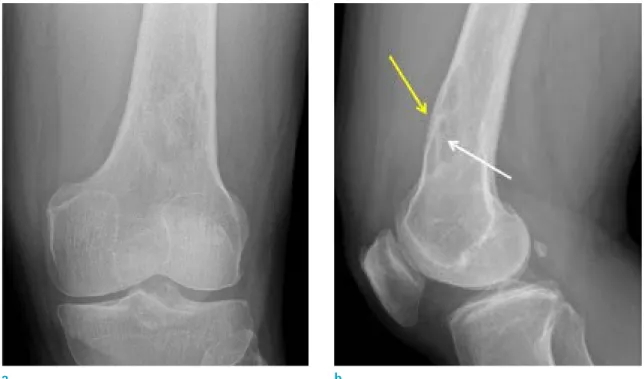
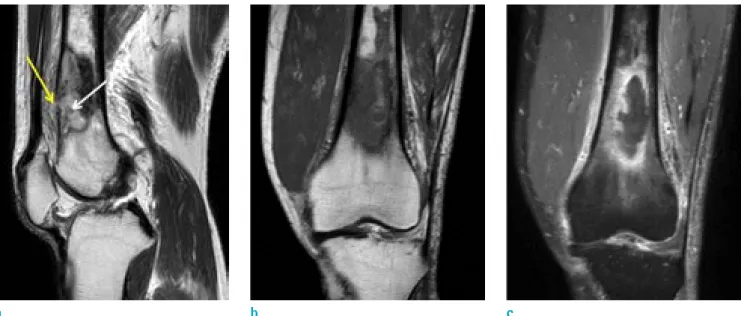
Plain radiographs revealed an osteolytic lesion with moth-eaten bone destruction in the distal diaphysis of the left femur (Fig. 1). Similarly, the radiographs showed septae in the lesion and a solitary layer of periosteal bone formation adjacent to the lesion (Fig. 1b). The likelihood of both chronic osteomyelitis and malignant bone tumor could be considered and the application of enhanced MRI planned based on the plain radiograph findings. MR images revealed an irregular lesion made of probable necrosis, mainly in the distal diaphysis of the left femur (Fig. 2). In addition, there was an irregular, small bone fragment causing suspicious bony sequestrum in the lesion, while cortical perforation and periosteal bone formation also appeared (Figs. 2, 3).
The bone marrow adjacent to the lesion exhibited diffuse enhancement. However, majority of the regions’ trabecullae identified by enhanced bone marrow was preserved (Fig.
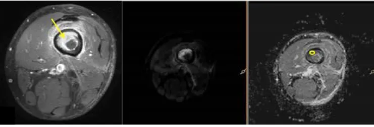
2). The mean ADC value was 0.84 x 10⁻³ mm2/s (b = 800) in the enhancing portion of the lesion which was located at peripheral region of the lesion and 1.87 x 10⁻³ mm2/s (b
= 800) in the non-enhancing region of the lesion which located at central portion and presumed to be necrosis (Fig. 4). Based on these imaging features, the initial and probable impression was suspected to be chronic osteomyelitis in addition to necrosis, intraosseous abscess, bony sequestrum, and cloaca. The differential diagnosis of the lesion was found to be malignant bone tumor such as bone metastasis, osteosarcoma and bone involvement of lymphoma which could include necrosis in the lesion.
The laboratory blood test findings also assisted to confirm the suspicion of osteomyelitis. The laboratory test results displayed slightly high levels of CRP and ESR (CRP, 7.67 mg/
dl; ESR, 26 mm/hr), but the WBC count was found to be in the normal range. Consequently, the patient was treated using antibiotics because of the initial impression of chronic osteomyelitis. However, pain in his left distal thigh failed to improve despite the treatment and he eventually underwent surgery. Based on the operative report, the orthopedic surgeon made a bone window to drain the pus. The swab culture for aspirated pus revealed negative culture result of bacteria, fungus and AFB (acid-fast bacilli). However,
Fig. 1. AP (a) and lateral (b) radiographs of the left knee shows an osteolytic lesion with moth-eaten bone destruction in the distal diaphysis of the left femur. The lateral view of the left knee shows suspected sequestrum (white arrow) in the lesion and a single layer of periosteal new bone formation (yellow arrow) adjacent to the lesion.
a b
some portion of the lesion in the medullary cavity, there was suspicion of a mass-like lesion and curettage of the suspected lesion was done. The curetted materials consistee some pale brown fragments. Pathological examination of the curettage specimen revealed that the tumor consisted of diffusely infiltrating neoplastic large lymphocytes, which tested positive for LCA and CD20, and negative for CD45RO, CD30, and ALK. Some necrotic areas were admixed using the tumor tissue. The diagnosis was diffuse large B-cell lymphoma with necrosis (Fig. 5). There was no pus in
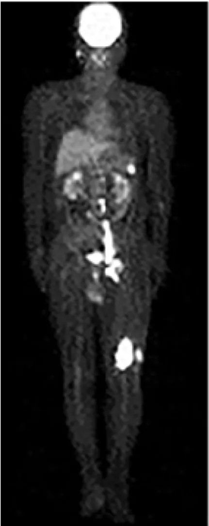
curetted material. Positron emission tomography (PET) CT, which was performed in sequence, revealed a suspiciously malignant lymphoma lesion in the left inguinal, femoral, iliac, and para-aortic areas (Fig. 6), and DLBCL was certainly diagnosed through excisional biopsy of the left inguinal lymph node. The patient is currently being treated by means of chemotherapy and is under observation.
Fig. 2. Sagittal T2-weighted MR image (a) shows cortical perforation suggesting cloaca (yellow arrow) and suspected bony sequestrum (white arrow) in the distal diaphysis of the left femur. On coronal T1-weighted MR image (b), the lesion of the left distal femur shows low signal intensity and on coronal contrast-enhanced fat-suppressed T1-weighted MR image (c), the lesion consisting of non-enhancing portion that has suspected necrosis and enhancing portion in the periphery of the lesion.
a b c
Fig. 3. Axial T2-weighted MR image (a) shows cortical perforation suggesting cloaca (yellow arrow) and axial contrast- e n h a n c e d f a t - s u p p r e s s e d T1-weighted MR image (b) demonstrates the drainage of necrotic material out of the bone (black arrow).
a b
DISCUSSION
DLBCL is the most common form of NHL in the world.
Even though it could occur in young adults and children, DLBCL generally prevalent with age and most of the patients are over the age of 60 at the time of their diagnosis. Generally, DLBCL manifests as a rapidly enlarging mass at a nodal or extra-nodal site anywhere in the body.
The extra-nodal site could include the gastrointestinal tract, skin, bone, brain and other tissues, and bone marrow involvement is relatively rare, usually occurring in the late stages of the disease (1). Even though DLBCL is fatal without treatment, it has a better response to therapy and a better
prognosis, hence, early diagnosis of bone involvement of DLBCL is crucial for patient survival. Since bone lymphoma may demonstrate varied appearances on imaging, such as plain radiograph and MR images, it is therefore important to distinguish between bone marrow involvement of DLBCL (NHL) and an infectious disease such as osteomyelitis or septic arthritis (2). In this case, a sequestrum could obtained from images. Sequestrum is detached or dead pieces of bone separated from surrounding bone during the process of necrosis and is commonly observed in long bones affected by osteomyelitis. Demonstration of sequestrum through imaging methods in the evaluation of malignant bone lesions is known to be rare. However, the imaging
Fig. 4. The measurement of ADC value at b 800 s/mm2 using axial contrast-enhanced fat-suppressed T1-weighted MR images, diffusion weighted images (DWI) and apparent diffusion coefficient (ADC) map. (a) The mean ADC value of 0.84 x 10⁻³ mm2/s (b = 800 s/mm2) was found in the enhancing portion of the lesion (yellow arrow, yellow region of interest). (b) The mean ADC value was 1.87 x 10⁻³ mm2/s (b = 800 s/mm2) for the non-enhancing portion of the lesion (white arrow, white region of interest).
b a
finding of chronic osteomyelitis, bony sequestrum, may be discovered in primary lymphoma of bone, radiation necrosis, eosinophilic granuloma, metastatic carcinoma or aggressive fibrous tumors. Therefore, the imaging diagnosis of bone involvement of lymphoma may sometimes be challenging.
When sequestrum was detected through imaging, there was need to consider such lesions at differential diagnosis. It is recommended that aggressive primary neoplasm of bone could likely lead to the formation of sequestrum as a result of the rapid destruction of bone and entrapment of partially destroyed bone in the tumor mass (3).
Usually, Acute-phase reactants, such as the erythrocyte sedimentation rate (ESR) and C-reactive protein (CRP) have
been used as markers for inflammation and as a measure of “sickness index” in infectious and non-infectious conditions. In this case, blood test revealed a slightly high level of ESR and CRP (4). However, the CRP level might be high in diseases associated with chronic inflammation, such as myocardial infarction, chronic hepatitis, chronic bronchitis, and arthritis (5). The ESR level could be increased in inflammation, pregnancy, anemia, autoimmune disorders such as rheumatoid arthritis and lupus, infections, some kidney diseases and some cancers such as lymphoma and multiple myeloma (6).
DWI is a recently introduced and applied MR sequence which provides qualitative and quantitative functional
Fig. 5. Photomicrograph (a, Hematoxylin & Eosin [H&E], x 40) showing curetted tumor tissue admixed with bony trabeculae (arrows) and photomicrograph (b, H&E, x 40) showing necrotic areas (arrowheads). Photomicrograph (c, H&E, x 400) demonstrates neoplastic large lymphocytes and photomicrograph (d, x 200) illustrate that they are positive for CD20.
a b
c d
information in regards to the microscopic movements of water at the cellular level. The application of DWI in bone marrow is expected to assist in the detection of bone marrow pathologies and the distinction between benign and malignant bone marrow lesions. Filograna et al. (7) established that the 95th percentile for ADC values in bone metastases was 1.21 x 10⁻³ mm2/s, and Wu et al. (8) established that the mean ADC value of DLBCL was 0.70 x 10⁻³ mm2/s. Ozgen et al. (9) reported that the average ADC value of skull base osteomyelitis was 1.26 x 10⁻³ mm2/s and the mean ADC value of skull lymphoma as 0.59 x 10⁻³ mm2/s. In addition, Abdel Razek et al. (10) reported the mean ADC value in diabetic osteomyelitis to be 0.86 ± 0.11 x 10⁻³ mm2/s and 0.85 ± 0.12 x 10⁻³ mm2/s and this result showed slightly low ADC value compared to the results
mentioned above. Based on previous studies, we could conclude that bone involvement of DLBCL could show a lower ADC value than that of bone metastasis or a benign lesion such as osteomyelitis and could possibly provide additional information. In cases which bone lymphoma resembles infectious disease such as osteomyelitis or septic arthritis, comprehensive and careful judgment coupled with advanced imaging methods such as DWI may provide useful information for accurate diagnosis.
REFERENCES
1. Kumar V, Abbas A, Fausto N, Aster J. Robbins and cotran pathologic basis of disease. 8th ed. Philadelphia, PA:
Saunders/Elsevier, 2010:606-607
2. Krishnan A, Shirkhoda A, Tehranzadeh J, Armin AR, Irwin R, Les K. Primary bone lymphoma: radiographic-MR imaging correlation. Radiographics 2003;23:1371-1383; discussion 1384-1377
3. Jennin F, Bousson V, Parlier C, Jomaah N, Khanine V, Laredo JD. Bony sequestrum: a radiologic review. Skeletal Radiol 2011;40:963-975
4. Markanday A. Acute phase reactants in infections:
evidence-based review and a guide for clinicians. Open Forum Infect Dis 2015;2:ofv098
5. Trichopoulos D, Psaltopoulou T, Orfanos P, Trichopoulou A, Boffetta P. Plasma C-reactive protein and risk of cancer:
a prospective study from Greece. Cancer Epidemiol Biomarkers Prev 2006;15:381-384
6. Wu S, Zhou Y, Hua HY, et al. Inflammation marker ESR is effective in predicting outcome of diffuse large B-cell lymphoma. BMC Cancer 2018;18:997
7. Filograna L, Magarelli N, Cellini F, et al. Diffusion weighted imaging (DWI) and apparent diffusion coefficient (ADC) values for detection of malignant vertebral bone marrow lesions. Eur Rev Med Pharmacol Sci 2018;22:590-597 8. Wu X, Pertovaara H, Dastidar P, et al. ADC measurements
in diffuse large B-cell lymphoma and follicular lymphoma:
a DWI and cellularity study. Eur J Radiol 2013;82:e158-164 9. Ozgen B, Oguz KK, Cila A. Diffusion MR imaging features
of skull base osteomyelitis compared with skull base malignancy. AJNR Am J Neuroradiol 2011;32:179-184 10. Abdel Razek AAK, Samir S. Diagnostic performance of
diffusion-weighted MR imaging in differentiation of diabetic osteoarthropathy and osteomyelitis in diabetic foot. Eur J Radiol 2017;89:221-225
Fig. 6. PET CT demonstrates increased uptake in the left distal femur and lymph nodes of left inguinal, femoral, iliac, and paraaortic areas.
![Fig. 5. Photomicrograph (a, Hematoxylin & Eosin [H&E], x 40) showing curetted tumor tissue admixed with bony trabeculae (arrows) and photomicrograph (b, H&E, x 40) showing necrotic areas (arrowheads)](https://thumb-ap.123doks.com/thumbv2/123dokinfo/5245675.132331/5.892.76.824.423.1028/photomicrograph-hematoxylin-curetted-trabeculae-photomicrograph-showing-necrotic-arrowheads.webp)