I. Introduction
The main goal for the treatment of periodontal diseases is the regeneration of lost periodontal liga- ment, cementum, and bone. Clinical and histologi- cal researches suggest that it is possible to restore periodontal structures (Becker et al., 1988;
Pontoriero et al., 1988; Schanllhorn and McClain, 1988; Bowers et al. 1985).
Normal healing of bone is characterized by the integrated actions of different cells, and its processes are divided into broad sequential phases of inflam- mation, proliferation, migration of osteogenic cells, and production and remodeling of trabecular bones (Branemark et al., 1964; Amsel et al., 1969; Patt and Maloney 1975; Watanabe et al., 1992; Furusawa 1993).
Recent in vivo and in vitro studies have suggested that various autocrine and paracrine growth factors present in serum play important roles in the wound healing of various tissues, including bone (Mustoe et al., 1987; Bab et al., 1988; Brown et al., 1988).
These growth factors appear to stimulate the early
phases of wound healing, cell differentiation, and increased primary matrix production, rather than remodeling and maturation processes (Mustoe et al., 1987; Brown et al. 1988).
The cells used in this study have the ability to dif- ferentiate into mature osteoblasts expressing the normal osteoblast phenotype. At the restrictive tem- peratures, cell division is slowed, differentiation is increased, and a more mature osteoblast phenotype is produced. The cells provide a homogenous, rapidly proliferating model system for studying nor- mal human osteoblast differentiation, osteoblast physiology, growth factor, and other cytokine effects on osteoblast function and differentiation.
Current bone and cartilage repair strategies utilize autogenous or allogeneic grafts, which are limited by donor site pain, morbidity, availability, or risk of disease transmission. Tissue engineering approach- es for bone and cartilage repair rely on the ability of growth and morphogenetic factors to exert host cell chemotaxis, proliferation, differentiation, and new tissue formation at the site of injury or defect when delivered in a suitable carrier (Kim et al., 2002).
The Effect of High Molecular Hyaluronic Acid on Bone Formation in Human Fetal Osteoblasts
Kwang-Soo Lee·Hyun-A Kim·Yun-Sang Kim·Hyung-Keun You·Hyung-Shik Shin Department of Periodontology, School of Dentistry, Wonkwang University
대한치주과학회지 : Vol. 32, No. 3, 2002
*이 논문은 2001년도 원광대학교 숭산연구비 지원에 의해서 수행됨.
Hyaluronic acid (HA) is a main ingredients of con- nective tissue intercellular matrix and exist between cell as adhesives role in form of salt as kind of amino-sugar-containing polysaccharide of gly- cosamino-glycans (GAG). Also, HA protects tissue surface because it has viscoelasticity such as jelly, and is used to arthritis treatment to act as lubricant (Balazs and Gibbs, 1970). HA may play an impor- tant role in the control of the migration of myogenic cells in vivoby its physicochemical properties (Veit et al., 1991). Although the biological function of hyaluronate is poorly understood, its relatively high concentration in a variety of different embryonic tis- sues (Loewi and Meyer, 1958; McConnachie and Ford, 1966; Kvist and Finnegan, 1970a,b; Breen et al., 1970) suggests that it may play an important role in early developmental events.
As one of the major secretory proteins of osteoblasts, BSP is believed to associated with events occurring during bone mineralization, and functions to regulate mineralization possibly by its direct interation with cell surface integrin receptors (Oldberg et al., 1988; Flores et al., 1992) and by ini- tiating nucleation of the bone mineral, hydroxyap- atite (Hunter and Goldberg, 1993).
It is well known about the role of HA in soft tis- sue, but yet little is investigated about bone induc- tive effect. The present study is designed to evaluate the bone formation by HA through differentiation of human fetal osteoblasts in vitro.
II. Materials and Methods
1. Culture of human fetal osteoblastsHuman fetal osteoblastic cell line (hFOB1 1.19 ; American Type Culture Collection, Manassas, VA) that have the ability of production and calcification of bone matrix protein were plated at 5×10⁴
cells/well of 6-well plate containing 2 ml of DMEM : F-12 HAM 1:1 Mixture (Sigma, St.Louis, MO, USA) , with 10% fetal bovine serum (FBS, GibcoBRL, Grand island, NY, USA) and 0.03mg/ml of G-418 (Duchefa, Netherlands). This cultured at 34℃ in a humidified atmosphere of 5% CO2, 95% air and 100% humidity.
When the cell cultures reached a confluence, culture medium exchanged every 2 days, and then subcul- tured in a ratio of 1:3. Cell cultures between the 4th and 9th passage were used in this study.
2. Cell number counting
Cultured hFOB1 were plated in 6-well culture dishes at 1×10⁴cells/well and allowed to attach.
After 24 h, cells were exposed to 0.063%, 0.125%, 0.25%, 0.5% of HA and distilled water in control group. High molecular HA that we used was gener- ously donated from Vacctec Co. (Korea). The num- ber of viable cells after trypan blue exclusion was counted at 2 and 4 days of incubation at 34℃.
There were four cultures in each group at each time.
3. Cell growth
Total viable cell numbers were assessed by a MTT (3-[4,5-dimethylthiazol-2-yl]-2.5 diphenyltetrazolim bromide) assay. hFOB1 were plated in 24-well plates (Falcon, Division of Becton Dickinson and Co., Lincoln Park, NJ, USA) at 2×10⁴cells/well. This cultured at 34℃ in a humidified atmosphere of 5%
CO2, 95% air and 100% humidity. And then unat- tached cells were removed by exchange culture medium. Each 0.063%, 0.125%, 0.25%, 0.5% of HA was treated, and cultured for 2 and 4 days. Control group was treated with normal saline. 300 μl of MTT was added to each well and then the plates were incubated for 4 h. The plates were agitated to
enhance the dissolution of formazan formed already. The cultured cells were transported on 96- well plate and then the absorbance was measured at 540 nm in ELISA reader (Spectra MAX 250, Molecular Devices Co., Sunnyvale, CA, USA).
4. Alkaline Phosphatase (ALP) assay
After hFOB1 were plated in 6-well plates at 1×10⁵ cells/well, it is cultured at 34℃ until a confluence in DMEM : F-12 HAM 1:1 Mixture containing 10% FBS, G-418, 50 μg/ml ascorbic acid, 10 mM sodium β- glycerophosphate. And 0.063%, 0.125%, 0.25%, 0.5% of HA were added in experimental group, 0.1
㎍/ml of dexamethasone was added in positive con- trol group, and normal saline was added in negative control group. And then each group was incubated for 3 days more. Cells were separated by trypsin/EDTA and centrifuged at 15,000rpm for 10 sec. Aliquot is removed and centrifuged again at 15,000 rpm for 10 sec. Aliquot was removed again and suspended with 0.5ml of sterilized distilled water. Each 0.1 ml of suspension was mixed with 0.1 M glycine NaOH buffer (pH 10.4) 0.2 ml, 15 mM pNPP (para-nitrophenyl phosphate : Sigma) 0.1 ml, 0.1% Triton X-100/saline 0.1 ml, and 0.1 ml of sterilized distilled water. The culture dishes were incubated at 37℃ for 30 min, and the reaction was stopped by 0.1 N NaOH 0.6 ml. The cultured cells were transported on 96-well plate and the absorbance was measured at 410 nm in ELISA read- er. And the standard concentrations of protein were calculated using BCA protein assay reagent (Pierce, USA). Results were expressed as nmol of para-nitro- phenol released per min/mg protein.
5. von Kossa staining
Cultured hFOB1 were plated in 6-well plates at 1
× 105cell/well. 0.063% HA for experimental group and 0.1 ㎍/ml dexamethasone for positive control were added in DMEM : F-12 HAM 1:1 Mixture con- taining 10% FBS, 50 μg/ml ascorbic acid and 10 mM sodium β-glycerophosphate. After incubation for 20 days, cells were fixed in 10% neutral formaldehyde, and stained with 2.5% silver nitrate. Then, cells were treated with sodium carbonate formaldehyde and washed with distilled water. The culture plates with stained nodules were photographed using a Nikon camera.
6. Collagen synthesis analysis
To measure the total collagen synthesis of hFOB1 indirectly, hydroxyproline contents were measured.
We modified the method Rojkind et al.(1979) pro- posed to measure hydroxyproline of each cells.
Briefly, hFOB1 were plated in 60-mm plates at 3×
105 cells with DMEM : F-12 HAM 1:1 Mixture contain- ing 10% FBS, G-4185, 50 ㎍/ml ascorbic acid, 10 mM sodium β-glycerophosphate. Cultures were main- tained at 34℃ in a humidified atmosphere. After cells were reached a confluence, 0,033%, 0.063%, 0.125%
of HA were added and cultured 3 days more. After hydrolyzing at 100℃ for 24 h, samples were filtered, collected, and completely dried at 60℃ and 50 μl methanol was added for removing of Hcl.
Remaining precipitates were dissolved with 1.2 ml of 50% isopropanol , and placed at room temperature for 10 minutes after mixing with 200㎕ chloramin-T solution (Sigma). 1.0 ml of Ehrlich reaction reagent (Sigma) was mixed and cultured at 50℃ for 90 min, and chilled at room temperature. Collagen synthesis was measured at 557 nm wave length using spec- trophotometer(Beckman, USA). And the standard concentrations of protein were calculated using BCA protein assay reagent (Pierce). Results were expressed as collagen/total protein(㎍/mg/ml).
7. Western blot analysis
hFOB1 were plated in 100-mm dishes at 1×10⁵ cells/well and cultured at 34℃ in DMEM : F-12 HAM 1:1 Mixture containing 10% FBS, G-418, 50 ㎍/ml ascorbic acid, 10 mM sodium β-glycerophosphate.
When these cells were reached a confluence, 0.063%
of HA were added in experimental group, 0.1 ㎍/ml of dexamethasone was added in positive control group, and normal saline was added in negative control group. And then each group was incubated for 3 days more. Protein was isolated form cells using lysis buffer under the conditions recommend- ed by the manufacturer. Protein concentration was determined by BCA solution. The denatured super- natant containing 100 ㎍ of protein was elec- trophoresed in a 15% SDS-polyacrylamide gel and transferred onto a PVDF (Immobilon-P membrane, Milipore, USA). To reduce nonspecific antibody binding, the membrane was incubated in a blocking solution (Zymed, San Francisco, CA, U.S.A.) for 1 h at room temperature. And then BSP as first antibody activated for 90 min. After washing with PBS, the membrane was treated with anti-mouse IgG-alkaline phosphatase conjugated secondary antibody for 1 h, and washed again with PBS. The membrane was then incubated in ECL Western Star Substrate reagent (Amersham, Buckinghamshire, UK), and exposed to Hyperfilm-MP (Amersham) for a few min. The mem- brane was stained with 1× Ponceau S.
8. Statistical analysis
Values were calculated as the mean ± standard deviation (S.D.). Statistical significance was evaluat- ed for by one way analysis of variance (ANOVA) using SPSS (v10.0, Chicago, IL, USA) program of computer.
III. Results
1. Cell proliferation assay of HA on hFOB1
1) Cell number counting
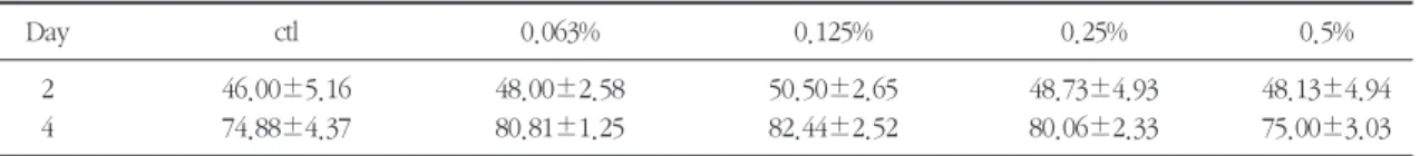
Cell proliferation by HA was determined by cell number counting (Zhang et al., 2000; Watson et al., 1998). HA-treated hFOB1 showed increasing ten- dency than control. All experimental groups showed higher proliferation rate than control and 0.125% HA-treated group was the highest at 2day.
But, there were no significant differences between the groups. At 4 day, experimental groups also showed increasing tendency than control group and 0.125% HA-treated group was the highest prolifera- tion rate. There were no significant differences between the groups (Table 1 and Figure 1) (P>0.05).
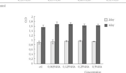
2) Cell growth
Cell growth of HA was determined using the MTT assay as described previously. Cell growth of all experimental groups showed the tendency of increase rate than control group, and showed high- er cell growth in 0.125% HA at 2 day. There were no significant differences between control group
Table 1. cell proliferation of hFOB1s treated with HA (Mean±S.D.) (×104)
Day ctl 0.063% 0.125% 0.25% 0.5%
2 46.00±5.16 48.00±2.58 50.50±2.65 48.73±4.93 48.13±4.94
4 74.88±4.37 80.81±1.25 82.44±2.52 80.06±2.33 75.00±3.03
ctl : control
and each HA-treated groups. Cell growth increased in experimental groups at 4 day, and 0.125% HA showed higher cell growth. There was no signifi-
cant difference compare with control group (Table 2, Figure 2).
Table 2. MTT assay of hFOB1s treated with HA (Mean±S.D.) (nmole/30min/mg)
Day ctl 0.063% 0.125% 0.25% 0.5%
2 0.89±0.06 0.92±0.08 0.95±0.02 0.95±0.02 0.94±0.01
4 1.53±0.09 1.65±0.10 1.66±0.06 1.59±0.07 1.61±0.06
ctl : control
Figure 1. Effect of HA on cell proliferation of hFOB1. hFOB1 were plated in a 6-well plate at 1×10⁴cells/well and cultured in F-12 HAM:DMEM containing 0.063%, 0.125%, 0.25%, 0.5% HA for 2 and 4 days.
Values represent averages from four independent experiments and standard deviation. (ctl : control) 2day
4day
ct1 0.063%HA 0.125%HA 0.25%HA 0.5%HA Concentration Cell Number(×104)
90 80 70 60 50 40 30 20 10 0
Figure 2. Effect of HA on cell growth of hFOB1. hFOB1 were plated in 24-well plates at 2×10⁴cells/well and cultured in F-12 HAM:DMEM containing 0.063%, 0.125%, 0.25%, 0.5% HA for 2 and 4 days. Values represent averages from four independent experiments and standard deviation. (ctl : control)
2day 4day
ct1 0.063%HA 0.125%HA 0.25%HA 0.5%HA Concentration
O.D
2 1.8 1.6 1.4 1.2 1 0.8 0.6 0.4 0.2 0
2. ALP activity of HA on hFOB1
The measurement of ALP synthesis is demonstrat- ed as table 3. Generally, activity is increased with the reduction of concentrations. 0.063% HA-treated group showed the highest ALP activity at 3 day. ALP activity of 0.063% HA is almost same with positive
control (dexamethasone- treated) group. There was significant difference between control group and 0.063% HA-treated group (Table 3, Figure 3).
3. Bone nodule formation of HA on hFOB1 Numerous mineralized nodules were identified as Figure 3. Effect of HA on ALP activity of hFOB1. hFOB1 were plated in 6-well plates at 1×10⁵cells/well and cultured in F-12 HAM:DMEM until cells reached a confluence. After that, 0.063%, 0.125%, 0.25%, 0.5% of HA and dexamethasone as a positive control were added and cultured for another 3 days more. Values represent averages from four independent experiments and standard deviation. (C-: negative control, C+: positive control, * : P<0.05)
Figure 4. Formation of calcification nodules in hFOB1.
3day
C- C+ 0.063% 0.125% 0.25% 0.50%
Concentration
nmole/min/mg of protein
0.3 0.25 0.2 0.15 0.1 0.05 0
Negative control positivie control 0.063%HA
Table 3. ALP activity of hFOB1s treated with HA (nmole/min/mg) (Mean±S.D.)
Day C- C+ 0.50% 0.25% 0.125% 0.063%
30.18±0.01 0.23±0.02* 0.14±0.00 0.16±0.01 0.19±0.01 0.23±0.02*
C- : negative control, C+ : positive control
* Statistically significant difference compared with the negative control (p<0.05).
darkly stained spots in 0.063% HA-treated group than two control groups, whereas a small number of mineralized nodules were showed in the positive control.
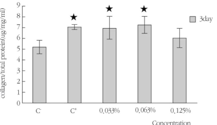
4. Collagen synthesis of HA on hFOB1
Collagen synthesis increased in all experimental groups (Table 4). 0.033% and 0.063% of HA were significantly increased collagen synthesis level com- pare with the negative control group. Especially, 0.063% HA showed the highest collagen synthesis than other groups.
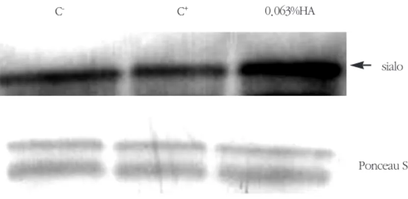
5. Expression of BSP on HA in hFOB1 Because BSP is one of the major markers of bone
formation in vitro, the expression level of BSP was investigated (Figure 6). The level of BSP protein in 0.063% HA notably increased when compared with negative control, and there was a slight change in positive control compare with negative control.
IV. Discussion
The regenerative processes that occur in bone marrow after ablation have been described precisely in previous morphological studies. Normal wound healing occurs in bone through three sequential phases: postoperative inflammation, proliferation and migration of mesenchymal cells with produc- tion of bone matrix, and bone remodeling (Branemark et al., 1964; Amsel et al., 1969;
Watanabe et al., 1992; Furusawa 1993).
Figure 5. Effect of HA on collagen synthesis of hFOB1. Cells were plated in 100-mm plates at 3×10⁵cells/well and cultured in F-12 HAM until cells reached a confluence. After that, 0.033%, 0.063%, 0.125% of HA and dexamethasone as a positive control were added and cultured for another 3 days more.
Values represent averages from four independent experiments and standard deviation (C-: negative control, C+: positive control, * : P<0.05).
3day
C- C+ 0.033% 0.063% 0.125%
Concentration
collagen/total protein(ug/mg/ml)
9 8 7 6 5 4 3 2 1 0
Table 4. Collagen synthesis of hFOB1 treated with HA (㎍/mg/ml) (Mean±S.D.)
Day C- C+ 0.033% 0.063% 0.125%
3 5.21±0.65 7.09±0.24* 7.06±1.05* 7.33±0.76* 6.09±0.89
C-: negative control, C+ : positive control
* Statistically significant difference compared with the negative control (p<0.05).
Bone marrow is a highly vascularized hematopoi- etic tissue and contains branching vascular sinuses lying in fibroblastic stromal cells(Weiss 1976). In this regard, stimulation of angiogenesis by partially degraded HA was reported to be due to the direct action of HA on endothelial cells, possibly via a receptor-mediated mechanism (West and Kumar 1989). If HA application did, in fact, increase blood- vessel invasion into the wounded bone marrow, it might have effected new bone formation indirectly during the process of bone wound healing.
HA is glycosaminoglycans (GAG) which is a kind of amino-sugar containing polysaccharide. This nat- ural material is easy to separate because it is not chemically bonded with protein. It was first collect- ed from fluid filling eyeball of animal, and it can be found from various tissues of animals. HA usually exists as salt, and this hyaluronate (salt of HA) acts as a binding material between cells. This hyaluronate or HA which has viscosity is like a jelly between cells, prevents invasion of bacteria and stops spreading of toxic materials, and is hydrolyzed by hyaluronidase (Soh 1988). HA had been much
examined for experimental and clinical study in soft tissue regeneration, but HA was used as a matrix or scaffold for bone regeneration. Kim et al (2002) sug- gested that HA scaffolds may be appropriate deliv- ery systems for rhBMP-2 in bone/cartilage repair because of their ability to retain rhBMP-2 to the local environment in a sustained manner, and stimulate differentiation of pluripotent stem cells.
Because there were seldom studies in relation to bone formation, we experimented to search bone inductive effect of HA on human osteoblastic cell line.
0.5%, 0.25%, 0.125%, and 0.063% of HA-treated groups showed increasing tendency in the cell pro- liferation rates than control group. In especially, most increased cell proliferation rate was showed at 0.125% HA-treated group in both the cell number counting and the cell growth for 2 and 4 day, but all experimental groups did not have significant differ- ence compare with control group (Table 1, 2, Figure 1,2). These results showed a high concentration of HA (0.5% in this study) was ineffective on hFOB1 and a low concentration of HA (0.125% and 0.063%
Figure 6. Western blot analysis for intracellular levels of BSP in hFOB1. First lane is negative control group and the second is dexamethasone group, and the last one is the experimental group supplemented with 0.063% HA. Cell extract equivalent to 100μg/ml of total cellular protein of hFOB1 was elec- trophoresed by 15% SDS-PAGE and transferred to a PVDF membrane. The intracellular protein lev- els of BSP in hFOB1s were probed with antibodies diluted by 1:1000. After probing, the membrane was stained with 1×Ponceus S for 10 min to reveal the total cellular protein loaded per each lane.
C- C+ 0.063%HA
sialo
Ponceau S
concentration of HA) may effect on proliferation of hFOB1. It was supposed that high viscosity of HA effected on cell survival rate. However, hFOB1 show only the increasing tendency than the remark- able increase on HA. We suggested that HA may effect on differentiation than proliferation in hFOB1.
ALP is a enzyme that increases the concentration of phosphoric acid at the area where calcification occurs by hydrolyzing organic phosphoric acid ester, and is known to induce calcification by accu- mulating calcium phosphate to extracellular matrix (Beertsen et al., 1989; Anderson, 1989; Bellows et al., 1991). De Bernard (1982) reported that ALP con- verts protein to phosphoprotein by increasing local phosphoric acid ion concentration, and phospho- protein in return has calcium binding tendency so acts as nucleus of calcification. It is said that dexam- ethasone differentiates osteoblastic cell resulting in increase of bone formation (Bellow et al., 1986;
Tenenbaum et al., 1985), and when dexamethasone collected from rat's skull is applied, bone specific indicator protein like osteopontin, ALP and osteocal- cin synthesis increases (Kasugai et al., 1992; Nagata et al., 1991; Owen et al., 1990). Meanwhile, culture medium with insufficient dexamethasone could not form osteoblastic cell as well as periodontal cells and bone nodule or anything similar(Arceo et al., 1991).
When we treated 0.063% of HA the amount of ALP synthesis increased significantly compare with the negative control, and increased as a dexametha- sone-positive control group. But the amount of ALP synthesis at 0.5% and 0.25% of HA groups decreased compare with control group (Table 3, Figure 3). And then we used lower concentration of HA (0.0315%, 0.0158%). ALP activity is appeared higher than the negative control in all group, but appeared lower than 0.063% of HA-treated group (data not shown). Also, we tested lows molecular
HA by same methods, there was no significant dif- ference in ALP activity (data not shown). This addi- tional test suggested the high-molecular HA have greater ALP activity than low-molecular HA (data not shown). So we determined that 0.063% of high molecular HA is appropriate for experiment of min- eralization, we used it to examine for bone nodule formation. Also, there was remarkable difference in 0.063% HA treated-group compare with both con- trol groups.
The carboxyl-terminal propeptide of typeⅠ colla- gen (c-propeptide) is a major protein secreted by osteoblast, and is present in bone(Mizuno et al., 1996). It is reported that 95% of collagen newly syn- thesized by MC3T3-E1 cells is type Ⅰ collagen (Hata et al., 1984). Type Ⅰ collagen is the major extracellu- lar matrix protein, accounting for approximately 85%
of the protein in bone (Eyre, 1980). Collagen synthe- sis is considered to be a reliable indicator of bone matrix synthesis, as it forms 90% of bone proteins.
The HA used was able to stimulate collagen syn- thesis in this study. Positive control, 0.033%, 0.063%
concentration of HA significantly increased collagen synthesis level compare with negative control group. Especially, 0.063% of HA showed the high- est collagen synthesis level than other groups.
Therefore, 0.063% of HA was understood as a opti- mal concentration for in vitro bone formation like ALP activity (Table 4, Figure 5). These finding indi- cate that collagen synthesis and collagen-osteoblast interaction are crucial for osteoblastic differentiation.
Bone sialoprotein(BSP) is a major noncollagenous protein that is expressed almost exclusively in bone and cementum (Butler, 1991). It contains a high level of sialic acid, phosphoserine and sulphotyro- sine, and the presence of two to three regions of glutamic acid residues and an RGD cell attachment motif (Oldberg et al., 1988; Fisher et al., 1990;
Shapiro et al., 1993). The presence of the cell
attachment domain indicates a potential role of bone sialoprotein in the attachment of bone cells to the mineralized matrix. However, the temporo-spa- tial expression of bone sialoprotein during de novo bone formation in vivo (Chen et al., 1992; McKee and Nanci, 1995) as well as in vitro (Kasugai et al., 1992; Yao et al., 1994) and its ability to nucleate hydroxyapatite crystal formation under steady-state conditions (Hunter and Goldberg, 1993) through the glutamate-rich sequences (Hunter and Goldberg, 1994) strongly suggest that bone sialoprotein is involved in the mineralization of bone (Sodek et al., 1992). Also, in previous study of the effects of OP- 1(BMP-7, osteogenic protein-1) on cell differentia- tion of PDL cells and preosteoblastic MC3T3 cells by comparing the synthesis and mineralization of matrix components (Zhumabayeva et al., 1988), while OP-1 dramatically stimulated ALP activity, and osteopontin as well as osteocalcin expression in both cell types, bone sialoprotein was induced only in MC3T3 cells. Moreover, only MC3T3 cultures demonstrated mineralization, indicating that bone sialoprotein may function as the nucleation sites for hydroxyapatite.
As one of the major secretory proteins of osteoblasts, BSP functions to regulate mineralization possibly by its direct interation with cell surface inte- grin receptors (Oldberg et al., 1988; Flores et al., 1992) and by initiating nucleation of the bone min- eral, hydroxyapatite (Hunter and Goldberg, 1993).
BSP is believed to associated with events occurring during bone mineralization. Additionally, in western blot analysis using BSP, this study also showed sig- nificant increase in expression of BSP in 0.063%
hFOB1 than other control group (Figure 6). In other words, 0.063% HA appears to be superior than other concentration in effect of ALP synthesis, bone nodule formation, BSP formation and secretion.
Therefore, in vitro, HA showed low increasing
tendency in stimulating osteoblastic cell proliferation but stimulates osteoblastic cell differentiation with an increase of ALP activity, collagen synthesis, and bone sialoprotein production. In conclusion, HA may effects bone mineralization progress of hFOB1 vastly, and close estimation of in vitro bone forma- tion related to applying HA are needed. Also, the biological interaction with various cytokine and sub- stances related to bone remodeling is needed. And further studies are required to find out whether HA can apply for clinical usage.
V. Conclusion
To evaluated the effect of HA on bone induction in vitro, we tested cell proliferation, ALP activity, col- lagen synthesis, bone nodule formation, protein expression of bone sialoprotein in human fetal osteoblast cell line, and the results were same as fol- lows.
1. 0.063% of HA-treated hFOB1 showed a signifi- cant increase in ALP synthesis at 3 day (P<0.05).
2. Bone nodule formation capability of HA-treated hFOB1s showed significantly difference com- pare with negative control and positive control group.
3. Collagen synthesis level of 0.063% of HA showed significant difference compare with negative control group and dexamethasone group (P<0.05).
4. The expression of BSP was increased compare with the control.
Taken together, this study indicated that 0.063%
of HA has an inductive effect on bone formation in vitro increasing with cell proliferation, ALP activity, collagen synthesis, bone nodule formation, and
expression of bone sialoprotein.
VI. References
1. Amsel, S., Maniatis, A., Tavassoli, M., and Crosby, W.H. : The significance of intramedullary bone formation in the repair of bone marrow tissue. Am J Anat 164: 101-112, 1969.
2. Anderson, H.C. : Mechanism of mineral forma- tion in bone. Lab Invest 60 : 320 - 330, 1989.
3. Arceo, N., Sauk, J.J., moehring, J., Foster, R.A., and Somerman, M.J. : Human periodontal cells initiate mineral - like nodules in vitro. J Periodontol 62: 499 - 503, 1991.
4. Bab, I., Gazit, D., Muhlrad, A., and Shteyer, A. : Renerating bone marrow produces a potent growth-promoting activity to osteogenic cells.
Endocrinology 123: 345-352, 1988.
5. Balazs E.A. and Gibbs D.A., "The Rheoligical Properties and Biological Function of Hyaluronic Acid" in " Chemistry and Molecular Biology of the Intercellular Matrix", volume 3, E. A. Balazs, ed., p.1241, Academic Press, N.Y. 1970.
6. Becker, W., Becker, B., Berg, L., Prichard, J., Caffessee, R., and Rosenberg, E.: New attach- ment after treatment with root isolation proce- dures : Report for treated classⅡ and classⅡ fur- cation and vertical osseous defects. Int J Perio Rest Dent 8(3): 2-16, 1988.
7. Beertsen, W. and Theo Van Den Bos. : Calcification of dentinal collagen by cultured rabbit periosteum : The role of alkaline phos- phatase. Matrix 9: 159-171, 1989.
8. Bellow, C.G., Aubin, J.E., Heersche, J.N.M., and Antosz, M.E. : Mineralized bone nodules formed in vitro from enzymatically released rat calvaria cell populations. Calcif Tissue Int 38: 143-154, 1986.
9. Bellows, C.G., Aubin, J.E., and Heershe, J.N.M.
: Initiation and progression of mineralization of bone nodules formed in vitro : the role of alka- line Phosphatase and organic phosphate. Bone and mineral 14: 27-40, 1991.
10. Bowers, G.M., Ganet, M., Stevens, M., Emerson, J., Corio, R., Mellonig, J., Lewis, S.B., Peltzman, B., Romberg, E., and Risdom, L. : Histological evaluation of new attachment in humans. J Priodontol 56: 381-396, 1985.
11. Branemark, P.I., Breine, U., Johansson, B., Royalance, P.J., Rockert, H., and Yoffey, J.M. : Regeneration of bone marrow: A clinical and experimental study following removal of bone marrow by curettage. Acta Anat 59: 1-46, 1964.
12. Breen. M., Weinstein, H.G., and Johnson, R.T. : Acidic glycosaminoglycans in human skin dur- ing fetal development and adult life. Biochem Biophys Acta 201: 54-60,1970.
13. Brown, G.L., Curtsinger, L.J., White, M., Michell, R.O., Pietsch, J., Nordquist, R., and Fraunhofer, A.; Schultz, G.S. : Acceleration of tensile strength of incisions treated with EGF and TGF-β. Ann Surg 208: 788-794, 1988.
14. Butler, W.T. : Sialoproteins of bone and dentin.
J. Biol. Buccale 19: 83-89, 1991.
15. Chen, J., Shapiro, H.S., and Sodek, J. : Developmental expression of bone sialoprotein mRNA in rat mineralized connective tissues. J Bone Min Res 7: 987-997, 1992.
16. De Bernard, B. : Glycoproteins in the local mechanism of calcification. Clin Orthop 162:
233-244, 1982.
17. Eyre D.R. : Collagen:molecular diversity in the body's protein scaffold. Science 207: 1315-1322, 1980.
18. Fisher, L.W., McBride, O.W., Termine, J.D., and Young, M.F.: Human bone sialoprotein : Deduced protein sequence and chromosomal
localization. J Biol Chem 265: 2347-2351, 1990.
19. Flores, M.E., Norgard, M., Heinegard, D., Reinholt, F.P., and Andersson, G.: RGD- Directed attachment of isolated rat osteoclasts to osteopontin, bone sialoprotein, and fibronectin.
Short note. Exp Cell Res 201: 526-530, 1992.
20. Furusawa, T. : The repaor process of intramedullary tissue after ablation of the tibial cavity of rat. Tokyo Jikeikai Med J 108: 591-607, 1993.
21. Hata R., Hori H., Nagai Y., Tanaka S., Kondo M., Kiramatsu M., Utsumi N., and Kumegawa M. ; Selective inhibition of type I collagen syn- thesis in osteoblastic cells by epidermal growth factor. Endocrinology 115: 867-876, 1984.
22. Hunter, G.K., and Goldberg, H.A.: Nucleation of hydroxyapatite by bone sialoprotein. Proc.
Natl Acad Sci USA 90: 8562-8565, 1993.
23. Hunter, G.K., and Goldberg, H.A. : Modulation of crystal formation by bone sialoprotein: role of glutamic acid-rich sequences in the nucleation of hydroxyapatite by bone sialoprotein. Biochem J 302: 175-179, 1994.
24. Kasugai, S., Nagata, T., and Sodek, J. : Temporal studies on the tissue compartmentalization of bone sialoprotein (BSP), osteopontin (OPN), and SPARC protein during bone formation in vitro. J Cell Physiol 152: 467-477, 1992.
25. Kim, H.D. and Valentini R.F. : Retention and activity of BMP-2 in hyaluronic acid-based scaf- folds in vitro, J Biomed Mater Res 59: 573-584, 2002.
26. Kvist, T.N. and Finnegan, C.V. : The distribution of glycosaminoglycans in the axial region of the developing chick embryo. I. Histochemical Analysis J Exp Zool 175: 221-240, 1970a.
27. Kvist, T.N. and Finnegan, C.V. : The distribution of glycosaminoglycans in the axial region of the developing chick embryo. Ⅱ. Biochemical
analysis J Exp Zool 175, 221-240, 1970b.
28. Loewi, G. and Meyer, K. : The acid mucopolysacchride of embryonic skin.
Biochemi Biophys Acta 27: 453-456, 1958.
29. McConnachie, P.R. and Ford, P. : Acid mucopolysacchrides in the development of the Pacific great skate, Raja binoculata. J Embryol Exp Morphol 16: 17-28, 1966.
30. McKee, M.D. and Nanci, A.: Postembedding colloidal-gold immunocytochemistry of noncol- lagenous extracellular matrix proteins in mineral- ized tissues. Microsc Res Tech 31: 44-62, 1995.
31. Mizuno M., Kitajima T., Tomita M., and Kuboki Y. : The osteoblastic MC3T3-E1 cells synthesize C-terminal propeptide of type I collagen, which promoted cell-attachment of osteoblasts.
Biochim Biophys Acta 1310: 97-102, 1996.
32. Mustoe, T.A., Piece, G.F., Thomason, A., Gramates, P., Sporn, M.B., and Deuel, T.F. : Accerated healing of incisional wound in rats induced by transforming growth factor-β.
Science 237: 1333-1336, 1987.
33. Nagata, T., Bellows, C,G., Kasugai, S., Butler, W.T., and Sodek, J. : Biosynthesis of bone pro- teins [ SPP - I ( secreted phosphoprotein - I, osteopontin ), BSP ( bone sialoprotein ) and SPARC ( osteonectin ) ] in association with min- eralized tissue formation by fetal rat - calcarial cells in culture. J Biochem 274: 513 - 520, 1991.
34. Oldberg, A., Franzen, A., and Heinegard, D., Pierschbacher, M. and Ruislahti, E. : The primary structure of bone sialoprotein. J Biol Chem 263:
19433-19436, 1988.
35. Owen, T,A., Aronow, M., Shalhoub, V., Barone, L.M., Wilming, L., Tassiinari, M.S., Kennedy, M.B, Pockwinse, S., Lian, J.B., and Stein, G.S. : Progressive development of the rat osteoblast in vitro : Reciprocal relationships in expression of genes associated with osteoblast proliferation
and differantiation during formation of the bone extracellular matrix. J Cell Physiol 143: 420-430, 1990.
36. Patt, H.M. and Maloney, M.A. : Bone marrow regeneration after local injury: A review. Exp Haematol 3: 135-146, 1975.
37. Pontoriero, R., Lindhe, J., Nyman, S., Karring, T., Rosenberg, E., and Sanavi, F. : Guided tissue regeneration in degreeⅡ furcation involved mandibular molars. J Clin Periodontol 15: 247- 254, 1988.
38. Rojkind M. : Chemistry and biosynthesis of colla- gen. A review. Bull Rheum Dis 30(1): 1006- 1010, 1979-80
39. Schanllhorn, R. and McClain, P. : Combined osseous composite grafting, root conditioning,and guided tissue regeneration. Int J Perio Rest Dent 8: 9-32, 1988.
40. Shapiro, H.S., Chen, J., Wrana, J.L., Zhang, Q., Blum, M., and Sodek, J.: Characterization of porcine bone sialoprotein: primary structure and cellular expression. Matrix 13: 431-440, 1993.
41. Sodek, J., Chen, J., Kasugai, S., Nagata, T., Zhang, Q., McKee, M.D., and Nanci, A.:
Elucidating the functions of bone sialoprotein and osteopontin in bone formation, pp. 297-306, In: H. Slavkin and P. Price (ed), Chemistry and Biology of Mineralized Tissues. B.V Elsevier Sciecne Publishers, 1992.
42. Soh, Y.S.: Hyaluronic Acid:Properties and Applications. Polymer(korea) 12(6):484-488, 1988.
43. Tenenbaum, H. C. and Heersche, J. N. M. : Dexamethasone stimulates osteogenesis in chick periosteum in vitro. Endocrinology 117: 2211- 2217, 1985.
44. Veit K., Beate B. S., and Franz W. : Hyaluronic
acid influences the migaration of myoblasts within the avian embryonic wing bud. American J Anatomy 192: 400-406, 1991.
45. Watanabe, C., Yamada, H., Watanabe, Y.,Kikuchi, E., Ito, H., Sonobe, H., and Sasaki, T. : A histological study of bone formayion stim- ulation in filling bone defects by fibrin adhesive material (Beriplast P). Jpn J Oral Maxillofac Surg 38: 353-365, 1992.
46. Watson, J.M., Parrish, E.A. and Rinehart, C.A.:
Selective potentiation of gynecologic cancer cell growth in vitro by electromagnetic fields.
Gynecol Oncol 71: 64-71, 1998.
47. Weiss, L. : The hematopoietic microenvironment of the bone marrow: An ultrastructuaral study of the stroma in rats. Anat Rec 186: 161-184; 1976.
48. West, D. C. and Kumar, S. : Hyaluronan and angiogenesis. The biology of hyaluronan, Chiba Foundation symposium. 143. Chichester Wiley 187-207, 1989.
49. Yao, K.L., Todescan, R., and Sodek, J.:
Temporal changes in matrix protein synthesis and mRNA expression during mineralized tissue formation by adult rat bone marrow cells in cul- ture. J Bone Min Res 9: 231-240, 1994.
50. Zhang, G.S., Tu, G.Q., Zhang, G.Y., Zhou, G.B., and Zheng, W.L.: Indomethacin induces apoptosis and inhibits proliferation in chronic myeloid leukemia cells. Leukemia Res 24: 385- 392, 2000.
51. Zhumabayeva, B.D., Lin, W.L., Choung, P.H., Chien, H.H., Sodek, J., Sampath, K.T., and Cho, M.I.: Differential induction of bone sialoprotein by dexamethasone and osteogenic protein-1 (OP-1, BMP-7) in rat periodontal ligament cells in vitro: relationship to the mineralization of tis- sue nodules. Int J Oral Bio 91-101, June 1988.
-국문초록-
사람 태아 골모세포에서 고분자 히알루론산의 골형성 유도에 관한 효과
이광수·김현아·김윤상·유형근·신형식 원광대학교 치과대학 치주과학교실
Hyaluronic acid (HA)는 중요한 glycosaminoglycan 중 하나로서 단백질과 화학적 결합을 하지 않기 때문에 분리가 쉽고 결합조직의 세포간 기질의 주요 성분이다. 우리는 점탄성 고분자 hyaluronic acid를 실험실상에서 사람 태아 골모세포의 골 형성 과정에 미치는 영향을 알아보고자 하였다. 우리는 여러 농도의 HA에 대한 사람 태아 골모세포에서의 세포증식, 염기성 인산분해효소 활성, 석회화 결절 형성능, 교원질 합성능 그리고 bone sialoprotein (BSP)의 발현 정도를 검사하였다. 세포증식에서 각 농도의 HA 처리군과 대조군 간에 2일과 4일간 의 결과에서 유의한 차이를 보이지 않았다. 염기성 인산분해효소 활성에서는 0.063% HA 처리군에서 음성 대 조군에 비해 가장 유의한 염기성 인산분해효소 활성을 보였다 (P<0.05). 0.063% HA 처리군은 교원질 합성능에 서도 가장 높은 수준을 보였다 (P<0.05). 석회화 결절 형성능에서는 0.063% HA 처리군에서 대조군에 비해 많 은 염색된 석회화 결절을 보였다. BSP의 발현 정도를 분석한 Western blot에서는 대조군에 비해 0.063% HA 처 리군에서 증가된 단백질 발현을 나타났다. 본 연구 결과 고분자 HA가 실험실상에서 사람 태아 골모세포의 분 화를 통해 새로운 골 형성을 유도할 수 있는 능력이 있음을 시사하였다.
주요어 : 고분자 히알루론산, 사람 태아 골모세포, Bone Sialoprotein, 골형성 유도