A case report about the reconstruction procedures of the previously failed cylinderical implants site using distraction osteogenesis
Jung-Tae Lee1, Shin-Young Park1, Yang-Jin Yi2, Young-Kyun Kim3, Hyo-Jung Lee1 Departments of 1Periodontology, 2Prosthodontics, 3Oral and Maxillofacial Surgery, Section of Dentistry, Seoul National University Bundang Hospital, Seongnam, Korea
Abstract(J Korean Assoc Oral Maxillofac Surg 2015;41:84-89)
We report the eventually successful treatment of a huge bone defect and peri-implantitis following reconstruction of a previously failed intra-mobile cylinder implant system (IMZ) implant site using distraction osteogenesis (DO). In the anterior mandible, two IMZ implants failed and surgical de- bridement was performed in accordance to the patient’s needs. Thereafter, mobility and suppuration were decreased and the patient visited the dental clinic on a regular basis for oral health maintenance. However, the inflammation did not resolve, and the bone destruction around the implants pro- gressed for 4 years. Finally, the implants failed and a severe bone defect remained after implant removal. To reconstruct the bone defects, we attempted bone graft procedures. Titanium mesh was unsuccessfully used to obtain bone volume regeneration. However, DO subsequently was used to obtain sufficient bone volume for implant placement. The new implants were then installed, followed by prosthetic procedures. In conclusion, progression of peri-implantitis could not be arrested despite surgical intervention and repeated maintenance care for 3 years. Reconstruction of the peri-implantitis site was complicated due to its horizontal and vertical bone defects. Lesions caused by implant failure require an aggressive regenerative strategy, such as DO. DO was successful in reconstruction of a peri-implantitis site that was complicated due to horizontal and vertical bone defects.
Key words: Peri-implantitis, Distraction osteogenesis, Implant
[paper submitted 2014. 10. 6 / revised 2014. 11. 24 / accepted 2014. 12. 9]
peri-implantitis have been proposed, including mechanical debridement, use of antibiotics, surgical procedures, and re- generative therapy. Distraction osteogenesis (DO) is a surgi- cal method to increase the height of the alveolar ridge using plates, screws, and lead rods. DO is indicated for patients with severe atrophy of alveolar bone. This case report de- scribes the successful treatment of peri-implantitis and recon- struction of alveolar bone by DO and eventual placement of new implants.
II. Case Report
A 53-year-old male patient has visited the Department of Dentistry, Seoul National University Bundang Hospital (Seongnam, Korea) since 2004. The chief reason for his first visit was to restore mandibular posterior missing teeth.
Two IMZ implants (Friatec, Mannheim, Germany) had been placed on the #31 and #41 areas in 1997 at another dental clinic. After finishing the restoration of the implants #31i and
#41i, the patient had no regular follow-up visits. Therefore, clinical symptoms related to peri-implantitis and radiolucent
I. Introduction
The incidence after implants after implant placement is 10%1. The intra-mobile cylinder implant system (IMZ) is one of the oldest and most widely used systems, and was de- veloped in the early 1970s. It is coated with plasma-sprayed titanium or hydroxyapatite2. However, it can cause various adverse reactions including peri-implantitis. Peri-implantitis is generally diagnosed by clinical symptoms, such as prob- ing depth, bleeding on probing, presence of pus, and thread exposure3-6. Various clinical protocols for the treatment of
Hyo-Jung Lee
Department of Periodontology, Section of Dentistry, Seoul National University Bundang Hospital, 82 Gumi-ro 173 Beon-gil, Bundang-gu, Seongnam 463-707, Korea
TEL: +82-31-787-2780 FAX: +82-31-787-4068 E-mail: [email protected]
ORCID: http://orcid.org/0000-0002-0439-7389
This is an open-access article distributed under the terms of the Creative Commons Attribution Non-Commercial License (http://creativecommons.org/licenses/by-nc/3.0/), which permits unrestricted non-commercial use, distribution, and reproduction in any medium, provided the original work is properly cited.
CC
Copyright Ⓒ 2015 The Korean Association of Oral and Maxillofacial Surgeons. All rights reserved.
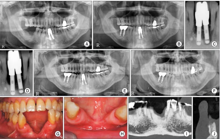
December 2012. In addition, the two lateral incisors adjacent to the implants were strategically removed in March 2013 for a prosthodontic issue.(Fig. 1. E, 1. F). Severe vertical and horizontal alveolar bone defects were observed after implant removal.
For bone augmentation and new implant placements, a bone grafting procedure using a titanium mesh and bovine bone was performed on the healed site after removal. Howev- er, the regenerated bone volume was insufficient to place im- plants both vertically and horizontally. Therefore, we planned vertical augmentation with DO. The exact height of bone loss was determined by computed tomography (CT).(Fig. 1. G-J)
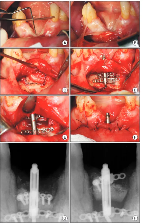
In September 2013, DO was performed under general anesthesia with naso-endotracheal intubation. A vestibular incision was placed after infiltration with local anesthesia.
The osteotomy site was exposed with subperiosteal dissec- tion, and trapezoidal osteotomy was made on the anterior mandible site. A minimum of 4 mm of bone was preserved to maintain sufficient blood supply. Before transporting the bone segment, a 2 mm hole was drilled through the crestal appearance around the implants were not observed.
Four years after his first visit to our clinic, where he was treated with IMZ at 46i and 47i, he complained of discharge of pus on #31i and #41i in Oct 2008. The probing pocket depth of #31i was 10 mm. A panoramic radiograph showed a radiolucent lesion at the apex of #31i. Although, the #31i was almost floating, there was no mobility of the #41i.(Fig.
1. A-D) The patient stated that pus and swelling occurred oc- casionally. He did not feel discomfort at the site. However, the implants were abnormal based on his symptoms and ra- diologic analysis. Since the patient strongly wished for only symptomatic improvement, surgical debridement was per- formed.
Periodontal flap surgery was performed at the inflamma- tory site in December 2008. Four months later, both mobility and the apical radiolucent lesion were decreased. Then, sup- portive maintenance therapy (SPT) with professional clean- ing was carried out every 6 months for 3 years. However, the patient visited our dental clinic with symptoms of pain and pus again in May 2011. Implant removal was decided in
Fig. 1. Panorama, periapical X-ray and clinical views. A. Initial visit. B. Four years after the initial visit. C. Before periodontal surgery. D. Two years after periodontal surgery. E. Before implant removal. F. Three months after bone grafting. G, H. Before distraction osteogenesis pro- cedure. I, J. Computed tomographic views.
Jung-Tae Lee et al: A case report about the reconstruction procedures of the previously failed cylinderical implants site using distraction osteogenesis. J Korean Assoc Oral Maxillofac Surg 2015
A B C
D E F
G H I J
4-0 vicryl sutures.(Fig. 2. A-F)
Postoperative instructions for the patient included a soft diet and oral hygiene with 0.2% chlorhexidine mouth rinse.
Sutures were removed 7 days after surgery. The distraction protocol involved a latency period of 7 days. Distraction was started at a daily rate of 0.6 mm (two activations of 0.3 mm a day). The consolidation period was 3 months.(Fig. 2. G, H) mucosa and bone for placement of the lead screw. The rods
were aligned according to the proposed distraction vector (Lead System, vertical alveolar distraction; KLS Martin, Freiburg, Germany). Furthermore, the distractors were ap- plied on the transport and basal segments with 2 micro plates and 8 screws. The transport segment was placed on the most basal portion, and the mucoperiosteal flap was closed with
Fig. 2. Clinical views and periapical X- ray. A. Measurement of vertical defect before the distraction osteogenesis (DO) procedure. B. Incision. C. Ex- posed osteotomy site. D. A trapezoidal osteotomy was made on the anterior mandible site. E. Distractors were ap- plied on the transport and basal seg- ments with microplates and screws.
The transport segment was placed on the most basal portion. F. Suture.
G. Ten days after DO. H. Twenty-one days after DO. Screw loosening and plate exposure occurred on the left side of the transport segment in this period.
Jung-Tae Lee et al: A case report about the recon- struction procedures of the previously failed cylind- erical implants site using distraction osteogenesis. J Korean Assoc Oral Maxillofac Surg 2015
A B
C D
E F
G H
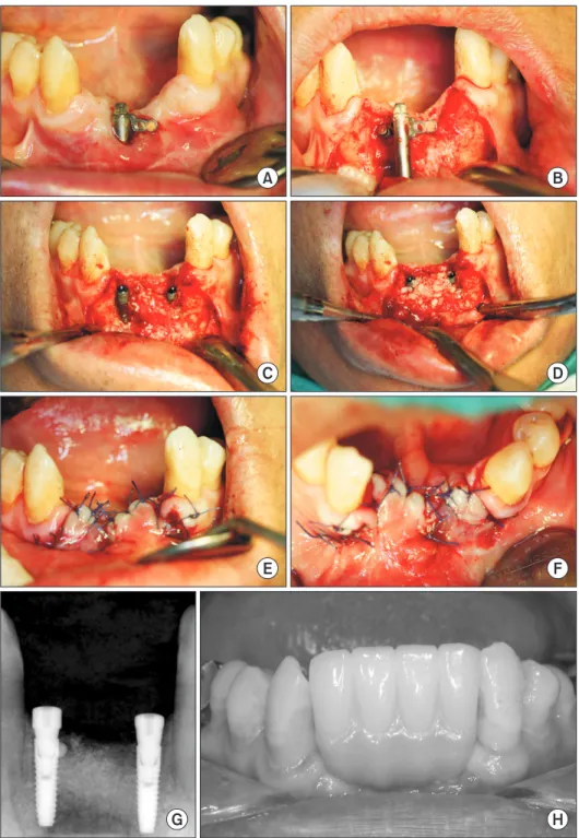
designed with computer-aided design and computer-aided manufacturing (CAD-CAM) to have equi-gingival margins for easier removal of residual cement. The pontic of the pros- thetic restoration was designed so that it did not place exces- sive pressure on peri-implant tissue to prevent inflammation.
To avoid food retention, the implant abutment-pontic embra- sure space was kept closed. For better oral hygiene, profes- sional plaque control was provided regularly using super- floss silk. Clinical and radiologic follow-up periods were 3 to 6 months.(Fig. 3. H)
After the consolidation period, a crestal incision was placed and the distractor was removed from the DO site with local anesthesia. Two implants (NR Line, Dentium, Seoul, Korea) were inserted on the previous lateral incisor area in December 2013. Bone graft materials (Osteon; Genoss, Suwon, Korea) were applied on the implants with a resorbable membrane (Bio-Arm; ACE Surgical Supply Company Inc., Brockton, MA, USA).(Fig. 3. A-G)
The prosthetic restoration of the implants was performed 4 months after the osseointegration. Implant abutments were
Fig. 3. Clinical views and periapical X- ray. A, B. Incision and flap reflection. C.
Implant placement. D. Bone grafting.
E, F. Suture. G. After implant insertion.
H. Final prosthetic restoration after 6 months.
Jung-Tae Lee et al: A case report about the recon- struction procedures of the previously failed cylind- erical implants site using distraction osteogenesis. J Korean Assoc Oral Maxillofac Surg 2015
A B
C D
E F
G H
associated with peri-implantitis. To regenerate the defect site, a titanium mesh and bovine bone were applied. Since, the results were not satisfactory due to insufficient alveolar bone gain, a DO procedure was employed on the same site. Suf- ficient bone height was obtained. New implants were placed on the #32 and #42 missing areas.
In this case, former implants (#31i and #41i IMZ) were eventually extracted due to peri-implantitis. There are several reasons for implant failure7. Although the majority of stud- ies observed that most failures occurred in the posterior sites, several studies showed a higher early implant failure rate in the anterior maxilla and mandible compared with the poste- rior region8,9. Poor hygiene is another key factor for implant failure. However, this patient visited the dental clinic regu- larly for SPT. Another report discussed the relative merit of implant designs using cylindrical or threaded implants. Cy- lindrical implants (IMZ) had a higher failure rate10. Another study found that the IMZ implant system showed favorable function during the first year, but that failure and marginal bone loss increased over time11. A possible contributor to peri-implantitis is the distance between two implants. In this case, the two implants were originally placed with <2 mm between #31i and #41i.
Clinical and experimental research has demonstrated that There was no clinical evidence of infection during the
distraction and consolidation period. After 1 month of DO, screw loosening and plate exposure occurred on the transport segment during the distraction period.(Fig. 2. H) Therefore, the two detached screws were removed. Except for screw loosening, there were no complications such as lingual dis- placement, fracture of the distracted segment, or intraopera- tive bleeding. The dental implants were therefore successfully inserted. Implant failure did not occur during the osseoin- tegration and loading periods. No severe alveolar bone loss around the implants was observed.
The subject was evaluated by radiograph and CT (1) dur- ing the follow-up period before removal of the old implants, (2) just before the DO, (3) after the DO procedure, and (4) at the end of the implant installation. The bone height was measured on the most mesial, distal, and apical areas of the inserted implants as the distance from the alveolar crest to the inferior margin of the mandible. To identify the criteria, the measurement line was perpendicular to the horizontal line connecting the inferior margin of the mandible.(Table 1)
III. Discussion
We set out to reconstruct a previously failed IMZ implant
Table 1. Alveolar bone height from the inferior border of the anterior mandible to the alveolar crest of the implants
Variable Alveolar bone height (mm)
Mesial Distal Apical
During the follow-up period before removal of former implants
#31i
#41i
Just before DO procedure
#31i extracted area
#41i extracted area One month after DO procedure
#31i extracted area
#41i extracted area
At the end of #32i, 42i installation
#31i extracted area
#41i extracted area
Three months after implant installation
#31i extracted area
#41i extracted area Gain in alveolar bone height1
#31i area
#41i area Relapse2, mm (%)
#31i area
#41i area
23.44 24.54 28.65 29.69 36.83 38.18 37.25 36.74 36.62 37.63 8.60 7.05 –0.21 (0.6) –0.55 (1.4)
23.42 24.05 29.30 32.16 36.65 41.38 36.54 37.98 33.36 37.91 7.24 5.82 –3.29 (9.0) –3.47 (8.4)
23.07 23.68 28.65 30.35 36.24 38.92 37.04 37.77 36.23 37.77 8.39 7.42 –0.01 (0.0) –1.15 (3.0) (DO: distraction osteogenesis; i: implant)
1Bone height difference at the end of implant installation compared with just before the DO procedure.
2Bone height difference between 3 months after implant installation and 1 month after the DO procedure.
Jung-Tae Lee et al: A case report about the reconstruction procedures of the previously failed cylinderical implants site using distraction osteogenesis. J Korean Assoc Oral Maxillofac Surg 2015
bono editing of this manuscript.
ORCID
Jung-Tae Lee, http://orcid.org/0000-0001-5383-3004 Shin-Young Park, http://orcid.org/0000-0002-3776-4130 Yang-Jin Yi, http://orcid.org/0000-0001-8341-4759 Young-Kyun Kim, http://orcid.org/0000-0002-7268-3870 Hyo-Jung Lee, http://orcid.org/0000-0002-0439-7389
References
1. Mombelli A, Müller N, Cionca N. The epidemiology of peri- implantitis. Clin Oral Implants Res 2012;23(Suppl 6):67-76.
2. Willer J, Noack N, Hoffmann J. Survival rate of IMZ implants: a prospective 10-year analysis. J Oral Maxillofac Surg 2003;61:691-5.
3. Rinke S, Ohl S, Ziebolz D, Lange K, Eickholz P. Prevalence of pe- riimplant disease in partially edentulous patients: a practice-based cross-sectional study. Clin Oral Implants Res 2011;22:826-33.
4. Leonhardt A, Renvert S, Dahlén G. Microbial findings at failing implants. Clin Oral Implants Res 1999;10:339-45.
5. Renvert S, Roos-Jansåker AM, Lindahl C, Renvert H, Rutger Pers- son G. Infection at titanium implants with or without a clinical di- agnosis of inflammation. Clin Oral Implants Res 2007;18:509-16.
6. Roccuzzo M, Bonino F, Aglietta M, Dalmasso P. Ten-year results of a three arms prospective cohort study on implants in periodon- tally compromised patients. Part 2: clinical results. Clin Oral Im- plants Res 2012;23:389-95.
7. Chrcanovic BR, Albrektsson T, Wennerberg A. Reasons for failures of oral implants. J Oral Rehabil 2014;41:443-76.
8. Machtei EE, Frankenthal S, Blumenfeld I, Gutmacher Z, Horwitz J.
Dental implants for immediate fixed restoration of partially eden- tulous patients: a 1-year prospective pilot clinical trial in periodon- tally susceptible patients. J Periodontol 2007;78:1188-94.
9. Levine RA, Clem D, Beagle J, Ganeles J, Johnson P, Solnit G, et al. Multicenter retrospective analysis of the solid-screw ITI implant for posterior single-tooth replacements. Int J Oral Maxillofac Im- plants 2002;17:550-6.
10. Spiekermann H, Jansen VK, Richter EJ. A 10-year follow-up study of IMZ and TPS implants in the edentulous mandible using bar- retained overdentures. Int J Oral Maxillofac Implants 1995;10:231-43.
11. Esposito M, Hirsch JM, Lekholm U, Thomsen P. Failure patterns of four osseointegrated oral implant systems. J Mater Sci Mater Med 1997;8:843-7.
12. Wolvius EB, Scholtemeijer M, Weijland M, Hop WC, van der Wal KG. Complications and relapse in alveolar distraction osteo- genesis in partially dentulous patients. Int J Oral Maxillofac Surg 2007;36:700-5.
13. Iizuka T, Hallermann W, Seto I, Smolka W, Smolka K, Bosshardt DD. Bi-directional distraction osteogenesis of the alveolar bone us- ing an extraosseous device. Clin Oral Implants Res 2005;16:700-7.
14. Polo WC, Cury PR, Sendyk WR, Gromatzky A. Posterior man- dibular alveolar distraction osteogenesis utilizing an extraosseous distractor: a prospective study. J Periodontol 2005;76:1463-8.
15. Cordaro L, Amadé DS, Cordaro M. Clinical results of alveolar ridge augmentation with mandibular block bone grafts in partially edentulous patients prior to implant placement. Clin Oral Implants Res 2002;13:103-11.
16. Kourtis SG, Sotiriadou S, Voliotis S, Challas A. Private practice results of dental implants. Part I: survival and evaluation of risk factors--Part II: surgical and prosthetic complications. Implant Dent 2004;13:373-85.
DO is an efficient method to treat severe alveolar ridge re- sorption. This procedure can generate new bone between the distracted and basal bone segments12-14. Therefore, DO was the proper method to overcome severe vertical and horizontal defects in this patient. The conventional bone graft has lim- ited vertical augmentation capacity when compared to DO in bone quantity and quality. In this case, conventional grafting procedures using bone substitutes and a barrier membrane were not effective prior to DO. Another study noted that on- lay bone grafting would implicate a large amount of initial bone resorption (42%)15.
This patient visited our dental clinic 63 times during treat- ment over 10 years and has undergone professional manage- ment of oral hygiene through regular checkups. Other reports mention that good oral hygiene and regular checkups are cru- cial in preventing implant failure16. In this case, surgical in- tervention as well as regular checkups with oral hygiene care were insufficient to arrest the progression of peri-implantitis.
The symptoms caused by the two implants (#31i, #41i) were included in diagnostic criteria of peri-implantitis5. Even the previous two implants were associated with peri-implantitis and were considered as clinical failures. It is possible that the former implants should have been extracted on the first visit, when the patient was diagnosed with severe peri-implantitis.
However, the opinion of the patient is important in the deci- sion for implant removal. The subject strongly objected to removal of the former implants, with the resulting delay in implant extraction. Consequently, surgical treatments were performed to salvage the implants. Radiographic evaluation may not permit observation of small bone changes in the crestal area. Therefore, it is not easy to determine when im- plants should be removed.
In conclusion, an aggressive regenerative strategy, such as DO, was successful in reconstruction of a peri-implantitis site that was complicated due to horizontal and vertical bone de- fects.
Conflict of Interest
No potential conflict of interest relevant to this article was reported.
Acknowledgements
The authors are indebted to J. Patrick Barron, Professor Emeritus, Tokyo Medical University and Adjunct Profes- sor, Seoul National University Bundang Hospital, for his pro