pISSN: 0378-6471⋅eISSN: 2092-9374
http://dx.doi.org/10.3341/jkos.2014.55.7.984
Original Article
에이엘 스캔, 자동각막굴절계, 펜타캠의 각막 굴절력 비교 분석
Comparative Analysis of Corneal Refractive Power Measured with AL‐Scan
®, Autokeratometer, and Pentacam
®박성진1⋅임성협1⋅이호영2
Sung Jin Park, MD1, Sung Hyup Lim, MD1, Ho Young Lee, MD2
인제대학교 의과대학 부산백병원 안과학교실1, 신세계안과의원2
Department of Ophthalmology, Busan Paik Hospital, Inje University College of Medicine1, Busan, Korea Shinsegae Eye Clinic2, Busan, Korea
Purpose: To investigate clinical availability of AL-ScanTM (Nidek, Gamagori, Japan) by comparing corneal refractive power with AL-ScanTM, AutokeratometerTM (Topcon KR-1, Tokyo, Japan) and PentacamTM (Oculus, Wetzlar, Germany) devices.
Methods: Seventy-one patients (142 eyes) who visited our hospital for refractive surgery were tested using AL-Scan®, Autokeratometer and Pentacam® and corneal refractive power was compared among devices.
Results: When comparing measurements with AL-Scan®, Autokeratometer and Pentacam®, the mean corneal refractive power was 43.37 ± 1.32 D (2.4 mm zone), 43.35 ± 1.32 D (3.3 mm zone), 43.36 ± 1.35 D, and 43.35 ± 1.36 D respectively and showed no significant differences. Corneal refractive power had strongly positive linear correlation (p < 0.001) and Bland-Altman plots showed high degree of agreement among AL-Scan®, Autokeratometer and Pentacam® devices.
Conclusions: Because measuring ocular biometry with AL-Scan® including axial length, intraocular lens power calculation and topography simultaneously is possible, clinical use is convenient. Corneal refractive power was not different when compared with autokeratometer and Pentacam® devices, thus, AL-Scan® can be used in the clinical environment.
J Korean Ophthalmol Soc 2014;55(7):984-990
Key Words: AL-Scan®, Autokeratometer, Corneal refractive power, Pentacam®
■Received: 2014. 1. 25. ■ Revised: 2014. 3. 28.
■Accepted: 2014. 6. 24.
■Address reprint requests to Ho Young Lee, MD
Shinsegae Eye Clinic, #783-1 Gaya-daero, Busanjin-gu, Busan 614-849, Korea
Tel: 82-51-808-8288, Fax: 82-51-808-8238 E-mail: [email protected]
ⓒ2014 The Korean Ophthalmological Society
This is an Open Access article distributed under the terms of the Creative Commons Attribution Non-Commercial License (http://creativecommons.org/licenses/by-nc/3.0/) which permits unrestricted non-commercial use, distribution, and reproduction in any medium, provided the original work is properly cited.
각막 굴절력은 각막 두께, 전방 깊이, 안축장 등의 다른 생체 계측과 더불어 각막굴절교정수술 및 백내장수술 시에 중요한 요소이다. 각막굴절교정수술 시에는 정확한 각막 굴절력 측정이 시력의 예후를 좌우하며, 수술 후의 경과 관 찰 시에도 진행성 근시, 난시 및 기타 각막 합병증 발생 등 을 관찰할 때 중요한 지표가 된다. 백내장수술 시에도 각막
굴절력을 비롯한 생체 계측이 정확한 인공수정체 도수 계 산에 중요한 요소이며, 많은 논문들에서 각막 굴절력과 전 방 깊이, 안축장에 관한 측정 기기별 정확성을 비교하였 다.1-5 각막 굴절력은 자동 및 수동각막굴절계, 부분결합간 섭계, 파면분석기, 비디오각막경 등의 다양한 기기를 통해 측정될 수 있으며, 전통적으로 인공수정체 도수 계산에서 는 자동각막굴절계를 이용하였고, 각막굴절교정수술 시에 는 회전 샤임플러그 카메라(rotating scheimpflug tomog- raphy) 방식의 각막지형도 검사계인 Pentacam®이 많이 사 용되고 있다.6,7
최근 개발된 AL-Scan®은 안축장 측정을 통해 백내장 수 술 시 인공수정체 도수 계산에 주로 이용되고 있으며, 몇 가지 전안부 계측이 가능하다. 하지만 IOL master®와 유사
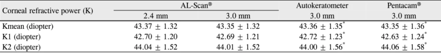
Table 1. Results of corneal refractive power (K) of AL-Scan®, Autokeratometer and Pentacam®
Corneal refractive power (K) AL-Scan® Autokeratometer Pentacam®
2.4 mm 3.0 mm 3.0 mm 3.0 mm
Kmean (diopter) 43.37 ± 1.32 43.35 ± 1.32 43.36 ± 1.35* 43.35 ± 1.36*
K1 (diopter) 42.70 ± 1.20 42.69 ± 1.21 42.72 ± 1.23* 42.63 ± 1.24*
K2 (diopter) 44.04 ± 1.52 44.01 ± 1.52 44.00 ± 1.56* 44.06 ± 1.58*
Values are presented as mean ± SD.
*Corneal refractive power (K) with Autokeratometer and Pentacam® was measured only 3.0 mm zone.
Table 2. Comparison of mean corneal refractive power (K)
among devicesDevices p-value
Autokeratometer* AL‐Scan® (2.4 mm zone) 0.595† AL‐Scan® (3.3 mm zone) 0.450†
Pentacam®* 0.349†
AL‐Scan® (2.4 mm zone) AL‐Scan® (3.3 mm zone) 0.009†
Pentacam®* 0.199†
AL‐Scan® (3.3 mm zone) Pentacam®* 0.794†
*Corneal refractive power (K) with Autokeratometer and Pentacam®
was measured only 3.0 mm zone; †Paired t-test.
한 방식의 부분결합간섭계(partial coherence interferometry) 임에도 불구하고, 전안부 계측은 Pentacam®과 같은 샤임플 러그 카메라를 통해서 이루어진다는 특징이 있다. 특히 AL-Scan®의 경우 830 μm 파장의 적외선 광선을 이용한 한 번의 비접촉식 측정으로 각막 굴절력, 각막 두께, 전방 깊 이, 안축장, 동공 크기, 각막 크기(white to white diameter) 등을 수 초 이내의 짧은 시간 안에 측정하는 장점이 있다.
최근 IOL master®와 비교한 AL-Scan®의 전안부 계측 및 정 확성에 대한 보고를 비롯하여,8 AL-Scan®에 대한 연구가 활발히 진행되고 있다. 하지만 기존의 장비와 비교한 AL-Scan®에서의 각막 굴절력 측정의 정확성 및 임상적 유 용성에 대해서는 아직까지 국내에서 보고된 바가 없다.
이에 본 연구에서는 정상 각막을 가진 각막굴절교정수술 예정 환자를 대상으로 하여 AL-Scan®을 통한 각막 굴절력 을 기존의 자동각막굴절계 및 Pentacam®의 결과와 비교 분 석하여 AL-Scan®의 임상적 유용성을 평가하고자 하였다.
대상과 방법
2013년 4월부터 10월까지 각막굴절교정수술을 위해 본 원을 방문한 71명 142안을 대상으로 의무기록을 후향적으 로 분석하였다. 환자들은 근시, 난시, 경도의 백내장을 제외 하고는 다른 안과적 질환을 가지고 있지 않았으며, 이전의 각막 수술, 안내 수술, 검사 결과에 영향을 미칠 수 있는 각 막 질환이 있는 경우는 제외하였다.
수술 전 같은 날 대상 환자들에게 같은 검사자에 의해서 연속적으로 자동각막굴절계(Topcon KR-1, Tokyo, Japan), AL-Scan® (Nidek, Japan)과 Pentacam® (Oculus, Wetzlar, Germany)을 시행하여 각막 굴절력을 측정하였으며, 검사 결과로 나온 측정값을 비교하였다.
모든 환자에서 자동각막굴절계를 먼저 시행하고 AL-Scan®, Pentacam®의 순서로 시행하였으며, 세 기계 모두 3번씩 검사 를 시행하였다. 검사 전에 눈을 깜박거리게 하여 눈물층을 균일하게 하였으며, 검사 도중 환자가 눈을 깜박이거나 움 직이지 않도록 주의하고 검사자에 의해 안구가 눌리지 않 도록 주의하며 검사를 시행하였다. 각 대상자는 측정 후, 뒤로 물러나 앉게 하여 기기가 다음 측정 전 재조정될 수
있게 하였다. 세 기계로부터 측정된 결과 중에 AL-Scan®은 2.4 mm와 3.3 mm 구역의 각막 굴절력, 자동각막굴절계와 Pentacam®은 3.0 mm 구역의 각막 굴절력을 측정하여 분석 하였다.
통계적인 분석은 SPSS v18.0 for Windows (SPSS Inc., Chicago, IL, USA)를 사용하였다. 각각의 측정에서 각막 굴 절력은 flat K와 steep K의 평균값으로 하였고, 이 수치는 3번의 각각의 측정값마다 적용되어 이 3회의 측정값을 평 균한 값으로 기기 간 비교를 하였다. Paired t-test를 통하여 기기에 따른 측정값의 평균의 차이를 비교하였고, p<0.05를 유의한 차이가 있다고 보았다. 또한 Pearson correlation을 이용하여 각 방법 간의 상관계수를 구하였으며, p<0.01를 유의한 상관관계가 있다고 보았다. 기기 간 측정값의 차이 를 알기 위해서 측정값의 차이의 평균을 구하여 비교하였 으며, 기기 간의 일치성을 평가하기 위해 Bland-Altman plot을 사용하였다.
결 과
총 71명 142안을 대상으로 하였으며, 남자는 28명, 여자 는 43명이었다. 환자들의 평균 나이는 29.73 ± 7.28세였으 며, 우안, 좌안 모두 71안이었다. AL-Scan®, 자동각막굴절 계, Pentacam®으로 측정한 각막 굴절력을 표로 나타내었다 (Table 1).
AL-Scan®은 2.4 mm 구역과 3.3 mm 구역 두 군데에서 각막 굴절력을 구분하여 측정하게 되며, 2.4 mm 구역에서 의 각막 굴절력은 가로축이 42.70 ± 1.20D, 세로축이 44.04
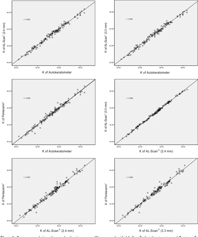
Figure 1. Pearson correlation of corneal refractive power (K) measured with AL-Scan®, Autokeratometer and Pentacam®.
± 1.52D, 평균 43.37 ± 1.32D로 측정되었으며, 3.3 mm 구 역에서의 각막 굴절력은 가로축이 42.69 ± 1.21D, 세로축이 44.01 ± 1.52D, 평균 43.35 ± 1.32D로 측정되었다. 자동각 막굴절계와 Pentacam®은 3.0 mm 구역의 각막 굴절력을 측 정하였다. 자동각막굴절계의 각막 굴절력은 가로축 42.72 ± 1.23D, 세로축 44.00 ± 1.56D, 평균 43.36 ± 1.35D로 측정
되었고, Pentacam®의 각막 굴절력은 가로축 42.63 ± 1.24D, 세로축 44.06 ± 1.58D, 평균 43.35 ± 1.36D로 측정되었다.
AL-Scan®에서 2.4 mm 구역과 3.3 mm 구역 간에는 유의한 차이가 있었으나, AL-Scan®, 자동각막굴절계, Pentacam® 세 기기 간의 평균 각막 굴절력은 통계적으로 유의한 차이 가 없었다(Table 2).
K of AL-Scan (2.4 mm)® K of AL-Scan (3.3 mm)® K of AL-Scan (3.3 mm)®
K of Pentacamn® K of Pentacamn® K of Pentacamn®
K of Autokeratometer K of Autokeratometer
K of Autokeratometer K of AL-ScanⓇ (2.4 mm)
K of AL-ScanⓇ (2.4 mm) K of AL-ScanⓇ (3.3 mm)
Figure 2. Bland-Altman plots of corneal refractive power (K) measured with AL-Scan®, Autokeratometer and Pentacam®.
Pearson correlation으로 두 기기 간의 평균 각막 굴절력을 비교했을 때, p-value는 0.001 이하로 모든 기기에서 통계적으 로 유의한 선형 상관 관계를 가지고 있었다. 그리고 Pearson correlation coefficient의 경우, 자동각막굴절계와 AL-Scan® (2.4 mm) (r=0.992), 자동각막굴절계와 AL-Scan® (3.3 mm) (r=0.992), 자동각막굴절계와 Pentacam® (r=0.989), AL-Scan® (2.4 mm)과 AL-Scan® (3.3 mm) (r=0.998), AL-Scan® (2.4 mm)
과 Pentacam® (r=0.987), AL-Scan® (3.3 mm)과 Pentacam® (r=0.988)으로 측정되어 모든 측정치에서 강한 양의 선형 상관 관계가 있었다. 측정치 중 AL-Scan®에서 2.4 mm 구 역과 3.3 mm 구역 간에는 통계적으로 유의한 차이를 보였 으나, Pearson correlation에서는 의미 있는 상관 관계를 보 였다(Fig. 1).
Bland-Altman plot으로 나타낸 두 기기 간 일치도에서 측
Mean K of Autokeratometer and AL-ScanⓇ (2.4 mm) Mean K of Autokeratometer and AL-ScanⓇ (3.3 mm)
Mean K of Autokeratometer and PentacamⓇ Mean K of AL-ScanⓇ (2.4 mm) and AL-ScanⓇ (3.3 mm)
Mean K of AL-ScanⓇ (2.4 mm) and PentacamⓇ Mean K of AL-ScanⓇ (3.3 mm) and PentacamⓇ
Difference of K
Difference of K Difference of K
Difference of K Difference of K
Difference of K
정값의 95% limits of agreement (LoA)는 각각 자동각막굴 절계와 AL-Scan® (2.4 mm)은 -0.35~0.33D, 자동각막굴절 계와 AL-Scan® (3.3 mm)은 -0.35~0.37D, 자동각막굴절계 와 Pentacam®은 -0.38~0.42D, AL-Scan® (2.4 mm)과 AL-Scan® (3.3 mm)은 -0.16~0.20D, AL-Scan® (2.4 mm)과 Pentacam®은 -0.42~0.46D, AL-Scan® (3.3 mm)과 Pentacam® 은 -0.42~0.42D로 측정되었다(Fig. 2).
고 찰
정확한 각막 굴절력의 측정은 각막굴절교정수술 및 백내 장수술 모두에서 시력 결과의 예측 및 수술 후 경과 관찰 등에 있어 매우 중요하고, 이는 환자의 수술 만족도에도 큰 영향을 미친다. 따라서 본 연구는 이전에 사용되고 있는 자 동각막굴절계, Pentacam®과 최근에 개발된 AL-Scan®에서 각막 굴절력을 비교하고자 시행되었다. 백내장수술 후의 굴절력의 정확한 예측을 위해서는 각막 굴절력, 안축장, 인 공수정체 도수 계산의 정확성 등의 여러 가지 임상적 변수 를 고려하게 된다.9,10 Olsen4은 백내장수술 후 요구되는 굴 절력에 이르지 못하는 이유로 안축장(54%), 각막 굴절력 (8%), 전방 깊이(38%)의 부정확한 측정을 들었고, Jo et al11 은 굴절 오차에 있어 각막 굴절력의 0.5D의 오차는 ±1.17D 의 인공수정체 도수 차이를 가져올 수 있다고 하였다.
최근 자동각막굴절계, 컴퓨터를 응용하여 개발된 비디 오각막경, 그리고 부분간섭계 등이 이용되면서 각막 굴절 력을 비롯한 생체 계측의 정확성은 많은 부분 증진되었으 며, 여러 연구에서 높은 정확성 및 재현성이 보고되었 다.4,9,10,12,13
하지만 아직도 이런 기기 간의 측정값은 다소 차이가 나고 있으며, 수술 후 결과에 대한 높은 기대감에 부응하기 위해서는 새로 개발된 기기들의 기존 기기들과의 정확성과 재현성에 대한 비교가 중요하다.
자동각막굴절계 중의 하나인 KR-1® (Topcon Inc., Tokyo, Japan)은 각막이 구형이라는 가정하에 3.0 mm의 직경 안에 서 카메라에 의해 받아지는 반사상으로 각막 굴절력을 측 정하게 된다. 각막에 대한 대강의 정보는 얻을 수 있으나 많은 정보를 얻기는 힘들다는 단점이 있다.13 Pentacam®은 세극등의 원리를 이용한 elevation-based system 방법으로 기존의 세극등 광선이 좌우로 지나가는 Orbscan®과는 다르 게 360도 회전하는 샤임플러그 카메라를 사용하여 2초 내 에 각막 전후면에 위치한 500개의 점을 포함하는 영상을 얻고, 중심부의 고정된 카메라는 동공의 모양을 감지하여 fixation을 담당하며, 한 번의 측정으로 전방의 구조를 3차 원적으로 재구성하게 되어 수정체 후면의 영상까지 얻을 수 있다.6,7 각막 굴절력 및 각막 두께, 각막 지형도, 전방 깊
이를 측정할 뿐만 아니라 전방각과 수정체의 혼탁과 두께의 정보를 추가로 얻을 수 있다.7,14 최근에 개발된 AL-Scan®은 IOL master®가 제공하는 각막 굴절력, 안축장, 전방 깊이, 각막 직경 외에도 중심부 각막 두께, 동공 크기 등의 정보도 같이 측정된다는 장점이 있다. 각막 굴절력 측정에 있어 AL-Scan®은 2.4 mm, 3.3 mm 구역의 이중 패턴 링 방식으 로 두 군데의 각막 굴절력을 측정하며, 각각의 구역에서 360개의 점을 분석한다는 특징이 있다. 또한 3D 추적 시스 템을 이용하여 눈의 움직임을 감지하여 자동으로 추적하여 측정하기 때문에 검사의 부정확성을 줄일 수 있다.
기존의 연구들에서 각막 굴절력은 측정기기 간에 차이가 나는 경우가 많았다. Butcher and O'Brien15은 자동각막굴절 계만을 대상으로 재현성이 좋음을 밝혔으며, Karabatsas et al16은 정상군 32안과 각막이식 후의 각막 33안을 대상으로 기존의 각막굴절계와 각막지형도를 측정한 논문에서 각막 굴절계가 각막지형도보다 재현성이 좋다고 발표하였다. Han et al17은 자동각막굴절계, IOL Master®, 각막지형도의 각막 굴절력을 비교 분석하였고, 그 결과 평균 각막 굴절력 은 각막지형도에서 높았고, 굴절력의 재현성은 각막지형도 에 비해 자동굴절검사와 IOL Master®의 측정값이 높았다고 하였다. 그러나 본 연구에서는 자동각막굴절계와 Pentacam® 의 3.0 mm 구역에서 측정한 각막 굴절력은 통계적으로 유 의한 차이가 없었다(p=0.349). 두 기기 간의 재현성의 차이 는 없었다. Lee et al7은 Pentacam®과 Sirius®의 전안부 측정 값을 비교하였으며, 평균 각막 굴절력은 두 기기 간 차이가 있었다고 하였다. 반면, Kim and Chung18은 자동각막굴절 계(RK-F1®), 수동각막굴절계(OM-2®), 부분결합간섭계(IOL master®), 파면분석기(KR-1W®), 비디오각막경(Orbscan II®) 에서의 각막 굴절력을 비교하였고, 다섯 기기 간의 재현성 은 서로 비슷했으나, KR-1W®를 이용할 때에는 수동각막굴 절계와 1D 이상 차이가 날 수 있다고 하였다. 또한 Savini et al19은 네 종류의 각막지형도(Pentacam®, Sirius®, TMS-5®, Keratron®) 측정값을 비교하였으며 각막 굴절력을 포함한 대 부분의 측정값에서 차이를 보였으나 예외적으로 Pentacam® 과 Sirius®로 측정한 각막 굴절력은 두 기기 간 유의한 차이 가 없었다고 하였으며, 이는 본 연구의 결과와 유사하였다.
Pentacam®, Sirius®, AL-Scan®의 세 기기는 각각 약간의 측 정 방식과 기계적인 차이는 있지만 기본적으로 모두 샤임 플러그 카메라를 사용한다는 공통점이 있다. 본 연구에서 도 Pentacam®의 3.0 mm 구역의 각막 굴절력과 비교하여 AL-Scan®의 2.4 mm와 3.3 mm 구역의 각막 굴절력은 통계 적으로 유의한 차이가 없었다(p=0.199, p=0.794). 임상적으 로 Pentacam®의 3.0 mm 구역의 측정값과 상호 호환 가능 할 것으로 보인다.
측정값에 대한 새로운 기기의 신뢰도를 판단하기 위해서 는 그 기기의 재현성을 측정하고, 높은 재현성을 확인하여 기존의 기기와 비교하는 과정이 필요하다. 위에서도 언급 했듯이 자동각막굴절계와 Pentacam®의 높은 재현성은 이 전의 많은 연구에서 확인되었고, 이를 바탕으로 임상에서 오랜 기간 사용되어 왔다.20,21 본 연구에서는 AL-Scan®의 재현성 확인을 위해 각막 굴절력을 3회 측정 후 평균값을 이용하였으며, 국내외에 보고되지 않은 첫 시도라는 점에 서 본 연구가 갖는 의의가 크다 하겠다. 추후 AL-Scan®의 재현성과 반복성, 정확성에 대한 추가 연구가 이루어진다 면, 임상적으로 보다 유용할 것으로 생각한다.
본 연구는 새로 개발된 AL-Scan®과 기존의 자동각막굴 절계 및 Pentacam®에서의 각막 굴절력을 비교 분석하였다.
AL-Scan®의 각막 굴절력은 자동각막굴절계, Pentacam®과 비교하여 유의한 차이가 없었으며, 세 기기 간의 서로 간의 분석에서도 유의한, 강한 양의 상관 관계를 보였다. 이러한 측정 결과를 통해 AL-Scan®으로 측정한 각막 굴절력은 기 존 장비에 비해 차이가 없었으므로 임상적으로 유용하게 사용할 수 있다.
REFERENCES
1) Koranyi G, Lydahl E, Norrby S, Taube M. Anterior chamber depth measurement: a-scan versus optical methods. J Cataract Refract Surg 2002;28:243-7.
2) Maeng HS, Ryu EH, Chung TY, Chung ES. Effects of anterior chamber depth and axial length on refractive error after intraocular lens implantation. J Korean Ophthalmol Soc 2010;51:195-202.
3) Norrby S. Sources of error in intraocular lens power calculation. J Cataract Refract Surg 2008;34:368-76.
4) Olsen T. Prediction of the effective postoperative (intraocular lens) anterior chamber depth. J Cataract Refract Surg 2006;32:419-24.
5) Qazi MA, Cua IY, Roberts CJ, Pepose JS. Determining corneal power using Orbscan II videokeratography for intraocular lens cal- culation after excimer laser surgery for myopia. J Cataract Refract Surg 2007;33:21-30.
6) Rufer F, Schroder A, Arvani MK, Erb C. [Central and peripheral corneal pachymetry--standard evaluation with the Pentacam sys-
tem]. Klin Monbl Augenheilkd 2005;222:117-22.
7) Lee DM, Ahn JM, Seo KY, et al. Comparison of corneal measure- ment values between two types of topography. J Korean Ophthalmol Soc 2012;53:1584-90.
8) Kim SI, Kang SJ, Oh TH, et al. Accuracy of ocular biometry and postoperative refraction in cataract patients with AL-Scan(R). J Korean Ophthalmol Soc 2013;54:1688-93.
9) Holladay JT, Prager TC, Ruiz RS, et al. Improving the predict- ability of intraocular lens power calculations. Arch Ophthalmol 1986;104:539-41.
10) Mamalis N. Complications of foldable intraocular lenses requiring explanation or secondary intervention--1998 survey. J Cataract Refract Surg 2000;26:766-72.
11) Jo DH, Oh JY, Kim MK, et al. Corneal power estimation using Orbscan II videokeratography in eyes with previous corneal re- fractive surgeries. J Korean Ophthalmol Soc 2009;50:1730-4.
12) Findl O, Drexler W, Menapace R, et al. Improved prediction of in- traocular lens power using partial coherence interferometry. J Cataract Refract Surg 2001;27:861-7.
13) Speicher L. Intra-ocular lens calculation status after corneal re- fractive surgery. Curr Opin Ophthalmol 2001;12:17-29.
14) Rosa N, Lanza M, Borrelli M, et al. Comparison of central corneal thickness measured with Orbscan and Pentacam. J Refract Surg 2007;23:895-9.
15) Butcher JM, O'Brien C. The reproducibility of biometry and kera- tometry measurements. Eye 1991;5:708-11.
16) Karabatsas CH, Cook SD, Papaefthymiou J, et al. Clinical evalua- tion of keratometry and computerised videokeratography: intra- observer and interobserver variability on normal and astigmatic corneas. Br J Ophthalmol 1998;82:637-42.
17) Han JM, Choi HJ, Kim MK, et al. Comparative analysis of corneal refraction and astigmatism measured with autokeratometer, IOL Master, and topography. J Korean Ophthalmol Soc 2011;52:1427-33.
18) Kim S, Chung SK. Comparison of corneal curvatures obtained with different devices. J Korean Ophthalmol 2012;53:618-25.
19) Savini G, Carbonelli M, Sbreglia A, et al. Comparison of anterior segment measurements by 3 Scheimpflug tomographers and 1 Placido corneal topographer. J Cataract Refract Surg 2011;37:1679-85.
20) Kawamorita T, Uozato H, Kamiya K, et al. Repeatability, reprodu- cibility, and agreement characteristics of rotating Scheimpflug photography and scanning-slit corneal topography for corneal power measurement. J Cataract Refract Surg 2009;35:127-33.
21) Jain R, Dilraj G, Grewal SP. Repeatability of corneal parameters with Pentacam after laser in situ keratomileusis. Indian J Ophthalmol 2007;55:341-7.
= 국문초록 =
에이엘 스캔, 자동각막굴절계, 펜타캠의 각막 굴절력 비교 분석
목적: AL‐ScanⓇ (Nidek, GAMAGORI, Japan)과 자동각막굴절계, PentacamⓇ(Oculus, Wetzlar, Germany)을 이용한 각막 굴절력을 비교함으로써 AL‐ScanⓇ의 임상적 유용성을 평가하고자 한다.
대상과 방법: 각막굴절교정수술을 목적으로 방문한 환자 71명(142안)을 대상으로 하여, AL‐ScanⓇ 과 자동각막굴절계, PentacamⓇ으로 각각 각막 굴절력을 측정하고 그 측정치를 비교하였다.
결과: AL‐ScanⓇ과 자동각막굴절계, PentacamⓇ으로 측정한 평균 각막 굴절력은 각각 43.37 ± 1.32D (2.4 mm zone), 43.35 ± 1.32D (3.3 mm zone), 43.36 ± 1.35D, 43.35 ± 1.36D로 각 기기 간 유의한 차이를 보이지 않았고, 통계적으로 강한 양의 선형 상관 관계가 있었다(p<0.001). 또한 이 세 기기를 Bland‐Altman plot으로 각각 분석하였을 때 기기 간의 높은 일치도를 보였다.
결론: AL‐ScanⓇ은 한 번의 측정으로 각막 굴절력, 중심 각막 두께, 전방 깊이 등의 전안부 계측, 안축장 측정과 인공수정체 도수 계산 까지 이루어지기 때문에 임상에서 사용 시 편리하다. 이번 연구를 통해 AL‐ScanⓇ으로 측정한 각막 굴절력은 기존 장비에 비해 차이가 없었으므로 임상적으로 유용하게 사용할 수 있다.
<대한안과학회지 2014;55(7):984-990>