ISSN 0378-6471 (Print)⋅ISSN 2092-9374 (Online)
http://dx.doi.org/10.3341/jkos.2016.57.10.1542
Original Article
중심각막두께 측정에 대한 파장가변 빛간섭단층촬영계의 유용성
Utility of the Swept Source Optical Coherence Tomography for Measurements of Central Corneal Thickness
김임규1⋅이종은2⋅이준석1⋅서 샘1⋅이규원1
Im Gyu Kim, MD1, Chong Eun Lee, MD, PhD2, Jun Seok Lee, MD1, Sam Seo, MD, PhD1, Kyoo Won Lee, MD, PhD1
제일안과병원1, 계명대학교 의과대학 안과학교실2 Cheil Eye Hospital1, Daegu, Korea,
Department of Ophthalmology, Keimyung University School of Medicine2, Daegu, Korea
Purpose: To evaluate the efficacy of swept source optical coherence tomography (SS-OCT) by comparing the measurement of central corneal thickness (CCT) to the measurement obtained using Orbscan II, anterior segment optical coherence tomography (AS-OCT) and ultrasound pachymetry.
Methods: One examiner measured the CCT in 65 eyes of 65 healthy subjects using Orbscan II, AS-OCT, SS-OCT and ultra- sound pachymetry. The mean values and correlations were analyzed.
Results: The average CCT measurements obtained using Orbscan II, AS-OCT, SS-OCT and ultrasound pachymetry were 534.83 ± 38.46, 517.80 ± 32.48, 528.22 ± 33.71 and 528.02 ± 34.90 μm, respectively. A significant linear correlation was ob- served among Orbscan II, AS-OCT, SS-OCT and ultrasound pachymetry (r > 0.894, p < 0.001). There was no significant differ- ence between the SS-OCT and ultrasound pachymetry (p = 0.782).
Conclusions: The results of the 4 methods were significantly correlated and the SS-OCT reached a high level of agreement when CCT was determined using ultrasound pachymetry. The CCT measurements using SS-OCT is a better alternative for ultra- sound pachymetry than Orbscan II and AS-OCT.
J Korean Ophthalmol Soc 2016;57(10):1542-1548
Keywords: Anterior segment optical coherence tomography, Central corneal thickness, Orbscan II, Swept source optical coher- ence tomography, Ultrasound pachymetry
■Received: 2016. 7. 14. ■ Revised: 2016. 8. 8.
■Accepted: 2016. 9. 20.
■Address reprint requests to Sam Seo, MD, PhD
Cheil Eye Hospital, #1 Ayang-ro, Dong-gu, Daegu 41196, Korea Tel: 82-53-959-1751, Fax: 82-53-959-1758
E-mail: [email protected]
ⓒ2016 The Korean Ophthalmological Society
This is an Open Access article distributed under the terms of the Creative Commons Attribution Non-Commercial License (http://creativecommons.org/licenses/by-nc/3.0/) which permits unrestricted non-commercial use, distribution, and reproduction in any medium, provided the original work is properly cited.
중심각막두께의 측정은 여러 안과 질환의 평가 및 수술 전 검사에 있어서 중요한 지표이다. 각막두께는 각막 내피 세포의 상태와 기능을 반영하고, 각막굴절교정수술의 가능 여부 및 수술 방법을 결정하는 데 있어 가장 중요한 역할을 하며, 수술 후 합병증의 발생과도 연관이 있다.1,2 녹내장에
서는 각막두께에 따라 안압 측정치를 보정하며, 중심각막두 께가 얇을수록 시신경의 손상이 빠르게 진행된다는 보고가 있다.3,4 각막이식술 후에도 중심각막두께의 측정은 이식된 각막의 상태를 나타내는 유용한 지표이다.5
각막두께의 측정에는 접촉식 방법인 초음파 각막두께측 정계(ultrasound pachymetry)와 비접촉식 방법인 비접촉경 면현미경(noncontact specular microscopy), 전안부 빛간섭 단층촬영계(anterior segment optical coherence tomography, AS-OCT), 동일초점현미경(confocal microscopy), 레이저 간섭계(laser interferometer), 초음파 생체현미경(ultrasonic biomicroscopy), Orbscan tomography system (Orbscan II) 등
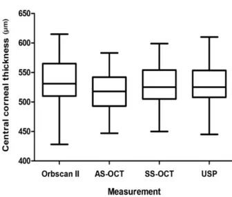
Figure 1. Box plots showing mean central corneal thickness
measured by Orbscan II, anterior segment optical coherence to- mography (AS-OCT), swept source optical coherence tomog- raphy (SS-OCT), and ultrasound pachymetry (USP). The hori- zontal lines within the box represent medians, the ends of the boxes represent the first and third quartiles, and the whiskers represent the smallest and largest non-outlier values.다양한 방법이 이용되고 있다. 이 중 초음파 각막두께측정 계(ultrasound pachymetry)가 가장 대표적이고 널리 이용되 고 있으나, 소식자를 접촉하는 측정방법으로 인한 각막상 피손상이나 감염 등의 위험 및 안구 함입으로 인한 측정값 오차에 대해 우려가 제기되어 왔다.6,7 이에 각막에 직접 접 촉하지 않고 각막두께를 측정할 수 있는 방법들이 대두되 고 있다.8 특히 최근에 도입된 IOLMaster 700 (Carl Zeiss Medited AG, Jena, Germany)는 파장가변 빛간섭단층촬영 (swept source optical coherence tomography, SS-OCT)을 이 용하여 높은 해상도 및 민감도로 안구 내부 구조를 정량적 으로 평가할 수 있으며, 정확한 각막두께 및 전안부 생체계 측치를 측정할 수 있다고 제시되었다.9
지금까지 국내에서 접촉식의 초음파 각막두께측정계와 비접촉 각막두께 측정방법들과의 측정치 차이를 보고하고 이를 교정하는 방법들을 제시한 연구들이 많았지만, 정상안 에서 파장가변 빛간섭단층촬영방법을 이용하여 측정한 각 막두께와의 비교는 아직 보고된 바가 없다. 따라서 본 연구 는 정상인을 대상으로 하여 초음파 각막두께측정계와 Orb- scan II, AS-OCT, SS-OCT를 이용하여 중심각막두께를 측정 하여 비교해 보고자 하였다.
대상과 방법
본 연구는 2016년 6월 1일부터 30일까지 본원을 방문한 전신질환 및 안과적 과거력이 없는 건강한 성인 65명 65안 을 대상으로 하였다. 본 연구는 본원 연구윤리심의위원회의 승인을 받아 시행하였고 헬싱키 선언을 준수하였으며 연구 및 검사에 대한 충분한 설명을 한 뒤 동의를 얻은 환자를 대상으로 하였다. 실험안은 양안 중 무작위로 선택하였다.
모든 대상 환자에서 교정시력측정 및 굴절검사, 안압검사, 세극등 현미경검사를 시행하였으며, 최대교정시력 0.8 이 상, 골드만 안압 21 mmHg 미만이며 세극등 현미경 검사에 서 각막, 홍채 및 전방에 이상 소견이 없는 경우를 대상으로 하였다. 안외상, 안내수술, 각막질환의 과거력이 있는 경우 는 대상에서 제외하였다. 모든 검사는 같은 날 동일한 한 명 의 검사자에 의해 연속적으로 진행되었고, 일중변동의 영향 을 최소화하기 위해 모든 검사는 오후 3시에서 5시 사이에 이루어졌다. 초음파 각막두께 측정 시 발생할 수 있는 각막 의 손상을 최소화하기 위해 비접촉성 검사인 Orbscan II (Bausch & Lomb, Rochester, NY, USA)과 전안부 빛간섭단 층촬영계(Spectralis HRA-OCT, Heidelberg, Germany), 파장 가변 빛간섭단층촬영계(IOLMaster 700) 검사를 먼저 시행 하고 초음파 각막두께측정검사(Pachymeter-SP3000, Tomey corporation, Nagoya, Japan)를 가장 마지막에 실시하였다.
검사 전에 눈을 깜박거리게 하여 눈물층을 균일하게 하 였으며, 검사 시 주시광선을 보면서 환자가 눈을 깜박이거 나 움직이지 않도록 하였다. 속눈썹이 각막을 가리지 않도 록 눈을 크게 뜨도록 지시한 후 안구에 압박이 가해지지 않 도록 조심스럽게 눈꺼풀을 벌리면서 측정하였다. 전안부 빛 간섭단층촬영 검사는 Spectralis HRA-OCT의 Cornea 영상을 찍은 후 Spectralis 소프트웨어에서 제공하는 Caliper를 이용 하여, 각막 중심부의 외측경계에서부터 내측경계까지 수직 선을 그어 측정하였다. Orbscan II의 측정치는 제작사에서 추천한 보정인자(acoustic equivalent factor) 0.92를 사용하여 보정하였다. 초음파 각막두께측정계를 이용한 측정은 각막 을 0.5% proparacaine hydrochloride 점안약으로 마취한 후 초음파 소식자를 가능한 중심각막에 수직으로 접촉하여 5 회 측정한 후 평균값을 측정값으로 하였다.
통계적인 분석은 SPSS 21.0 for Windows (IBM Corpora- tion, Armonk, NY, USA)를 사용, 4가지 검사기기로 측정된 중심각막두께의 값을 비교하여 검사기계 간의 차이를 분석 하였다. Repeated-measures ANOVA와 Pearson correlation coefficient를 통해 각 기기의 계측치 평균의 차이를 비교하 고, 상관관계를 분석하였다. 각 기기 간의 측정 일치도는 Bland-Altman plots를 이용하여 분석하였고
p
값의 유의수준 은 0.05 미만으로 하였다.Table 1. Pairwise comparison of central corneal thickness measurements
Comparison Mean difference ± SD
p
-value*Ultrasound pachymetry and Orbscan II 6.82 ± 15.92 0.001
Ultrasound pachymetry and AS-OCT 10.22 ± 8.18 <0.001
Ultrasound pachymetry and SS-OCT 0.20 ± 5.80 0.782
SS-OCT and Orbscan II 6.62 ± 16.26 0.002
SS-OCT and AS-OCT 10.42 ± 7.68 <0.001
Orbscan II and AS-OCT 17.03 ± 17.34 <0.001
Values are presented as mean ± SD unless otherwise indicated.
SD = standard deviation; AS-OCT = anterior segment optical coherence tomography; SS-OCT = swept source optical coherence tomography.
*Paired-
t
test,p
< 0.05: statistically significantp
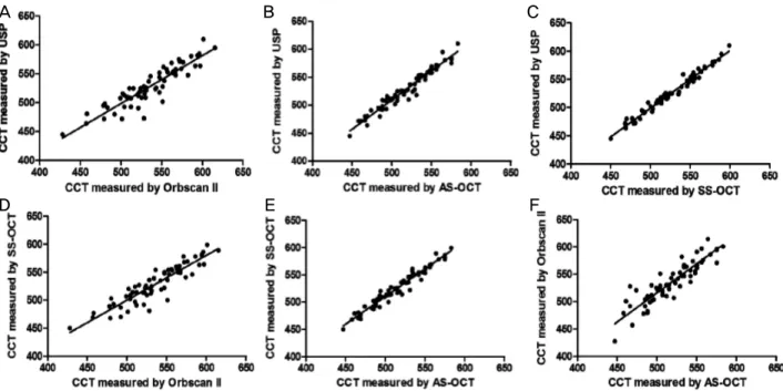
-value.Figure 2. The correlation plot of the central corneal thickness (CCT) measured by ultrasound pachymetry (USP), Orbscan II, ante-
rior segment optical coherence tomography (AS-OCT), and swept source optical coherence tomography (SS-OCT). (A) Correlation between USP and Orbscan II (r2 = 0.834, p < 0.001), The best-fit line (y = 0.83x + 83.28) is designated by solid line. (B) Correlation between USP and AS-OCT (r2 = 0.947, p < 0.001), The best-fit line (y = 1.05x -13.48) is designated by solid line.(C) Correlation between USP and SS-OCT (r2 = 0.973, p < 0.001), The best-fit line (y = 1.02x - 11.42) is designated by solid line.
(D) Correlation between SS-OCT and Orbscan II (r2 = 0.830, p < 0.001), The best-fit line (y = 0.80x + 99.58) is designated by solid line. (E) Correlation between SS-OCT and AS-OCT (r2 = 0.948, p < 0.001). The best-fit line (y = 1.01x + 4.87) is des- ignated by solid line. (F) Correlation between Orbscan II and AS-OCT (r2 = 0.899, p < 0.001). The best-fit line (y = 1.06x - 14.66) is designated by solid line.
결 과
전체 대상군은 총 65명 65안이었으며, 평균나이는 48.05
± 20.84세였다. 실험안은 우안이 32안, 좌안이 33안이었다.
Orbscan II, AS-OCT, SS-OCT와 초음파 각막두께측정계를 이용하여 측정한 평균 중심각막두께는 각각 534.83 ± 38.46, 517.80 ± 32.48, 528.22 ± 33.71, 528.02 ± 34.90 μm였다.
Orbscan II로 측정한 값이 가장 두꺼웠고 AS-OCT의 측정치 가 가장 얇게 나타났으며, 각 측정치 사이에 유의한 차이를 보였다(repeated-measures ANOVA,
p
<0.001, Fig. 1).네 검사기기 중 어떠한 검사기기 사이에서 통계적으로 유의한 일치율의 차이를 보였는지를 각각 Paired
t
test를 이 용하여 알아 보았다. 초음파 각막두께측정계와 SS-OCT로 측정한 중심각막두께를 비교하였을 때 통계적으로 일치율 이 높은 것으로 나타났으며(p
=0.782), 이를 제외한 각 검사 기기 간 중심각막두께 측정치 비교 결과는 모두 통계적으 로 일치율이 없는 것으로 나타났다(Table 1).각 검사기기 간 상관관계 분석에서는 초음파 각막두께측 정계와 Orbscan II, AS-OCT, SS-OCT 사이, SS-OCT와 Orb- scan II, AS-OCT 사이, Orbscan II와 AS-OCT 사이 각각 모
A B C
D E F
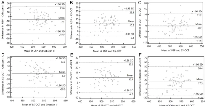
Figure 3. Bland-Altman plots of the difference in central corneal thickness (CCT, μm). Bland-Altman plots comparing measurement
of CCT (A) between ultrasound pachymetry (USP) and Orbscan II, (B) between USP and anterior segment optical coherence tomog- raphy (AS-OCT), (C) between USP and swept source optical coherence tomography (SS-OCT), (D) between SS-OCT and Orbscan II, (E) between SS-OCT and AS-OCT, (F) between Orbscan II and AS-OCT. The middle solid line represents the mean difference in CCT values and the upper and lower dashed lines represent the 95% limits of agreement. SD = standard deviation.두에서 통계적으로 강한 양의 상관관계를 보였다(Pearson correlation, r=0.986, r=0.973, r=0.910, r=0.974, r=0.907, r=0.894,
p
<0.001 in all groups) (Fig. 2).또한 4가지 검사기기 중 2가지 기기 사이의 각각의 차이 에 대해 알아보기 위해서 Bland-Altman plots를 만들어 일치 도 범위를 알아 보았다. 일치도 분석에서는 초음파 각막두 께측정계와 SS-OCT 간의 95% 일치도 범위가 22.8 μm (-11.6~11.2)로 가장 작았으며, Orbscan II와 AS-OCT 간의 95% 일치도 범위가 66.2 μm (-15.8~50.4)로 가장 컸다(Fig. 3).
고 찰
중심각막두께의 측정은 녹내장, 각막굴절교정 수술 등 안 과 질환의 진단과 치료에 필수적인 검사로, 정확한 측정의 필요성이 증대되고 있다. 초음파 각막두께측정계는 1950년 대부터 각막두께 측정에 사용되었으며, 사용방법이 간단하 고 검사 시간이 짧은 장점과 함께 높은 재현성과 신뢰도를 보여 현재까지 널리 이용되고 있다.10-12 그러나 초음파 각막 두께측정계는 초음파 소식자를 각막의 동일한 위치에 적절 한 힘을 가해 위치시켜야 하고 검사하는 동안 피검자가 주 시를 일정하게 유지하여야 하므로 검사자의 숙련도에 영향 을 받기 쉽다.13-15 또한 국소마취제 점안이 필요하고 직접
각막에 접촉해야 하므로 각막 상피의 손상, 감염의 전파의 위험이 있다.8
이러한 문제점을 보완하기 위한 비접촉 각막두께 측정법 들이 개발되어 사용되고 있는데, 가장 널리 사용되고 있는 Orbscan II는 Slit scanning 방식을 이용하여 전체 각막의 두 께뿐만 아니라 굴절률 및 각막 후면의 만곡을 측정 가능하 며, 정상안에서 각막두께 측정에 높은 신뢰도를 가지는 것 으로 알려져 있다.16-18 또 다른 비접촉 각막두께 측정법 중 한 가지인 AS-OCT는 적외선 파장을 이용한 기기로 후방 산란된 빛의 강도 차이를 통해 각막, 전방, 홍채 및 수정체 구조의 분석 및 측정이 가능하다.19 최근 개발된 비접촉 각 막두께 측정법들과 비교한 여러 연구에서 초음파 각막두께 측정계는 검사자 간, 검사자 내 더 높은 일치도를 보이고 결 과치에 변동이 적어 접촉성인 단점에도 불구하고 현재까지 각막두께 측정에 있어 표준 측정방법으로 간주되고 있 다.20-23
IOLMaster는 1990년대에 부분결합간섭계 원리를 이용하 여 개발된 장비로 760 nm 파장의 적외선과 슬릿광원을 이 용하여 각막곡률값, 수평각막직경, 전방깊이 등의 정보를 제공하였다. 최근 도입된 IOLMaster 700은 1,050 nm의 중심 파장과 약 100 nm의 band width를 가지는 파장가변 빛간섭 단층촬영 원리를 이용하여 안구 전체 구조의 계측이 가능
A B C
D E F
650 650
650
한 최초의 장비로, 이전의 기기에서는 제공하지 않았던 각 막두께의 측정이 가능하여 그 활용폭이 점차 확대되고 있 다.9,24 따라서 새로운 파장가변 빛간섭단층촬영 장비를 이 용한 각막두께 측정치의 정확성을 평가하고, 널리 사용되고 있는 다른 각막두께측정계와 측정치를 비교해 보는 것이 필수적이라 생각된다.
본 연구에서는 총 4가지 장비를 이용하여 정상안을 대상 으로 중심각막두께를 측정한 결과, SS-OCT를 이용한 측정 치만이 표준 측정방법으로 간주되고 있는 초음파 각막두께 측정계의 측정치와 통계적으로 유의한 차이가 없는 것으로 나타났다. AS-OCT와 Orbscan II로 측정한 중심각막두께는 초음파 각막두께측정계의 측정치와 유의한 상관관계가 있 었지만, 각각 측정치가 평균 10.22 ± 8.18 μm 두껍게, 6.82
± 15.92 μm 얇게 측정되었다.
각막두께측정계들은 측정방식에 따라 오차가 발생하게 되는데, 본 연구와 유사하게 Orbscan II를 이용한 중심각막 두께 측정은 초음파 각막두께측정계보다 두껍게 측정된다 는 보고들이 있다.11,25 Yaylali et al25과 Kang et al26은 정상 인에서 Orbscan II로 측정한 중심각막두께가 초음파 각막두 께측정계를 이용한 측정치보다 각각 28.0 μm, 24.0 μm 두껍 게 측정된다고 보고하였다. Orbscan II가 초음파 각막두께 측정계보다 더 두껍게 측정되는 이유는 각막표면의 수화된 점액층까지 측정하기 때문일 것으로 생각된다.27
AS-OCT는 이전의 연구들에서 초음파 각막두께측정계보 다 정도는 다양하나 더 얇게 측정됨이 보고되었다.28,29 Bechmann et al28과 Shim et al30은 정상인에서 AS-OCT로 측정한 중심 각막두께가 초음파 각막두께측정계를 이용한 측정치보다 각각 51 μm, 16.7 μm 얇게 측정되었음을 보고하였다. 이러 한 오차의 원인은 AS-OCT는 적외선이 조직 내에서 반사되 는 빛의 시간 차이를 이용하여 계측하는 원리를 이용하므 로 눈물층, 각막, 전방 등에서의 적외선 반사지수의 부정확 성이 각막두께 계산에 영향을 줄 수 있다는 보고가 있다.31 본 연구에서 이용한 SS-OCT인 IOLMaster 700은 정상인에 서 각막 굴절률, 전방깊이, 수정체두께, 안구길이 등의 측정 시 반복성 및 재현성이 뛰어남이 보고되어 있다.24 Fukuda et al32은 파장가변 전안부 빛간섭단층촬영계(SS-1000 CASIA, Tomey, Japan)와 초음파 각막두께측정계를 이용하여 측정한 중심각막두께를 비교하였을 때 높은 일치도를 보임을 보고 한 바 있으나, 같은 원리를 이용한 기기인 IOLMaster700으로 측정한 각막두께를 다른 장비와 비교한 보고는 아직 없다.
본 연구를 통해 정상인에서 중심각막두께를 측정할 때 SS-OCT로 측정한 중심각막두께가 초음파 각막두께측정계 와 통계적으로 유의한 일치율이 높은 값을 나타냄을 확인 하였다. SS-OCT는 기존의 빛간섭단층촬영계에 비해 검사
시간이 단축되었으며, 긴 파장을 이용하여 높은 해상도의 정확한 생체계측치를 얻을 수 있다는 장점이 있다. 또한 한 번의 검사로 전안부뿐만 아니라 안구 전체에 걸쳐 여러 가 지 생체계측 정보를 동시에 얻을 수 있어 잦은 검사가 불필 요하므로 환자의 불편을 줄일 수 있다.
SS-OCT는 같은 원리를 이용한 AS-OCT와 비교하였을 때 측정치에 유의한 차이가 있게 나타났는데, 이는 2가지 검사 기기 간 검사 소요시간 차이가 원인으로 생각된다. 이전의 연구에서도 검사 원리가 같은 2가지 기계에서 측정시간의 차이가 중심각막두께에 영향을 미침을 보고한 바 있다. 측 정 시간이 짧은 스펙트럼영역 빛간섭단층촬영계로 측정 시 시간영역 빛간섭단층촬영계로 측정 시보다 중심각막두께가 두껍게 나타났는데, 이는 검사 소요시간이 짧은 경우 눈물 층 두께에 미치는 영향이 적어 더 두껍게 측정될 수 있다고 하였다.33 또한 SS-OCT는 AS-OCT에 비하여 투과성이 더 높은 긴 파장을 이용하기 때문에 빛의 산란효과를 덜 받아 더 높은 해상도의 이미지를 얻을 수 있으므로 해상도에 따 라 측정치에 차이가 있을 것으로 생각된다.34
Orbscan II는 환자의 눈꺼풀 틈새가 작거나, 자주 깜박거 리는 경우, 건성안이 심한 경우, 각막부종이 있거나 각막의 앞뒤 표면이 균일하지 않은 경우 등에서 검사 신뢰도가 떨 어진다는 보고가 있으므로 이러한 경우 검사시간이 상대적 으로 짧고 눈물층 두께의 영향을 덜 받는 SS-OCT가 유용하 게 이용될 수 있을 것이다.18,29
결론적으로 SS-OCT는 비접촉 각막두께 측정기기들 중 기존에 사용되고 있는 초음파 각막두께측정계와 비교해 일 치도가 가장 높음을 확인하였다. SS-OCT는 각막접촉으로 인한 감염위험성이 없고, 한 번의 검사로 여러 가지 생체계 측정보를 동시에 얻을 수 있으며 안구 구조 전체를 영상으 로 파악할 수 있는 장점이 있어 널리 이용될 수 있으리라 생각된다.
REFERENCES
1) Ou RJ, Shaw EL, Glasgow BJ. Keratectasia after laser in situ kera- tomileusis (LASIK): evaluation of the calculated residual stromal bed thickness. Am J Ophthalmol 2002;134:771-3.
2) Wang Z, Chen J, Yang B. Posterior corneal surface topographic changes after laser in situ keratomileusis are related to residual cor- neal bed thickness. Ophthalmology 1999;106:406-9; discussion 409-10.
3) Grieshaber MC, Schoetzau A, Zawinka C, et al. Effect of central corneal thickness on dynamic contour tonometry and Goldmann applanation tonometry in primary open-angle glaucoma. Arch Ophthalmol 2007;125:740-4.
4) Jonas JB, Stroux A, Velten I, et al. Central corneal thickness corre- lated with glaucoma damage and rate of progression. Invest
Ophthalmol Vis Sci 2005;46:1269-74.
5) Kim DH, Kim MS, Kim JH. Early corneal-thickness changes after penetrating keratoplasty. J Korean Ophthalmol Soc 1997;38:1355-61.
6) Solomon OD. Corneal indentation during ultrasonic pachometry.
Cornea 1999;18:214-5.
7) Choi KS, Nam SM, Lee HK, et al. Comparison of central corneal thickness after the instillation of topical anesthetics: proparacaine versus oxybuprocaine. J Korean Ophthalmol Soc 2005;46:757-62.
8) Li EY, Mohamed S, Leung CK, et al. Agreement among 3 methods to measure corneal thickness: ultrasound pachymetry, Orbscan II, and Visante anterior segment optical coherence tomography.
Ophthalmology 2007;114:1842-7.
9) Akman A, Asena L, Güngör SG. Evaluation and comparison of the new swept source OCT-based IOLMaster 700 with the IOLMaster 500. Br J Ophthalmol 2016;100:1201-5.
10) Baum G, Greenwood I. The application of ultrasonic locating tech- niques to ophthalmology. II. Ultrasonic slit lamp in the ultrasonic visualization of soft tissues. AMA Arch Ophthalmol 1958;60:263-79.
11) Marsich MW, Bullimore MA. The repeatability of corneal thick- ness measures. Cornea 2000;19:792-5.
12) Miglior S, Albe E, Guareschi M, et al. Intraobserver and interob- server reproducibility in the evaluation of ultrasonic pachymetry measurements of central corneal thickness. Br J Ophthalmol 2004;88:174-7.
13) Ling T, Ho A, Holden BA. Method of evaluating ultrasonic pachometers. Am J Optom Physiol Opt 1986;63:462-6.
14) Copt RP, Thomas R, Mermoud A. Corneal thickness in ocular hy- pertension, primary open-angle glaucoma, and normal tension glaucoma. Arch Ophthalmol 1999;117:14-6.
15) Al-Farhan HM, Al-Otaibi WM. Comparison of central corneal thickness measurements using ultrasound pachymetry, ultrasound biomicroscopy, and the Artemis-2 VHF scanner in normal eyes.
Clin Ophthalmol 2012;6:1037-43.
16) Khaja WA, Grover S, Kelmenson AT, et al. Comparison of central corneal thickness: ultrasound pachymetry versus slit-lamp optical coherence tomography, specular microscopy, and Orbscan. Clin Ophthalmol 2015;9:1065-70.
17) Jonuscheit S, Doughty MJ. Regional repeatability measures of corneal thickness: Orbscan II and ultrasound. Optom Vis Sci 2007;84:52-8.
18) Choi SH, Kim JH, Han NS, et al. Comparison of corneal thickness measurements with optical low coherence reflectometry, orbscan system and ultrasound pachymeter. J Korean Ophthalmol Soc 2006;47:19-24.
19) Thomas J, Wang J, Rollins AM, Sturm J. Comparison of corneal thickness measured with optical coherence tomography, ultra- sonic pachymetry, and a scanning slit method. J Refract Surg 2006;22:671-8.
20) Bao F, Wang Q, Cheng S, et al. Comparison and evaluation of cen- tral corneal thickness using 2 new noncontact specular microscopes and conventional pachymetry devices. Cornea 2014;33:576-81.
21) Nam SM, Im CY, Lee HK, et al. Accuracy of RTVue optical co- herence tomography, Pentacam, and ultrasonic pachymetry for the measurement of central corneal thickness. Ophthalmology 2010;117:2096-103.
22) Uçakhan OO, Ozkan M, Kanpolat A. Corneal thickness measure- ments in normal and keratoconic eyes: Pentacam comprehensive eye scanner versus noncontact specular microscopy and ultrasound pachymetry. J Cataract Refract Surg 2006;32:970-7.
23) Christensen A, Nárvaez J, Zimmerman G. Comparison of central corneal thickness measurements by ultrasound pachymetry, konan noncontact optical pachymetry, and orbscan pachymetry. Cornea 2008;27:862-5.
24) Kunert KS, Peter M, Blum M, et al. Repeatability and agreement in optical biometry of a new swept-source optical coherence to- mography-based biometer versus partial coherence interferometry and optical low-coherence reflectometry. J Cataract Refract Surg 2016;42:76-83.
25) Yaylali V, Kaufman SC, Thompson HW. Corneal thickness meas- urements with the Orbscan Topography System and ultrasonic pachymetry. J Cataract Refract Surg 1997;23:1345-50.
26) Kang PS, Yang YS, Kim JD. Comparison of corneal thickness measurements with the orbscan and ultrasonic pachymetry. J Korean Ophthalmol Soc 2000;41:1697-703.
27) Liu Z, Huang AJ, Pflugfelder SC. Evaluation of corneal thickness and topography in normal eyes using the Orbscan corneal top- ography system. Br J Ophthalmol 1999;83:774-8.
28) Bechmann M, Thiel MJ, Neubauer AS, et al. Central corneal thick- ness measurement with a retinal optical coherence tomography de- vice versus standard ultrasonic pachymetry. Cornea 2001;20:50-4.
29) Wong AC, Wong CC, Yuen NS, Hui SP. Correlational study of cen- tral corneal thickness measurements on Hong Kong Chinese using optical coherence tomography, Orbscan and ultrasound pachy- metry. Eye (Lond) 2002;16:715-21.
30) Shim HS, Choi CY, Lee HG, et al. Utility of the anterior segment optical coherence tomography for measurements of central corneal thickness. J Korean Ophthalmol Soc 2007;48:1643-8.
31) Zhao PS, Wong TY, Wong WL, et al. Comparison of central cor- neal thickness measurements by visante anterior segment opti- cal coherence tomography with ultrasound pachymetry. Am J Ophthalmol 2007;143:1047-9.
32) Fukuda R, Usui T, Miyai T, et al. Corneal thickness and volume measurements by swept source anterior segment optical coherence tomography in normal subjects. Curr Eye Res 2013;38:531-6.
33) Prakash G, Agarwal A, Jacob S, et al. Comparison of four- ier-domain and time-domain optical coherence tomography for as- sessment of corneal thickness and intersession repeatability. Am J Ophthalmol 2009;148:282-90.e2.
34) Unterhuber A, Povazay B, Hermann B, et al. In vivo retinal optical coherence tomography at 1040 nm-enhanced penetration into the choroid. Opt Express 2005;13:3252-8.
= 국문초록 =
중심각막두께 측정에 대한 파장가변 빛간섭단층촬영계의 유용성
목적: 파장가변 빛간섭단층촬영계(swept source optical coherence tomography, SS-OCT)로 측정한 중심각막두께를 초음파 각막두 께측정계(ultrasound pachymetry), Orbscan II, 전안부 빛간섭단층촬영계(anterior segment optical coherence tomography, AS-OCT) 로 측정한 중심각막두께와 비교하여 SS-OCT의 임상적 일치율의 유용성을 알아보고자 하였다.
대상과 방법: 한 명의 검사자가 65명 65안의 정상안을 대상으로 Orbscan II, AS-OCT, SS-OCT, 초음파 각막두께측정계의 순서로 중심 각막두께를 측정하여 상관관계를 비교, 분석하였다.
결과: Orbscan II, AS-OCT, SS-OCT와 초음파 각막두께측정계를 이용하여 측정한 각각의 평균 중심각막두께는 각각 534.83 ± 38.46, 517.80 ± 32.48, 528.22 ± 33.71, 528.02 ± 34.90 μm였다. 4가지 검사기기에서 높은 상관관계를 보였으며(r>0.894, p<0.001 in all groups), SS-OCT와 초음파 각막두께측정계를 이용한 측정치에서 통계적으로 유의한 일치율을 보였다(p=0.782).
결론: 정상인에서 SS-OCT로 측정한 중심각막두께가 초음파 각막두께측정계로 측정한 값에 높은 일치율을 보여, 정상인의 중심각막 두께 측정에 SS-OCT가 초음파 각막두께측정계를 대체할 수 있는 유용한 방법이라 생각된다.
<대한안과학회지 2016;57(10):1542-1548>