pISSN: 0378-6471 eISSN: 2092-9374 http://dx.doi.org/10.3341/jkos.2012.53.5.707
= 증례보고 =
눈꺼풀처짐을 동반한 위눈꺼풀올림근에 국한된 외안근염 1예
윤제환1⋅문현승2⋅지미정1
가천대학교 길병원 안과학교실1, 서울 밝은세상안과2
목적: 위눈꺼풀올림근을 침범한 외안근염을 경험하였고 이는 드문 것이기에 보고하고자 한다.
증례요약: 27세 남자 환자가 1주일 전부터 발생한 좌안 위눈꺼풀의 발적, 부종 및 압통으로 내원하였다. 환자는 안와 전산화단층촬영상 상직근-위눈꺼풀올림근 복합체의 비후소견이 관찰되었으나 모든 방향에서 안구운동은 정상이었고 좌안 위눈꺼풀처짐만이 관찰되었 다. 환자는 내원일 이후 3세대 세팔로스포린 주사제를 사용하여 3일간 치료하였으나 증상의 호전이 없었다. 이후 특발성 외안근염으로 진단하고 부신 피질 호르몬을 1개월간 경구 투약하였고 위눈꺼풀의 발적, 부종 및 위눈꺼풀처짐은 호전되었다.
결론: 특발성외안근염은 외안근을 침범하는 비특이적인 안와 염증으로 원인이 확실히 규명되어 있지 않지만 면역이상과 관련되어 발생 될 수 있다고 알려졌다. 외안근염의 침범부위는 내직근이 가장 흔하며 위눈꺼풀올림근에만 국한되어 발생하는 경우는 매우 드물다.
저자들은 위눈꺼풀올림근에만 발생한 특발성 외안근염을 경험하였기에 보고하며, 눈꺼풀처짐을 동반한 눈꺼풀부종의 감별진단 시 염 두에 두어야 할 것으로 생각한다.
<대한안과학회지 2012;53(5):707-711>
■ 접 수 일: 2011년 8월 12일 ■ 심사통과일: 2011년 11월 15일
■ 게재허가일: 2012년 4월 7일
■ 책 임 저 자: 지 미 정
인천광역시 남동구 남동대로 774번길 21 가천대길병원 안과
Tel: 032-460-3751, Fax: 032-460-3358 E-mail: [email protected]
* 이 논문의 요지는 2011년 대한안과학회 제105회 학술대회에서 포스터로 발표되었음.
특발성안와염은 판별할 수 있는 국소적 혹은 전신적 원 인이 없는 환자에서 나타나는 염증성 변화로 임상적, 영상 의학적, 필요한 경우 조직학적 검사를 통해 배제진단이 필 요한 질환이다.1 특발성안와염은 전체 안와 질환의 약 4.7%에서 6.3%를 차지하며, 1900년대 초 안구돌출이 있 었으나 신생물의 흔적이 없고 저절로 호전된 환자 30명을 분석하여 안와위종양으로 처음 명명하며 분류가 이루어졌 다.2-5 이러한 특발성안와염은 발생위치에 따라서 눈물샘 염, 외안근염, 앞안와염, 뒤안와염, 미만성안와염으로 분류 할 수 있으며 이 중 외안근염은 염증성 변화가 1개 이상의 외안근을 침범한 경우 지칭한다.6특발성외안근염의 침범부 위는 내직근이 가장 흔하며 그 외의 근육에도 다양한 빈도 로 발생하고 상직근-위눈꺼풀올림근 복합체에 발생하는 경우 복시와 눈꺼풀처짐이 함께 발생할 수 있다.7,8저자들 은 특발성외안근염이 위눈꺼풀올림근에만 발생하여 복시 없이 눈꺼풀처짐만 있었던 환자 1예를 경험하였기에 문헌
고찰과 함께 보고하는 바이다.
증례보고
27세 남자 환자가 1주일 전부터 발생하여 점차 심화된 좌안 위눈꺼풀의 발적, 부종 및 압통을 주소로 내원하였다.
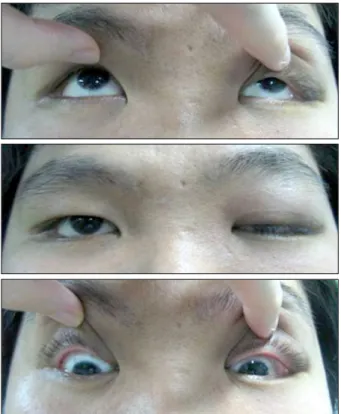
과거력이나 가족력상 특이 사항은 없었고 안과적 검사상 나안 시력은 양안 1.0이었으며 안구운동검사는 정상이었고 원거리 및 근거리 사시각은 교대가림법으로 측정시 정위였 다. 눈꺼풀각막반사간거리(MRD1)는 우안 3.0 mm, 좌안 0.5 mm로 좌안 눈꺼풀처짐이 있었으며 눈꺼풀올림근 기능 은 우안 15 mm, 좌안 10 mm로 좌안에서 감소되어 있었다 (Fig. 1). 안구돌출은 없었으며 전안부 검사에서 좌안 결막 충혈이 관찰되었다. 발열이나 근육통 등의 전신증상은 없었 으며 전혈구 수, 전해질, 갑상샘기능을 포함한 혈액 검사는 정상이었다. 안와 전산화단층촬영상 상직근-위눈꺼풀올림 근 복합체의 비후소견이 관찰되었으며 부비동의 염증은 없 었다(Fig. 2). 안와봉와직염 의심하에 내원일부터 3세대 세 팔로스포린(Ceftriaxone)을 1 g 하루 2회씩 3일간 주사하 였으나 증상의 호전은 없었다. 특발성외안근염으로 배제진 단하고 스테로이드(Methylprednisolon) 경구제제를 48 mg 하루 1회로 1주일간 복용하였으며 12 mg씩 1주일 간 격으로 점차 감량하였다. 치료 1개월 후 눈꺼풀의 발적, 부 종 및 압통은 호전되었으며 눈꺼풀각막반사간거리 2.0
Figure 3. One month later, the patient shows improvement of
left upper blepharoptosis.Figure 2. Coronal and sagittal computed tomographic scans show enlargement of the left superior rectus/levator muscle complex.
Figure 1. At first day, the patient shows left upper eyelid pto-
sis and normal movement of eyes.mm,눈꺼풀올림근기능 15 mm로 호전되었다(Fig. 3). 내 원 2개월째 환자는 눈꺼풀의 발적 및 부종은 없으며 눈꺼풀 처짐도 소실되었고, 1년 후까지 재발소견은 없었다.
고 찰
특발성외안근염은 특발성안와염의 일종으로 특징적으로 외안근에 염증을 보이며 증상으로 눈꺼풀부종, 안구돌출,
안통, 복시 및 눈꺼풀처짐 등을 보일 수 있고 영상검사상 흔히 힘줄까지 침범된 외안근의 비후를 관찰할 수 있다. 18 세에서 40세에 많이 발생하나 3세에서 84세까지 발생한 경 우가 보고되었다.9-12 특발성외안근염의 원인으로 Atabay et al13은 외안근염 환자 31명의 혈액검사를 통해 외안근에 자가항체로 작용하는 인자가 존재하였음을 보고하여 특발 성외안근염의 원인이 자가면역에 의한 손상에 있음을 설명 하였다. 그러나 이 자가항체들이 외안근에 특별하게 작용하
는지 명확하지 않고 대부분의 특발성외안근염이 단안에 많 이 발생하는 이유를 설명하지 못하는 등의 맹점이 있다.
특발성외안근염 환자에서 외안근의 침범 빈도에 관해 Stevenet al7은 내직근, 외직근, 상직근, 하직근 순이라 하 였으며 Trokel et al14은 내직근(43%), 상직근(19%), 외직 근(17%), 사근들(5-10%)순이었고 국내의 Lee et al15은 외안근염 환자 14명을 분석하였을 때 상직근(64.3%), 내 직근(64.3%), 외직근(42.9%), 하직근(42.9%)의 순으로 발생하였음을 보고하였다. 이렇게 침범된 근육들로 인해 안 구운동장애와 눈움직임시의 통증이 특발성 외안근염 환자 의 주요한 증상으로 나타나며 이러한 증상은 시기에 따라 진행하게 된다.16 Siatkowski et al17에 따르면 외안근염의 발생 초기 10일간은 외안근이 특별한 기능이상을 보이지 않으나 이후 11-14일 사이에는 마비증상이 나타나고 그 이후에는 제한성 운동장애가 나타난다고 하였다. 특히 특발 성외안근염이 상직근에 침범하는 경우 상직근의 움직임에 영향을 주어 상하전 장애를 일으킬 뿐만 아니라 상직근과 외안근막으로 구분되어 연접해있는 위눈꺼풀올림근에도 영 향을 주어 안구운동장애와 눈꺼풀처짐이 동반되어 나타날 수 있다.18저자들이 경험한 환자의 경우 안와 전산화 단층 촬영검사상 상직근-위눈꺼풀올림근 복합체의 비후가 관찰 되었으며 상직근의 기능저하 없이 위눈꺼풀올림근의 기능 저하만이 나타났다. 상직근-위눈꺼풀올림근 복합체 모두 에 외안근염이 발생하였다면 두 근육이 유사한 기능저하의 정도를 보일 것이며, 상직근에만 발생하였다면 상직근의 기 능장애가 먼저 나타나야 하나 환자는 위눈꺼풀올림근의 기 능장애만이 관찰되어 상직근이 아닌 위눈꺼풀올림근만의 이상을 추측할 수 있었다.19-21위눈꺼풀올림근을 침범하여 눈꺼풀처짐을 일으킨 환자에 대한 보고는 매우 드물게 있 었으며 침범부위의 정확한 판단을 위하여 자기공명영상 촬 영을 사용할 수 있다.22
이러한 특발성외안근염은 외안근이 비대되는 갑상샘눈 병증, 안와봉와직염, 안와 신생물, 동정맥 기형 등과 감별이 필요하며 급성 안와봉와직염의 경우 대개 통증, 발열, 백혈 구 증가 등이 동반되고 항생제 치료로 증상이 호전되는 것 이 특징이다.23,24저자들이 경험한 환자의 경우 통증, 발열, 백혈구 증가 등은 없었으나 우선 안와봉와직염으로 의심하 고 3일간의 3세대 세팔로스포린을 사용하였으며 그럼에도 불구하고 증상의 호전이 없었다. 그 외에 감별해야 할 질환 으로 안와 신생물은 전형적으로 만성적으로 나타나고 동정 맥 기형과 더불어 안통이 대부분 없다는 점에서 특발성외 안근염과 차이가 있으나 정확한 감별을 위해서는 조직검사 가 필요하다. 저자들이 경험한 환자의 경우 발병기간이 1주 일 이내로 급성으로 나타났으며 스테로이드에 신속히 반응
하여 증상이 소실되고 1년 후까지 재발소견 없이 유지되어 림프종 등의 안와 신생물 감별을 위한 조직검사는 시행하 지 않았다. 해면정맥동 혈전은 자주 양측에서 발생되며 발 열, 오심 구토가 동반되고 의식의 변화가 있을 수 있다.25 갑상샘눈병증은 특발성외안근염과 가장 흔하게 혼동되는 질환으로 특발성외안근염보다 더 증상이 천천히 나타나고 통증이 약하며 스테로이드에 대한 반응이 느리다. 또한 눈 꺼풀후퇴가 갑상샘눈병증에서 흔히 나타나는 증상이며 특 발성외안근염에서는 눈꺼풀처짐이 보다 흔하다.26 이외에 도갑상샘눈병증에서는 갑상선자가항체의 증가가 나타난다 는 점, 외안근 중 하직근에 주로 발생하는 점, 외안근의 힘줄 침범은 드문 점 등이 외안근염과의 감별점이라 할 수 있다.9
특발성외안근염은 보통 발생 후 자연 호전되는 경우가 많지만 안통과 복시를 감소시키고 근육의 섬유화 및 변성 을 막기 위해 조기 치료가 추천되고 있다.27,28특발성외안근 염은 2일에서 5일간의 고용량 스테로이드의 전신적인 투여 나 약 2주간의 고용량 스테로이드의 경구 투여 후 극적인 반응을 보이며 이후 수주에서 수개월간 감량하게 된다.10,29 더 좋은 결과를 얻기 위해 스테로이드제제의 투여 이전에 비스테로이드성 항염증제의 투여가 고려되기도 한다.11 면 역억제제는 스테로이드에 반응이 부족한 환자나 스테로이드 의 부작용이 심한 환자의 경우 사용하게 되며 사용효과와 이 에 따른 부작용의 비를 조율하기 어렵다.1,30사용가능한 약제 로는 항대사제, T림프구 억제제, 알킬화 약물 등이 있으나 이들은 일반적으로 스테로이드의 부족함을 보충해주는 약제 로 사용되며 스테로이드를 대체하는 약물로 사용되지는 못 한다. Immunobiologic 제제인 alefacept, enternacept 등도 사용될 수 있으며 최근 각광받고 있는 Infliximab, Efalizumab 등의 단클론항체 약제 또한 사용을 고려해 볼 수 있다.31
방사선치료는 스테로이드제재를 장기간 사용했음에도 반응이 없는 경우나 스테로이드의 부작용으로 인해 치료를 지속하기 어려운 경우, 재발이 잘 되는 경우에 적응증이 되 며 사용되는 방사선 조사량은 2주 동안 약 10번에 걸쳐 총 1000-3000 cGy를 조사한다. 방사선을 조사하는 경우 백 내장, 안구건조증, 방사선 망막증 등이 발생할 수 있으므로 유의해야 하며 방사선 조사의 치료효과는 일반적으로 약 50-70%로 알려졌다.32,33
저자들은 안와전산화단층촬영상 상직근-위눈꺼풀올림 근 복합체의 비후가 관찰되는 환자에서 특징적으로 눈꺼풀 처짐은 있었으나 안구운동장애는 없는 점으로 미루어 위눈 꺼풀올림근을 침범한 외안근염으로 판단하였으며 조기에 고용량의 스테로이드를 경구투여하여 증상의 호전을 보였 다. 따라서 안구운동장애 없이 눈꺼풀처짐만을 보이는 환자 에서도 특발성외안근염의 가능성을 생각하여야 하며 또한
기타 외안근염의 환자에서와 마찬가지로 경구 고용량 스테 로이드요법이 조기치료로서 유용할 것으로 생각한다.
참고문헌
1) Yuen SJ, Rubin PA. Idiopathic orbital inflammation: distribution, clinical features, and treatment outcome. Arch Ophthalmol 2003;121:491-9.
2) Kennerdell JS, Dresner SC. The nonspecific orbital inflammatory syndromes. Surv Ophthalmol 1984;29:93-103.
3) Garrity JA, Henderson JW, Cameron JD. Henderson's Orbital Tumors, 4th ed. Philadelphia: Williams & Wilkins, 2007;343-60.
4) Rootman J, Nugent R. The classification and management of acute orbital pseudotumors. Ophthalmology 1982;89:1040-8.
5) Birch-Hirschfeld A. Zur diagnostic and pathologic der orbital tumoren. Ber Zusammenkunft Dtsch Ophthalmol Ges 1905;32:
127-35.
6) Lutt JR, Lim LL, Phal PM, Rosenbaum JT. Orbital inflammatory disease. Semin Arthritis Rheum 2008;37:207-22.
7) Dresner SC, Rothfus WE, Slamovits TL, et al. Computed tomog- raphy of orbital myositis. AJR Am J Roentgenol 1984;143:671-4.
8) Sekhar GC, Mandal AK, Vyas P. Non specific orbital inflammatory diseases. Doc Ophthalmol 1993;84:155-70.
9) Scott IU, Siatkowski RM. Idiopathic orbital myositis. Curr Opin Rheumatol 1997;9:504-12.
10) Mombaerts I, Koornneef L. Current status in the treatment of orbi- tal myositis. Ophthalmology 1997;104:402-8.
11) Mannor GE, Rose GE, Moseley IF, Wright JE. Outcome of orbital myositis, clinical features associated with recurrence. Ophthalmology 1997;104:409-13.
12) Slavin ML, Glaser JS. Idiopathic orbital myositis: a report of six cases. Arch Ophthalmol 1982;100:1261-5.
13) Atabay C, Tyutyunikov A, Scalise D, et al. Serum antibodies re- active with eye muscle membrane antigens are detected in patients with nonspecific orbital inflammation. Ophthalmology 1995;102:
145-53.
14) Trokel SL, Hilal SK. Recognition and differential diagnosis of en- larged extraocular muscles in computed tomography. Am J Ophthalmol 1979;87:503-12.
15) Lee JS, Oum BS, Choi CH, Kim HJ. Clinical study of the idiopathic orbital myositis. J Korean Ophthalmol Soc 1999;40:1109-15.
16) Volle E, Levy R, Miléa D, et al. [Idiopathic orbital myositis].
[Article in French] Rev Neurol 2001;157:430-2.
17) Siatkowski RM, Capó H, Byrne SF, et al. Clinical and echographic findings in idiopathic orbital myositis. Am J Ophthalmol 1994;15:343-50.
18) Jomura Y, Jokura K, Kuroiwa Y. Orbital myositis with ptosis.
Neurological Medicine 2001;55:84-6.
19) Lee SK, Song JG. Effect of corticosteroid on orbital pseudotumor caused by orbital myositis. J Korean Ophthalmol Soc 1991;32:482-8.
20) Wheatcroft S, Elston J. Unilateral ptosis due to isolated involve- ment of the levator muscle in acute orbital myositis. Br J Ophthalmol 1999;83:631-2.
21) Nikola S, Jasenka B, Ivana KS. Rectus superior and levator palpe- brae superioris (RS+LPS) muscle complex myositis. Acta Clin Croat 2007;46:317-20.
22) Almekhlafi MA, Fletcher WA. Levator palpebrae myositis.
Neurology 2008;7:1202.
23) Serop S, Vianna RN, Claeys M, De Laey JJ. Orbital myositis sec- ondary to systemic lupus erythematosus. Acta Ophthalmol (Copenh) 1994;72:520-3.
24) Barnes C. Orbital myositis--a case report. Aust N Z J Ophthalmol 1990;18:251-5.
25) Kim CH, Lim JY, Ahn JH, Jang JW. A case of idiopathic orbital myositis involving all extraocular muscles of both eyes. J Korean Ophthalmol Soc 2001;42:1615-20.
26) Bessant DA, Lee JP. Management of strabismus due to orbital myositis. Eye (Lond) 1995;9:558-63.
27) Mombaerts I, Koornneef L. Current status in the treatment of orbi- tal myositis. Ophthalmology 1997;104:402-8.
28) Hankey GJ, Silbert PL, Edis RH, Nicoll AM. Orbital myositis: a study of six cases. Aust N Z J Med 1987;17:585-91.
29) Moorman CM, Elston JS. Acute obital myositis. Eye (Lond) 1995;9:96-101.
30) Jacobs D, Galetta S. Diagnosis and management of orbital pseudotumor. Curr Opin Ophthalmol 2002;13:347-51.
31) Harris GJ. Idiopathic orbital inflammation: a pathogenetic con- struct and treatment strategy. Ophthalmic Plast and Reconstr Surg 2006;22:79-86.
32) Leone CR Jr, Lloyd WC 3rd. Treatment protocol for orbital in- flammatory disease. Ophthalmology 1985;92:1325-31.
33) Fraunfelder FT, Roy FH. Current Ocular Therapy, 4th ed.
Philadelphia: W.B Saunders Company, 1995;745-8.
=ABSTRACT=
Unilateral Ptosis Due to Isolated Levator Myositis
Je Hwan Yoon, MD1, Hyun Seung Moon, MD2, Mijung Chi, MD, PhD1
Department of Ophthalmology, Gachon University Gil Hospital1, Incheon, Korea Bright World Eye Center2, Seoul, Korea
Purpose: To present a rare case of idiopathic orbital myositis involving levator palpebrae superioris.
Case summary: A 27-year-old male presented with a 1-week history of redness, discomfort, swelling, and drooping of his left upper eyelid. A computed tomography scan showed isolated enlargement of the right superior rectus/levator muscle complex. On examination, there was a left blepharoptosis, although eye movements were normal. The authors treated the patient with 3rd-generation cephalosporin; however, after 3 days, the symptoms did not improve. Subsequently, the pa- tient was diagnosed with idiopathic orbital myositis and treated with oral corticosteroids for 1 month; the symptoms gradu- ally resolved.
Conclusions: Idiopathic orbital myositis is a subtype of nonspecific orbital inflammation primarily involving the extraocular muscles. Although the exact cause of orbital myositis is unknown, an immune-mediated pathophysiologic mechanism ap- pears to be one of the causes. Medial rectus myositis is the most common, and isolated levator muscle myositis is very rare. The authors of the present study reported a case of orbital myositis involving the levator palpebrae superioris which should be considered a differential diagnosis of blepharoptosis with eyelid swelling.
J Korean Ophthalmol Soc 2012;53(5):707-711
Key Words: Corticosteroid, Levator muscle, Orbital myositis
Address reprint requests to Mijung Chi, MD, PhD
Department of Ophthalmology, Gachon University Gil Hospital
#21 Namdong-daero 774beon-gil, Namdong-gu, Incheon 405-760, Korea Tel: 82-32-460-3751, Fax: 82-32-460-3358, E-mail: [email protected]